Predictors of the Response to an Epidural Blood Patch in Patients with Spinal Leakage of Cerebrospinal Fluid
- PMID: 31942752
- PMCID: PMC6974819
- DOI: 10.3988/jcn.2020.16.1.1
Predictors of the Response to an Epidural Blood Patch in Patients with Spinal Leakage of Cerebrospinal Fluid
Abstract
Background and purpose: An epidural blood patch (EBP) is a highly effective therapy for spinal cerebrospinal fluid (CSF) leakage. However, the factors predicting the response to an EBP have not been fully elucidated. The aim of this study was to elucidate factors predicting the response to an EBP.
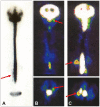
Methods: We retrospectively examined the relationship between the response to an EBP and clinical variables of 118 patients with spinal CSF leakage, such as patient age, sex, etiology, interval from the onset to EBP application, CSF opening pressure (OP), radioisotope (RI) cisternography findings, rate of RI remaining in the CSF space, computed tomography (CT) myelography findings, magnetic resonance imaging (MRI) findings, and subjective symptoms (headache, vertigo/dizziness, visual disturbance, nausea, numbness, nuchal pain, back pain/lumbago, fatigability, photophobia, and memory disturbance). The correlations between these variables and the responses to EBPs were analyzed statistically.
Results: A positive response to an EBP was significantly (p<0.05) correlated with the following variables: <1.5 years from the onset to EBP application, age <40 years, CSF OP <7 cm H₂O, epidural CSF leakage in RI cisternography, epidural CSF collection in MRI, <20% RI remaining after 24 hours, orthostatic headache, nausea, nuchal pain, and photophobia. The other variables did not show significant correlations with the responses to EBPs.
Conclusions: It might be prudent to take the following variables into account when applying an EBP to treat spinal CSF leakage: the interval from the onset to EBP application, age, CSF OP, epidural CSF leakage in RI, epidural CSF collection in MRI, rate of remaining RI, orthostatic headache, nuchal pain, photophobia, and nausea.
Keywords: cerebrospinal fluid volume depletion; epidural blood patch; predictor; spinal cerebrospinal fluid leakage.
Copyright © 2020 Korean Neurological Association.
Conflict of interest statement
The authors have no potential conflicts of interest to disclose.
Figures


References
-
- Schievink WI. Spontaneous spinal cerebrospinal fluid leaks and intracranial hypotension. JAMA. 2006;295:2286–2296. - PubMed
-
- Mokri B. Spontaneous low pressure, low CSF volume headaches: spontaneous CSF leaks. Headache. 2013;53:1034–1053. - PubMed
-
- Mokri B, Atkinson JL, Piepgras DG. Absent headache despite CSF volume depletion (intracranial hypotension) Neurology. 2000;55:1722–1724. - PubMed
-
- Mokri B, Schievink WI. Headache associated with abnormalities in intracranial structure and function: low cerebrospinal fluid pressure headache. In: Silberstein SD, Lipton RB, Dodick DW, editors. Wolff's Headache and Other Head Pain. New York: Oxford University Press; 2000. pp. 513–531.
Grants and funding
LinkOut - more resources
Full Text Sources
Miscellaneous

