. 2020 Mar 13;29(4):635-648.
doi: 10.1093/hmg/ddz318.

A VPS13D spastic ataxia mutation disrupts the conserved adaptor-binding site in yeast Vps13

Affiliations

A VPS13D spastic ataxia mutation disrupts the conserved adaptor-binding site in yeast Vps13

Samantha K Dziurdzik et al. Hum Mol Genet. .

Abstract

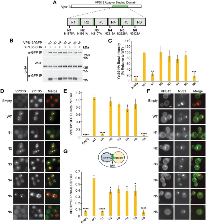
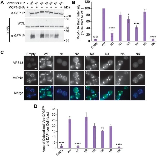
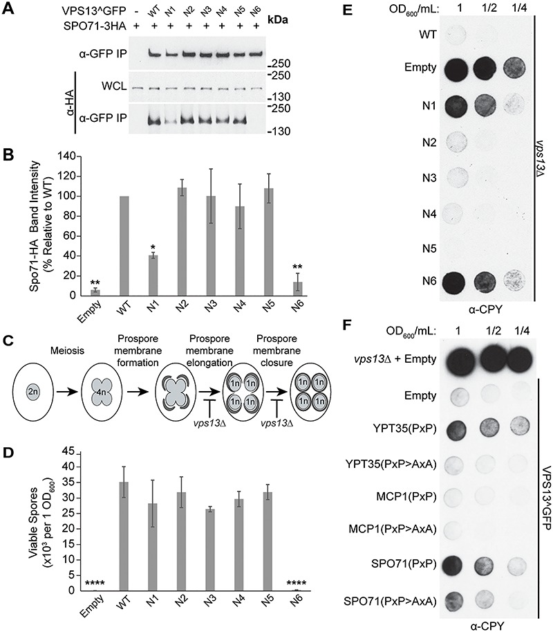
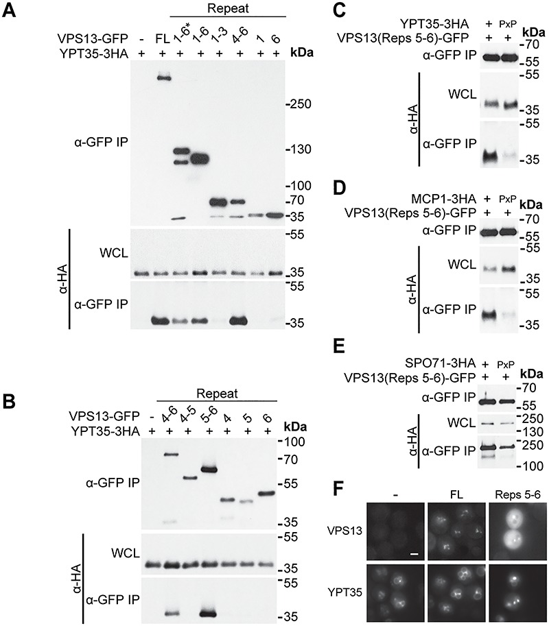
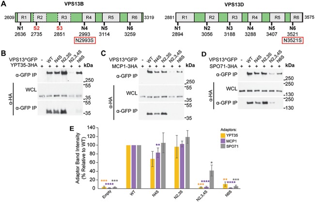
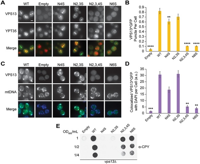
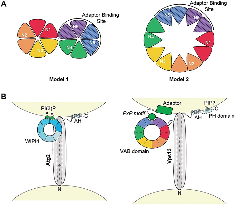
Mutations in each of the four human VPS13 (VPS13A-D) proteins are associated with distinct neurological disorders: chorea-acanthocytosis, Cohen syndrome, early-onset Parkinson's disease and spastic ataxia. Recent evidence suggests that the different VPS13 paralogs transport lipids between organelles at different membrane contact sites. How each VPS13 isoform is targeted to organelles is not known. We have shown that the localization of yeast Vps13 protein to membranes requires a conserved six-repeat region, the Vps13 Adaptor Binding (VAB) domain, which binds to organelle-specific adaptors. Here, we use a systematic mutagenesis strategy to determine the role of each repeat in recognizing each known adaptor. Our results show that mutation of invariant asparagines in repeats 1 and 6 strongly impacts the binding of all adaptors and blocks Vps13 membrane recruitment. However, we find that repeats 5-6 are sufficient for localization and interaction with adaptors. This supports a model where a single adaptor-binding site is found in the last two repeats of the VAB domain, while VAB domain repeat 1 may influence domain conformation. Importantly, a disease-causing mutation in VPS13D, which maps to the highly conserved asparagine residue in repeat 6, blocks adaptor binding and Vps13 membrane recruitment when modeled in yeast. Our findings are consistent with a conserved adaptor binding role for the VAB domain and suggest the presence of as-yet-unidentified adaptors in both yeast and humans.


Figures

Figure 1
Figure 1
Mutations in the Vps13 VAB domain disrupt its interaction with the endosomal adaptor Ypt35. (A) Schematic of Vps13 VAB domain indicating the position of the mutated conserved asparagines in repeats 1–6 (R1–R6). (B) Co-IP of WT or mutant Vps13^GFP with Ypt35-3HA overexpressed from the ADH1 promoter. Proteins were expressed on plasmids in vps13Δ strains. WCL, whole-cell lysate. (C) Densitometry analysis of Ypt35-HA co-IP bands shown in (B); one-way ANOVA with Dunnett-corrected post hoc test; n = 4; ***P < 0.001; **P < 0.01. (D) Localization of WT or mutant Vps13^GFP to bright puncta colocalizing with ADH1pr-driven Ypt35-RFP. Proteins were expressed on plasmids in ypt35Δ strains. (E) Automated quantitation of Vps13^GFP puncta from (D). One-way ANOVA with Dunnett-corrected post hoc test; n = 3; cells/strain/replicate ≥1370; ****P < 0.0001; *P < 0.05. (F) Localization of WT or mutant Vps13^GFP to NVJ1-RFP-labeled NVJs in acetate-based media. Proteins were expressed on plasmids in vps13Δ strains. (G) Automated quantitation of the number of Vps13^GFP-labeled NVJs, from (F). The diagram shows the location of the NVJ. One-way ANOVA with Dunnett-corrected post hoc test; n = 3, cells/strain/replicate ≥495; ****P < 0.0001; *P < 0.05. Error bars indicate SEM. Bars, 2 μm.
Figure 2
Figure 2
VAB domain mutations disrupt Vps13 Mcp1-mediated mitochondrial localization. (A) Co-IP of ADH1pr-driven Mcp1-3HA with WT or mutant Vps13^GFP. WCL, whole-cell lysate. (B) Densitometry analysis of Mcp1-HA co-IP bands shown in (A); one-way ANOVA with Dunnett-corrected post hoc test; n = 4; ****P < 0.0001; *P < 0.05. (C) Colocalization of WT or mutant Vps13^GFP and DAPI-stained mtDNA in cells overexpressing Mcp1-3HA from an ADH1 promoter. (D) Automated quantitation of colocalized Vps13^GFP and mtDNA area/cell from (C). One-way ANOVA with Dunnett-corrected post hoc test; n = 3, cells/strain/replicate ≥1012; ****P < 0.0001; **P < 0.01. Error bars indicate SEM. Bar, 2 μm.
Figure 3
Figure 3
VAB domain mutations disrupt Spo71-mediated sporulation and CPY sorting. (A) Co-IP of WT or mutant Vps13^GFP with Spo71-3HA overexpressed from a TEF1 promoter. WCL, whole-cell lysate. (B) Densitometry analysis of Spo71-HA co-IP bands shown in (A); one-way ANOVA with Dunnett-corrected post hoc test; n = 3; **P < 0.01; *P < 0.05. (C) Model of yeast sporulation depicting prospore membrane formation, elongation and closure. Red bars indicate steps in sporulation that are defective in vps13Δ strains. (D) Mutation in the sixth VAB domain repeat prevents viable spore formation. Sporulated vps13Δ diploids carrying WT or mutant VPS13^GFP on plasmids were plated on selective media and colonies counted after 3 days of growth at 30°C. One-way ANOVA with Dunnett-corrected post hoc test; n = 3; ****P < 0.0001. (E) Mutations in the VAB domain result in CPY secretion. vps13Δ strains carrying WT or mutant VPS13^GFP on plasmids were spotted at the indicated dilutions, covered in nitrocellulose, grown overnight at 30°C and blotted with α-CPY antibody to detect secretion. Representative image shown; n = 3. (F) Overexpression of adaptor PxP-motifs results in CPY secretion that is rescued by mutating the conserved prolines within the motifs. The following fragments were expressed as RFP fusions from the strong TEF1 promoter: Ypt35(1–48), Mcp1(1–58) and Spo71(359–411) with PxP > AxA fragments containing Ypt35(P10,12A), Mcp1(P9,11A) and Spo71(P390,392A) substitutions. Error bars indicate SEM. Representative image shown; n = 3.
Figure 4
Figure 4
VAB domain repeats 5–6 are sufficient for binding all three adaptors. (A) VAB domain repeats 4–6 are sufficient for interaction with Ypt35. Co-IP of ADH1pr-driven Ypt35-3HA with ENVY-tagged Vps13 VAB domain truncations. 1–6* indicates an N-terminally extended VAB domain sequence. (B) VAB domain repeats 5–6 are sufficient for interaction with Ypt35. Co-IP of Ypt35-3HA with ENVY-tagged Vps13 VAB domain repeats 4–6 truncations. (CE) Adaptor interaction with VAB domain repeats 5–6 is PxP motif dependent. Co-IP of WT or PxP motif mutant overexpressed Ypt35-3HA, Mcp1-3HA and Spo71-3HA with ENVY-tagged Vps13 VAB repeats 5–6. WCL, whole-cell lysate. (F) GFP-tagged Vps13 VAB repeats 5–6 are sufficient for colocalization with ADH1pr-driven Ypt35-RFP. Representative images selected from n = 3. Bar, 2 μm.
Figure 5
Figure 5
Causative Cohen syndrome and spastic ataxia mutations result in perturbed adaptor binding in yeast. (A) Schematic of the VAB domains of VPS13B and VPS13D indicating the six repeats and position of conserved asparagines. VPS13B has a divergent VAB domain with serines in place of asparagines in repeats 2 and 3 indicated by red text. The causative Cohen syndrome mutation in VPS13B and spastic ataxia mutation in VPS13D are indicated in red boxes. (BD) Co-IP of overexpressed Ypt35-3HA, Mcp1-3HA and Spo71-3HA with WT or mutant Vps13^GFP modeling the human mutations. WCL, whole-cell lysate. (E) Densitometry of immunoprecipitated HA-tagged adaptors from (B)–(D). One-way ANOVA with Dunnett-corrected post hoc test; n = 3; ****P < 0.0001; ***P < 0.001; **P < 0.01; *P < 0.05. Error bars indicate SEM.
Figure 6
Figure 6
Causative Cohen syndrome and spastic ataxia mutations result in loss of yeast Vps13 localization and CPY secretion. (A) Disease-causing mutations in the Vps13 VAB domain block recruitment to ADH1pr-driven Ypt35-RFP. Proteins were expressed on plasmids in ypt35Δ cells. (B) Automated quantitation of Vps13^GFP puncta from (A). One-way ANOVA with Dunnett-corrected post hoc test; n = 3, cells/strain/replicate ≥1164; ****P < 0.0001; **P < 0.01. (C) Disease-causing mutations in the Vps13 VAB domain block recruitment to mitochondria. Colocalization of Vps13^GFP mutants and DAPI-stained mtDNA in cells overexpressing Mcp1-3HA from an ADH1 promoter. (D) Automated quantitation of colocalized Vps13^GFP and mtDNA area/cell from (C). One-way ANOVA with Dunnett-corrected post hoc test; n = 3, cells/strain/replicate ≥1200; ***P < 0.001; **P < 0.01. (E) Human disease-causing mutations in the VAB domain result in CPY secretion. vps13Δ cells carrying WT or mutant VPS13^GFP were blotted with α-CPY antibody to detect secretion as described in Figure 3E. Error bars indicate SEM. Bar, 2 μm.
Figure 7
Figure 7
Model of the proposed VAB domain structure. (A) Proposed models of the VPS13 VAB domain with repeats 1–6 shown from red to purple. Each repeat contributes two blades to a β-propeller fold with the conserved asparagines in the first blade of each repeat. The adaptor-binding site, indicated by stripes, is within repeats 5–6. In model 1, the VAB domain forms two 6-bladed β-propellers, and in model 2, this domain forms a single 12-bladed β-propeller. (B) Model of Atg2 and Vps13 recruitment to membranes through interaction with WIPI4 or the VAB domain, respectively. In Atg2, the 7-bladed WD-40 protein, WIPI4, binds two molecules of PI(3)P at the isolation membrane via blades 5 and 6 (46,47) and interacts with Atg2 via blade 2 (45) to target it to membranes. In Vps13, the VAB β-propeller structure binds PxP motif-containing adaptors through an interaction with the binding pocket in repeats 5–6. Other conserved C-terminal domains, such as amphipathic helices (AHs) and a predicted PH domain, may help position these proteins at membranes. Arrows indicate the putative direction of lipid transport along a hydrophobic groove shown by dotted lines. The N- and C-termini of both proteins are indicated. Models are not to scale.

References

    1. Scorrano L., De Matteis M.A., Emr S., Giordano F., Hajnóczky G., Kornmann B., Lackner L.L., Levine T.P., Pellegrini L., Reinisch K. et al. (2019) Coming together to define membrane contact sites. Nat. Commun., 10, 1287. - PMC - PubMed
    1. Wu H., Carvalho P. and Voeltz G.K. (2018) Here, there, and everywhere: the importance of ER membrane contact sites. Science, 361, eaan5835. - PMC - PubMed
    1. Paillusson S., Stoica R., Gomez-Suaga P., Lau D.H.W., Mueller S., Miller T. and Miller C.C.J. (2016) There’s something wrong with my MAM; the ER-mitochondria axis and neurodegenerative diseases. Trends Neurosci., 39, 146–157. - PMC - PubMed
    1. Henne W.M. (2017) Discovery and roles of ER-Endolysosomal contact sites in disease. Adv. Exp. Med. Biol., 997, 135–147. - PMC - PubMed
    1. Krols M., van Isterdael G., Asselbergh B., Kremer A., Lippens S., Timmerman V. and Janssens S. (2016) Mitochondria-associated membranes as hubs for neuro-degeneration. Acta Neuropathol., 131, 505–523. - PMC - PubMed

Publication types

MeSH terms

Supplementary concepts

Grants and funding