

Preparation of placental tissue transplants and their application in skin wound healing and chosen skin bullous diseases - Stevens-Johnson syndrome and toxic epidermal necrolysis treatment
- PMID: 31943788
- PMCID: PMC7949443
- DOI: 10.1111/iwj.13305
Preparation of placental tissue transplants and their application in skin wound healing and chosen skin bullous diseases - Stevens-Johnson syndrome and toxic epidermal necrolysis treatment
Abstract
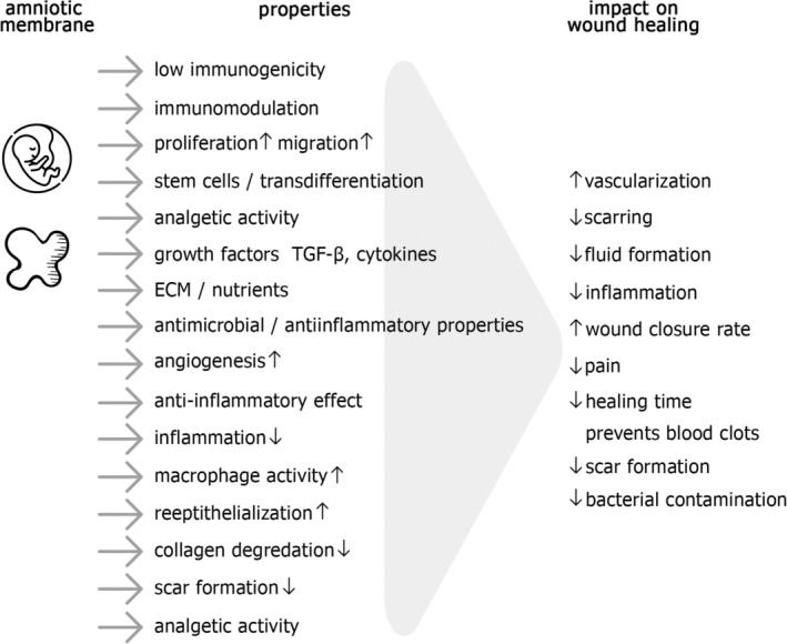
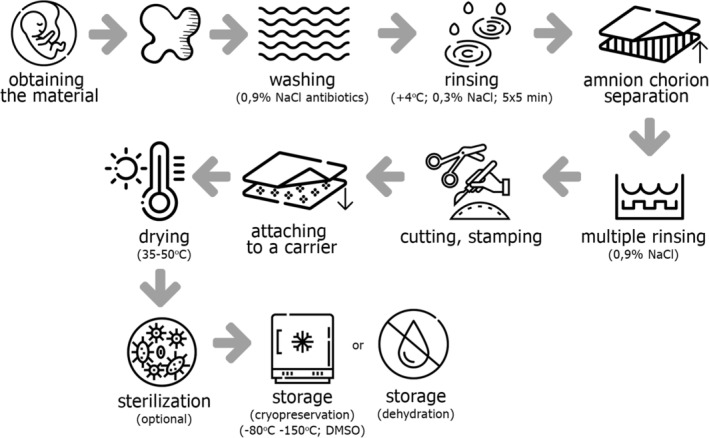
Unique properties of amniotic membrane make it a promising source for tissue engineering and a clinically useful alternative for patients suffering from chronic wounds including, for example, ulcers, burns, ocular surface damages and wounds occurring in the course of bullous diseases like stevens-johnson syndrome and toxic epidermal necrolysis. Its use has many advantages over standard wound care, as it contains pluripotent cells, nutrients, anti-fibrotic and anti-inflammatory cytokines, growth factors and extracellular matrix (ECM) proteins. Placental tissues can be prepared as a medical component, an advanced therapy medicinal product or a tissue graft. In addition to basic preparation procedures such as washing, rinsing, cutting, drying and sterilisation, there are many optional steps such as perforation, crosslinking and decellularisation. Finally, transplants should be properly stored-in cryopreserved or dehydrated form. In recent years, many studies including basic science and clinical trials have proven the potential to expand the use of amniotic membrane and amnion-derived cells to the fields of orthopaedics, dentistry, surgery, urology, vascular tissue engineering and even oncology. In this review, we discuss the role of placental tissues in skin wound healing and in the treatment of various diseases, with particular emphasis on bullous diseases. We also describe some patented procedures for placental tissue grafts preparation.
Keywords: amnion; healing; placenta; wound.
© 2020 Medicalhelplines.com Inc and John Wiley & Sons Ltd.
Conflict of interest statement
The authors declare no conflicts of interest.
Figures
References
-
- Benirschke K, Burton GJ, Baergen RN. Pathology of the Human Placenta. New York, NY: Springer‐Verlag Berlin Heidelberg; 2012.
-
- Gude NM, Roberts CT, Kalionis B, King RG. Growth and function of the normal human placenta. Thromb Res. 2004;114(5–6):397‐407. - PubMed
-
- Chen CP, Aplin JD. Placental extracellular matrix: gene expression, deposition by placental fibroblasts and the effect of oxygen. Placenta. 2003;24(4):316‐325. - PubMed
-
- Underwood MA, Gilbert WM, Sherman MP. Amniotic fluid: not just fetal urine anymore. J Perinatol. 2005;25(5):341‐348. - PubMed
-
- Nyman E, Huss F, Nyman T, Junker J, Kratz G. Hyaluronic acid, an important factor in the wound healing properties of amniotic fluid: in vitro studies of re‐epithelialisation in human skin wounds. J Plast Surg Hand Surg. 2013;47(2):89‐92. - PubMed
Publication types
MeSH terms
LinkOut - more resources
Full Text Sources
