. 2020 Feb;9(4):e1901419.
doi: 10.1002/adhm.201901419. Epub 2020 Jan 15.

Control of Astrocyte Quiescence and Activation in a Synthetic Brain Hydrogel

Affiliations

Control of Astrocyte Quiescence and Activation in a Synthetic Brain Hydrogel

Sualyneth Galarza et al. Adv Healthc Mater. 2020 Feb.

Abstract

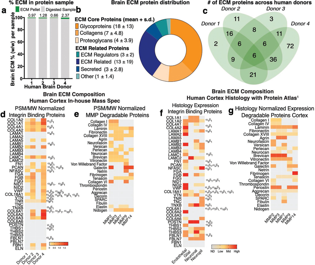
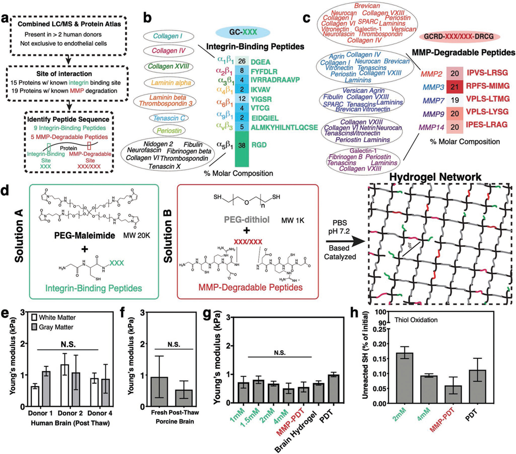
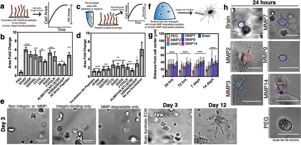
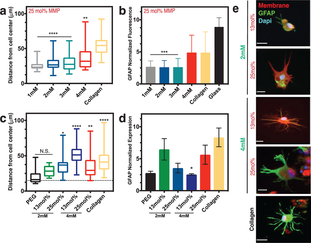
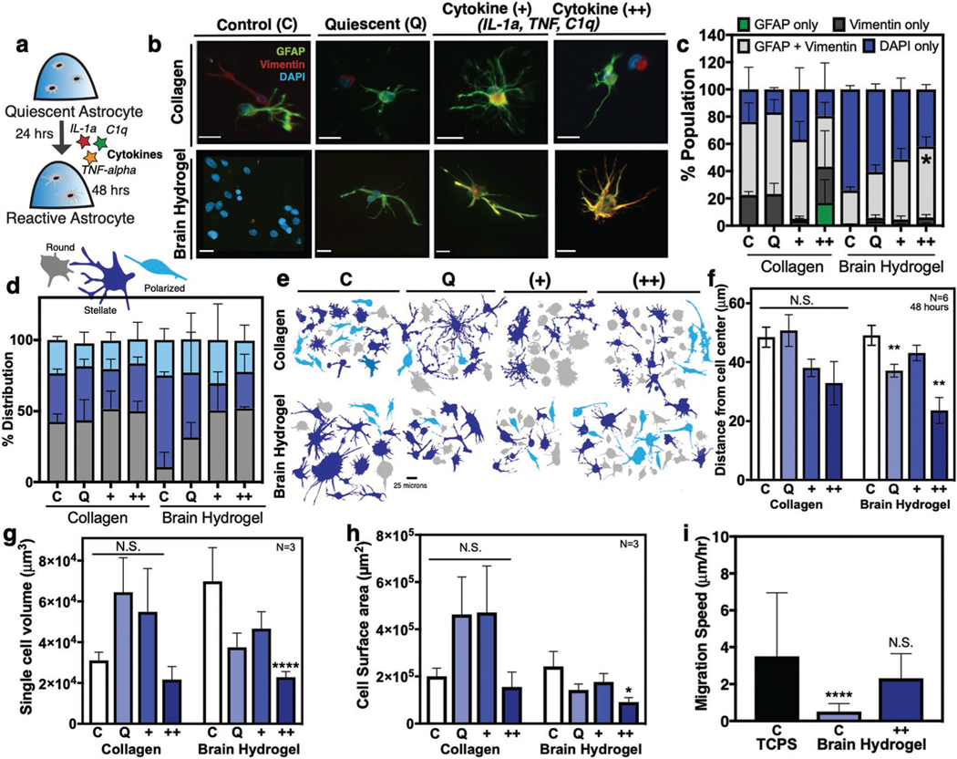
Bioengineers have designed numerous instructive brain extracellular matrix (ECM) environments with tailored and tunable protein compositions and biomechanical properties in vitro to study astrocyte reactivity during trauma and inflammation. However, a major limitation of both protein-based and synthetic model microenvironments is that astrocytes within fail to retain their characteristic stellate morphology and quiescent state without becoming activated under "normal" culture conditions. Here, a synthetic hydrogel is introduced, which for the first time demonstrates maintenance of astrocyte quiescence and activation on demand. With this synthetic brain hydrogel, the brain-specific integrin-binding and matrix metalloprotease-degradable domains of proteins are shown to control astrocyte star-shaped morphologies, and an ECM condition that maintains astrocyte quiescence with minimal activation can be achieved. In addition, activation can be induced in a dose-dependent manner via both defined cytokine cocktails and low molecular weight hyaluronic acid. This synthetic brain hydrogel is envisioned as a new tool to study the physiological role of astrocytes in health and disease.

Keywords: biomaterials; hydrogels; mass spectrometry; peptides; poly(ethylene glycol); tissue engineering.


Conflict of interest statement

Conflict of Interest

The authors declare no conflict of interest.

Figures

Figure 1.
Figure 1.
Characterization of the human brain cortex extracellular matrix. a) Right frontal cortex samples from four healthy human donors were decellularized and enriched for ECM proteins—resulting in an insoluble pellet that was solubilized, reduced, and digested into peptides identified via liquid chromatography-mass spectrometry (LC-MS). b) Protein hits were compared to the Human Matrisome Database and classified into ECM-core or ECM-related proteins. Data shown are average number of proteins found across the four donors + standard deviation (s.d.). c) Distribution of brain ECM signature proteins among donors. d) Proteins identified in at least two donors were screened for affinity to integrin binding and e) enzymatic degradation via matrix metalloproteinases (MMPs). Heat map depicts protein abundance based on peptide spectrum match (PSM) normalized to protein molecular weight (MW) for integrin-binding and MMP-degradable proteins. f) The Human Protein Atlas was screened for ECM proteins in the human brain cortex as identified via the provided histology. Heat map depicts protein abundance via histology expression of integrin binding and g) MMP-degradable proteins in the human brain cortex. Degree of expression denoted as ND (not detected), low, medium, or high.
Figure 2.
Figure 2.
Design of a synthetic brain ECM hydrogel. a) Proteins identified via in-house proteomics (LC/MS, present in >2 donors) and histology from the Protein Atlas (to exclude proteins specific to endothelial cells) were screened by integrin-binding and matrix metalloproteinase (MMP)-degradable peptide sequences. b) Integrin-binding peptides include 15 proteins, incorporated into the hydrogel via single cysteine. Heat map depicts molar concentration of each peptide in the final design. c) MMP-degradable peptides include 19 proteins, and were incorporated into the hydrogel by a cysteine at each end. Heat map depicts molar concentration of each peptide in the final design. d) A Michael-addition reaction is used to combine these peptides with PEG-maleimide to form a hydrogel network via two solutions: solution A containing integrin-binding peptides and the PEG polymer, and solution B with the MMP-degradable peptides and a nondegradable crosslinker. ξ is the mesh size of the hydrogel (≈20 nm). e) Young’s modulus of previously frozen human brain (3 donors), f) fresh and previously frozen porcine brain n = 6, and g) hydrogel tuned to the same Young’s modulus as brain tissue as measured via indentation. Modulus is maintained after incorporation of integrin-binding (green) or MMP-degradable (red) peptides. n = 8. h) Percent of unreacted thiols after hydrogel is formed, and then reduced with NaBH4, indicating high network formation efficiency. n = 8. All data are mean ± s.d. Data in (e)–(g) were analyzed using an ANOVA followed by a Tukey’s multiple comparison test with 95% confidence interval. N.S. is not significant.
Figure 3.
Figure 3.
Biological validation of brain integrin-binding and MMP-degradable peptides. a) Schematic depicting coverslip functionalization with integrin-binding peptides and cell seeding. b) Cell area fold change after seeding onto integrin-binding functionalized coverslips in comparison to a negative control (PEG). N = 2, n = 3. c) Schematic of competitive binding assay of integrin-binding peptides. d) Change in cell area after preincubation with peptides in solution, before seeding onto peptide-functionalized coverslips. Cell area is depicted as a fold change compared to a positive control (no preincubation). N = 2, n = 3. e) Representative human primary astrocyte morphology in the different hydrogel conditions. f) Schematic depicting encapsulation of cells in 3D hydrogels functionalized with MMP-degradable peptides and measurement of process length originating from the cell center. g) Astrocyte process length for individual and all brain MMP-degradable peptides in comparison to a PEG gel with no degradable crosslinks after 24 h, 72 h, 7 days, and 14 days of encapsulation. N = 2, n = 4. h) Representative images of astrocytes after 24 h of encapsulation. All data are mean + s.d. Statistical analyses were performed using Prism (GraphPad). Data in (b), (d), and (f) were analyzed using a one-way analysis of variance followed by a Dunnett’s multiple comparison test with 95% confidence interval. *, **, ***, and **** indicate P < 0.05, P < 0.01, P < 0.001, and P < 0.0001.
Figure 4.
Figure 4.
Astrocyte activation can be controlled via integrin-binding and MMP-degradable peptides in the brain hydrogel. a) Box and whisker plots show distance of process length from the cell center as a function of the integrin-binding peptide concentration (in mm) in the hydrogel, with collagen gels as a comparison. N = 2, n = 3. b) Normalized fluorescence intensity of glial fibrillary acidic protein (GFAP) as a function of integrin-specific peptide concentration, with both collagen gels and a glass coverslip for comparison (N = 3). All hydrogels in (a) and (b) had 25 mol% MMP-degradable peptides and the time point is 48 h after encapsulation. c) Box and whisker plots showing distance of process length from the cell center as a function of both integrin-binding peptide concentration and concentration of the MMP-degradable peptides. Statistics are in comparison to a PEG hydrogel with no peptides included (negative control). N = 2, n = 4. d) Normalized GFAP fluorescent intensity as a function of hydrogel integrin-binding and MMP-degradable peptide concentrations. N = 2, n = 4. Data in (c) and (d) are after 72 h of encapsulation. e) Representative images of astrocytes encapsulated in different hydrogel conditions after 72 h. Scale bar is 20 μm. Data in (a)–(c) are mean + s.d. Data in (d) are mean + SEM. Data in (a)–(d) were analyzed using a one-way analysis of variance (ANOVA) followed by a Dunnett’s multiple comparison test with 95% confidence interval. *, **, ***, and **** indicate P < 0.05, P < 0.01, P < 0.001, and P < 0.0001. N.S. is not significant.
Figure 5.
Figure 5.
Astrocyte activation can be controlled in the brain hydrogel. a) Human primary astrocytes were encapsulated in hydrogels for 24 h and subsequently dosed with cytokines IL-1α, TNF-α, and C1q for an additional 24 h. b) Representative images of astrocytes after 48 h in brain hydrogels and collagen and incubated with either standard culture medium (control), defined quiescent medium, dosed with cytokines (+) IL-1α (3 ng mL−1), TNF-α (30 ng mL−1), and C1q (400 ng mL−1), or dosed with a 2× dose of cytokines (++) IL-1α (6 ng mL−1), TNF-α (60 ng mL−1), and C1q (800 ng mL−1). Scale bar is 20 μm. c) Quantification of cells from (b) stained for GFAP, vimentin, and DAPI. N = 2, n = 250 cells per condition. d) Distribution of cell morphologies and e) representative cell morphologies in the different populations identified as round (gray), stellate (blue), or polarized (light blue). N = 3, n = 100 cells per condition. f) Quantification of astrocyte process length N = 6, n = 3. g) Single cell volume and h) single cell surface area for astrocytes encapsulated for 48 h. N = 3, n = 3. i) Comparison of migration speed astrocytes seeded on plastic (TCPS), or in the brain hydrogels with and without cytokines. N = 2. All plots show mean + SEM. Data in (c) were analyzed using a one-way analysis of variance (ANOVA). Data in (f)–(h) were analyzed using a two-way ANOVA followed by a Tukey’s multiple comparison test with 95% confidence interval. Data in (i) were analyzed using a one-way ANOVA followed by a Dunnett’s multiple comparison test with 95% confidence interval. *, **, and **** indicate P < 0.05, P < 0.01, and P < 0.0001, respectively; N.S., not significant.

References

    1. Liddelow SA, Barres BA, Immunity 2017, 46, 957. - PubMed
    1. Oberheim NA, Goldman SA, Nedergaard M, in Heterogeneity of astrocytic form and function, Astrocytes, Humana Press; 2012, pp. 23–45; - PMC - PubMed
    2. Oberheim NA, Takano T, Han X, He W, Lin JH, Wang F, Xu Q, Wyatt JD, Pilcher W, Ojemann JG, J. Neurosci 2009, 29, 3276. - PMC - PubMed
    1. Okada S, Hara M, Kobayakawa K, Matsumoto Y, Nakashima Y, Neurosci. Res 2018, 126, 39. - PubMed
    1. Farina C, Aloisi F, Meinl E, Trends Immunol. 2007, 28, 138. - PubMed
    1. Johnson KM, Milner R, Crocker SJ, Neurosci. Lett 2015, 600, 104; - PMC - PubMed
    2. Hsiao TW, Tresco PA, Hlady V, Biomaterials 2015, 39, 124; - PMC - PubMed
    3. Hara M, Kobayakawa K, Ohkawa Y, Kumamaru H, Yokota K, Saito T, Kijima K, Yoshizaki S, Harimaya K, Nakashima Y, Nat. Med 2017, 23, 818; - PubMed
    4. Placone AL, McGuiggan PM, Bergles DE, Guerrero-Cazares H, Quiñones-Hinojosa A, Searson PC, Biomaterials 2015, 42, 134; - PMC - PubMed
    5. Struve J, Maher PC, Li Y.-q, Kinney S, Fehlings MG, Kuntz Iv C, Sherman LS, Glia 2005, 52, 16. - PubMed

Publication types