. 2020 Apr 1;174(2):278-290.
doi: 10.1093/toxsci/kfaa006.

Wood Smoke Particles Stimulate MUC5AC Overproduction by Human Bronchial Epithelial Cells Through TRPA1 and EGFR Signaling

Affiliations

Wood Smoke Particles Stimulate MUC5AC Overproduction by Human Bronchial Epithelial Cells Through TRPA1 and EGFR Signaling

Tosifa A Memon et al. Toxicol Sci. .

Abstract

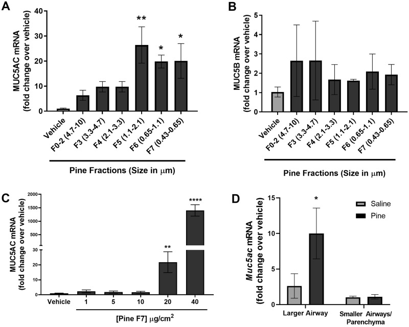
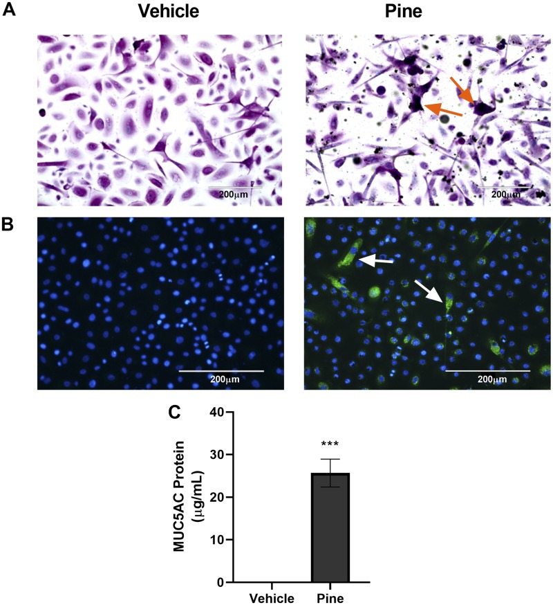
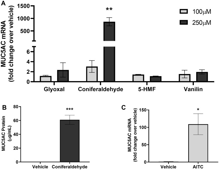
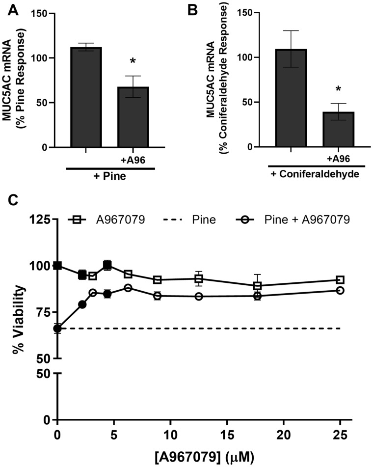
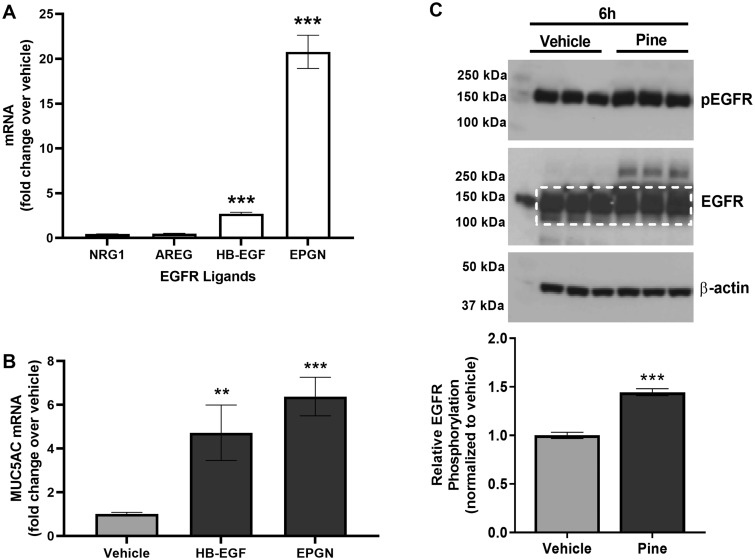
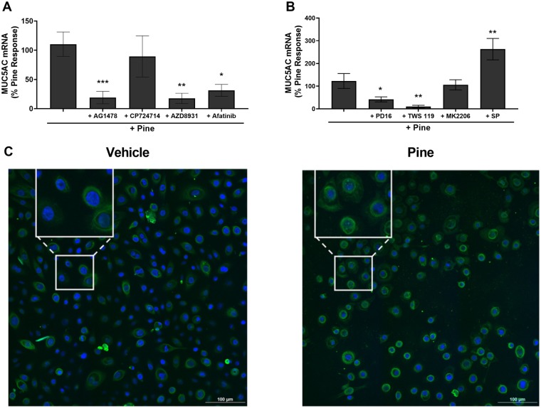
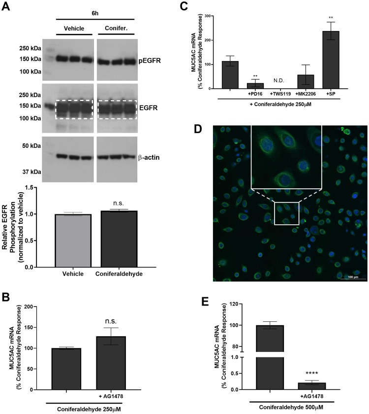
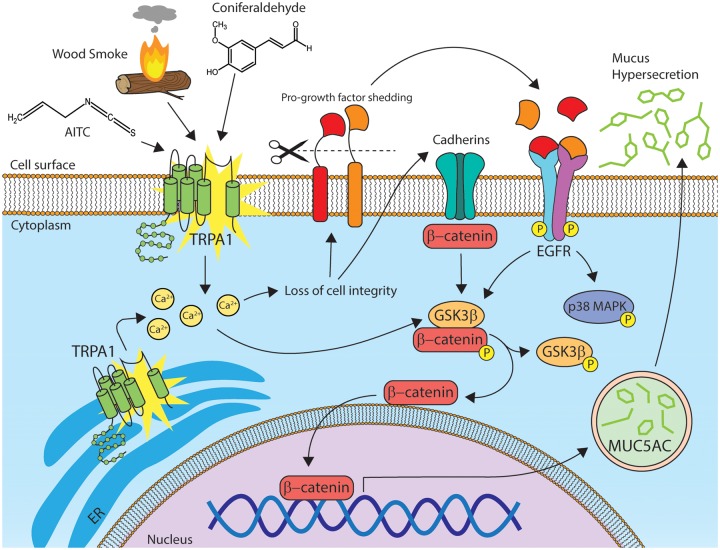
Mucus hypersecretion is a pathological feature of acute inflammatory and chronic obstructive pulmonary diseases. Exposure to air pollutants can be a cause of pathological mucus overproduction, but mechanisms by which different forms of air pollutants elicit this response are not fully understood. In this study, particulate matter (PM) generated from burning pine wood and other types of biomass was used to determine mechanisms by which these forms of PM stimulate mucin gene expression and secretion by primary human bronchial epithelial cells (HBECs). Biomass PM < 2.5 μm generated from pine wood and several other fuels stimulated the expression and secretion of the gel-forming glycoprotein MUC5AC by HBECs. Muc5ac gene induction was also observed in mouse airways following subacute oropharyngeal delivery of pine wood smoke PM. In HBECs, MUC5AC was also induced by the transient receptor potential ankyrin-1 (TRPA1) agonists' coniferaldehyde, a component of pine smoke PM, and allyl isothiocyanate, and was attenuated by a TRPA1 antagonist. Additionally, inhibition of epidermal growth factor receptor (EGFR/ErbB1) and the EGFR signaling partners p38 MAPK and GSK3β also prevented MUC5AC overexpression. Collectively, our results suggest that activation of TRPA1 and EGFR, paired with alterations to p38 MAPK and GSK3β activity, plays a major role in MUC5AC overproduction by bronchial epithelial cells exposed to biomass smoke PM. These results reveal specific processes for how biomass smoke PM may impact the human respiratory system and highlight potential avenues for therapeutic manipulation of lung diseases that are affected by air pollutants.

Keywords: EGFR; MUC5AC; TRPA1; mucin; particulate matter; wood smoke.


Figures

Figure 1.
Figure 1.
Pine wood smoke particulate matter (WSPM) induced MUC5AC mRNA expression. mRNA expression of the secreted gel-forming mucins, (A) MUC5AC and (B) MUC5B by human lobar bronchial epithelial cells (HLBECs) treated with either vehicle or size-fractionated pine WSPM (20 μg/cm2) for 24 h. C, Dose-dependent effect of pine WSPM F7 on MUC5AC expression by HLBECs. D, Muc5ac mRNA expression in larger and smaller airways/parenchyma isolated from 3 C57BL/6 mice following treatment with either saline or pine WSPM. Data are normalized to vehicle or saline treatment (fold change, y-axis) and are presented as the mean ± SEM from n ≥ 3 replicates. *p < .05, **p < .01, and ****p < .0001.
Figure 2.
Figure 2.
Pine WSPM induced MUC5AC protein expression. A, HLBECs treated with vehicle (left) or 10 μg/cm2 pine (right) for 24 h were stained with Alcian Blue and periodic acid-Schiff (PAS) to detect mucins. Violet color with orange arrows (online) or black color (print) corresponds to acidic mucins, whereas magenta (online) or gray (print) color corresponds to neutral mucins. B, Immunocytochemical detection of MUC5AC using a MUC5AC-specific antibody conjugated with Alexa-488 (green (online) or brighter spots (print); white arrows) in HLBECs treated with vehicle (left) or 20 μg/cm2 pine (right) for 24 h. Images were captured using an EVOS FL Auto Imaging System at 20× and positive cells in (A) and (B) are highlighted by arrows. C, Secreted MUC5AC protein measured by ELISA in supernatants collected 24 h post pine WSPM treatment. Data are presented as the mean ± SEM from n = 3 replicates. ***p < .001.
Figure 3.
Figure 3.
The pine WSPM chemical and TRPA1 agonist coniferaldehyde, and TRPA1 agonist allyl isothiocyanate (AITC), induced MUC5AC expression. A, MUC5AC induction in HLBECs treated with glyoxal, coniferaldehyde, 5-hydroxymethylfurfural (5-HMF), and vanillin at 100 and 250 μM for 24 h. MUC5AC expression for each treatment is represented as fold change over vehicle treatment. B, Secreted MUC5AC protein concentration measured by ELISA in supernatants of HLBECs treated with vehicle or 250 μM coniferaldehyde for 24 h. C, HLBECs treated with the TRPA1 agonist AITC (60 μM) for 24 h show significant upregulation of MUC5AC expression. Data are presented as fold change over vehicle treatment. Data are presented as the mean ± SEM from n ≥ 3 treatments. *p < .05, **p < .01, and ***p < .001.
Figure 4.
Figure 4.
TRPA1 inhibition partially reduced MUC5AC induction and pine particulate matter (PM) cytotoxicity. Effect of the TRPA1 antagonist, A967079 (A96; 20 μM) on (A) 20 μg/cm2 pine WSPM- and (B) 250 μM coniferaldehyde-induced MUC5AC expression in HLBECs. MUC5AC expression was normalized to pine WSPM or coniferaldehyde treatment. C, Effect of A967079 on cell viability of HLBECs in the presence (circles) and absence (squares) of pine WSPM (20 μg/cm2). Cell viability for pine only treatment is shown as the dotted line. Data are normalized to vehicle only treatment (ie, 100% residual viability). Data are presented as the mean ± SEM from n ≥ 3 replicates. *p < .05. In (C), black-filled circles and squares indicate ***p < .001.
Figure 5.
Figure 5.
Pine WSPM induced the expression of epidermal growth factor receptor (EGFR) ligands and increased EGFR phosphorylation by HLBECs. A, mRNA expression of EGFR ligands neuregulin 1 (NRG1), amphiregulin (AREG), heparin-binding epidermal growth factor (HB-EGF), and epigen (EPGN) by HLBECs treated with vehicle or 20 μg/cm2 pine WSPM for 24 h. B, The EGFR ligands HB-EGF (50 ng/ml) and EPGN (250 ng/ml) induced MUC5AC expression in HLBECs. MUC5AC mRNA expression was normalized to vehicle treatment and the data are presented as fold change relative to vehicle controls. C, EGFR and phospho-EGFR protein expression by HLBECs treated with vehicle or 20 μg/cm2 pine for 6 h. The ratio of p-EGFR to total EGFR (quantified within the white dotted box) was used to determine relative EGFR phosphorylation. Data are presented as the mean ± SEM from n ≥ 3 replicates. **p < .01 and ***p < .001.
Figure 6.
Figure 6.
Pine WSPM-induced MUC5AC overproduction involved EGFR and kinases downstream of EGFR. A, Effect of the inhibitors AG1478 (10 μM; EGFR/ErbB1), CP724714 (1 μM; ErbB2), AZD8931 (1 μM; EGFR, ErbB2, and ErbB3), and Afatinib (1 μM; EGFR, ErbB2, and ErbB4) on pine WSPM-induced MUC5AC expression. B, MUC5AC expression in the presence of the kinase inhibitors PD16936 (10 μM; p38 MAPK), TWS119 (5 μM; GSK3β), MK2206 (5 μM; AKT), and SP600125 (10 μM; JNK). MUC5AC mRNA expression was normalized to pine WSPM treatment and is presented as the percentage of the pine only response. Data are presented as the mean ± SEM from n ≥ 3 replicates. *p < .05, **p < .01, and ***p < .001. C, HLBECs treated with vehicle or pine WSPM for 24 h were stained with a primary antibody for β-catenin (green (online) or brighter rings and spots (print)) and nuclei were stained with Hoechst 33342 (blue (online) or darker center spots (print)). Images were captured using Nikon A1 confocal microscope at 60×. The insets show expanded images of representative cells exhibiting perinuclear and nuclear localization of β-catenin.
Figure 7.
Figure 7.
Coniferaldehyde-induced MUC5AC overproduction occurred via EGFR-dependent and EGFR-independent mechanisms. A, EGFR and phospho-EGFR protein (p-EGFR) expression by HLBECs treated with vehicle or 250 μM coniferaldehyde for 6 h. Relative EGFR phosphorylation (y-axis) as determined using the ratio of p-EGFR to total EGFR (quantified within the white dotted box). The full unmodified Western blot for vehicle, pine, and coniferaldehyde treatments is shown in Supplementary Figure 5. B, Effect of the EGFR/ErbB1 inhibitor AG1478 (10 μM) on coniferaldehyde (250 μM) induced MUC5AC expression by HLBECs. C, Effects of the kinase inhibitors PD16936 (10 μM; p38 MAPK), TWS119 (5 μM; GSK3β), MK2206 (5 μM; AKT), and SP600125 (10 μM; JNK) on coniferaldehyde (250 μM) induced MUC5AC expression by HLBECs. D, Immunocytochemical detection of β-catenin expression in HLBECs treated with 250 μM coniferaldehyde. The inset is an expanded view of representative cells exhibiting accumulation and perinuclear/nuclear localization of β-catenin; the vehicle control image is shown in Figure 6C. E, Effect of the EGFR/ErbB1 inhibitor AG1478 (10 μM) on MUC5AC expression by HLBECs treated with 500 μM coniferaldehyde. MUC5AC mRNA expression was normalized to the respective coniferaldehyde treatments and presented as a percentage of the coniferaldehyde response. Data are presented as the mean ± SEM from n ≥ 3 replicates. **p < .01 and ****p < .0001, N.D. stands for not detected.
Figure 8.
Figure 8.
Schematic representation of the proposed mechanism underlying Pine WSPM-induced MUC5AC expression by HLBECs. Pine WSPM, coniferaldehyde, and AITC activate TRPA1 causing increased cytosolic calcium. TRPA1 activation and intracellular calcium can disrupt cellular integrity, leading to shedding of existing EGFR ligands, induction of EGFR ligand synthesis, and the accumulation of β-catenin in the cytoplasm, perinuclear region, and nucleus. Epidermal growth factor receptor ligands such as EPGN and HB-EGF also activate EGFR/ErbB-1 and the downstream kinases p38 MAPK and GSK3β. Alternatively, TRPA1 activation, independent of EGFR activation, modulates GSK3β and/or p38 MAPK directly. Activation of GSK3β can further lead to accumulation and translocation of β-catenin to the nucleus. β-Catenin, a transcriptional coactivator has been shown by others to contribute MUC5AC overproduction and hypersecretion.

References

    1. Andersson D. A., Gentry C., Moss S., Bevan S. (2008). Transient receptor potential A1 is a sensory receptor for multiple products of oxidative stress. J. Neurosci. 28, 2485–2494. - PMC - PubMed
    1. Atkinson R. W., Kang S., Anderson H. R., Mills I. C., Walton H. A. (2014). Epidemiological time series studies of PM2.5 and daily mortality and hospital admissions: A systematic review and meta-analysis. Thorax 69, 660–665. - PMC - PubMed
    1. Bao Z., Lim S., Liao W., Lin Y., Thiemermann C., Leung B. P., Wong W. (2007). Glycogen synthase kinase-3β inhibition attenuates asthma in mice. Am. J. Respir. Crit. Care Med. 176, 431–438. - PubMed
    1. Bessac B. F., Jordt S.-E. (2008). Breathtaking TRP channels: TRPA1 and TRPV1 in airway chemosensation and reflex control. Physiology 23, 360–370. - PMC - PubMed
    1. Bonser L., Erle D. (2017). Airway mucus and asthma: The role of MUC5AC and MUC5B. J. Clin. Med. 6, 112. - PMC - PubMed

Publication types