Review
. 2020 Jan 17;126(2):280-293.
doi: 10.1161/CIRCRESAHA.119.316306. Epub 2020 Jan 16.

Role of Mitochondrial Calcium and the Permeability Transition Pore in Regulating Cell Death

Affiliations
Review

Role of Mitochondrial Calcium and the Permeability Transition Pore in Regulating Cell Death

Tyler M Bauer et al. Circ Res. .

Abstract

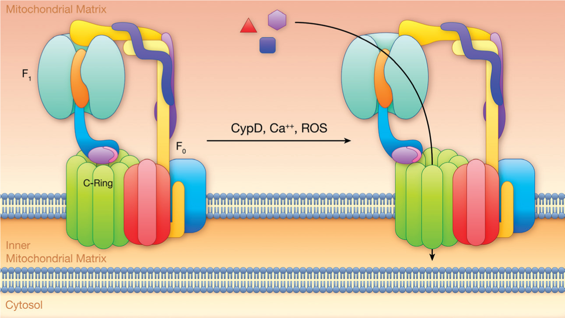
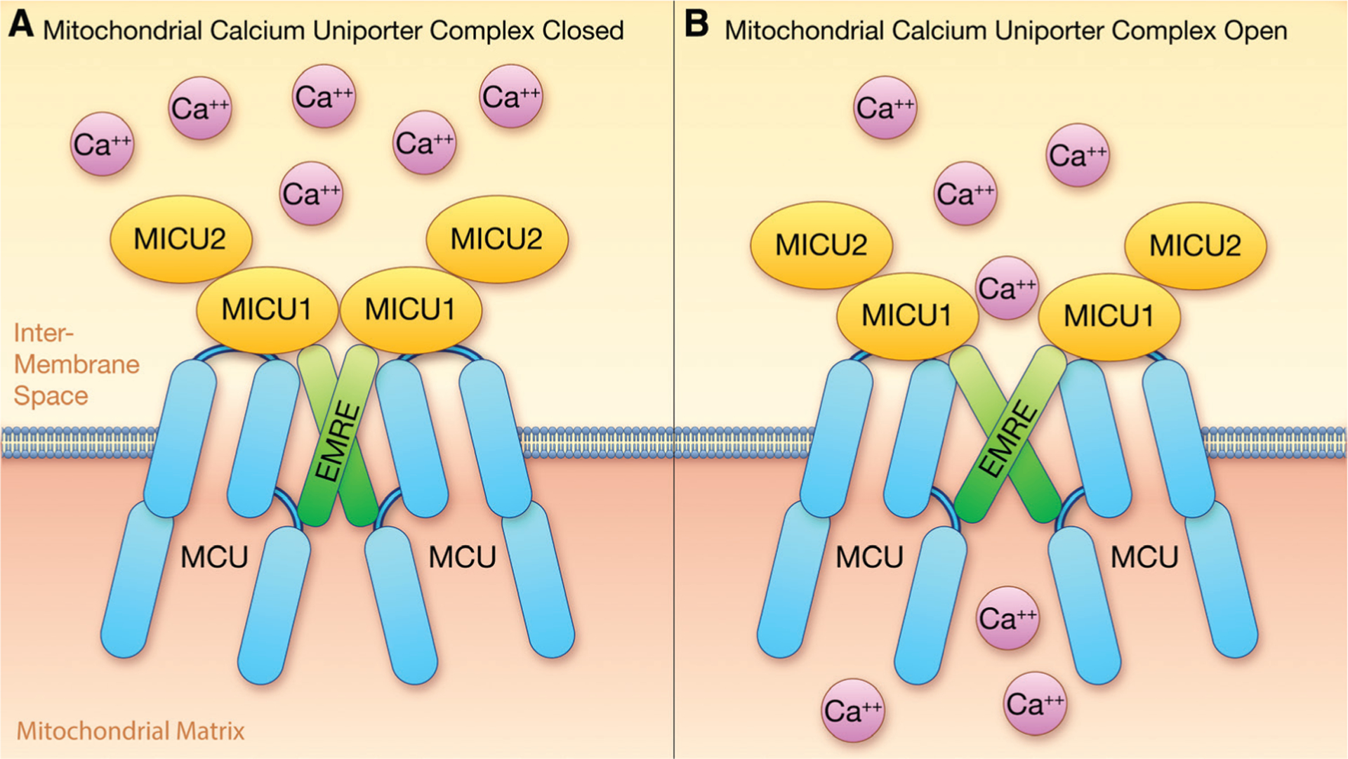
Adult cardiomyocytes are postmitotic cells that undergo very limited cell division. Thus, cardiomyocyte death as occurs during myocardial infarction has very detrimental consequences for the heart. Mitochondria have emerged as an important regulator of cardiovascular health and disease. Mitochondria are well established as bioenergetic hubs for generating ATP but have also been shown to regulate cell death pathways. Indeed many of the same signals used to regulate metabolism and ATP production, such as calcium and reactive oxygen species, are also key regulators of mitochondrial cell death pathways. It is widely hypothesized that an increase in calcium and reactive oxygen species activate a large conductance channel in the inner mitochondrial membrane known as the PTP (permeability transition pore) and that opening of this pore leads to necroptosis, a regulated form of necrotic cell death. Strategies to reduce PTP opening either by inhibition of PTP or inhibiting the rise in mitochondrial calcium or reactive oxygen species that activate PTP have been proposed. A major limitation of inhibiting the PTP is the lack of knowledge about the identity of the protein(s) that form the PTP and how they are activated by calcium and reactive oxygen species. This review will critically evaluate the candidates for the pore-forming unit of the PTP and discuss recent data suggesting that assumption that the PTP is formed by a single molecular identity may need to be reconsidered.

Keywords: calcium; cell death; mitochondria; permeability; reactive oxygen species.


Figures

Figure 1.
Figure 1.. Calcium and reactive oxygen species (ROS) lead to activation of the mitochondrial PTP (permeability transition pore) and initiation of cell death.
A, At the end of ischemia and the first few seconds of reperfusion, an increase in mitochondrial calcium can occur via MCU (mitochondrial calcium uniporter) which can activate PTP. There is also a buildup of succinate which upon conversion to fumarate by complex II, generates large levels of QH2 (ubiquinol) which drives reverse electron transport (RET) of complex I leading to ROS production. As shown in B, inhibition of PTP,. inhibition of MCU and inhibition of complex II (or complex I) are potential mechanisms to reduce PTP opening and reduce cell death. CypD indicates cyclophilin D. Illustration credit: Ben Smith.
Figure 2.
Figure 2.. ANT (adenine nucleotide transporter)/VDAC (voltage-dependent anion carrier) hypothesis.
CypD indicates cyclophilin D; and ROS, reactive oxygen species. Illustration credit: Ben Smith.
Figure 3.
Figure 3.. F1F0-ATPase dimer hypothesis.
In normal mitochondria, the F1F0ATPase is thought to self-assembly into a ribbon dimer structure. Bernardi et al, have proposed that PTP (permeability transition pore) channel formation could occur at a monomer-monomer interface following a calcium-dependant and reactive oxygen species–dependent change in conformation. Illustration credit: Ben Smith.
Figure 4.
Figure 4.. C-ring hypothesis.
Calcium, reactive oxygen species (ROS), and cellular stress signaling activation of the mitochondrial permeability transition pore and initiation of cell death through a potential pore in the c-ring. In this theory, the F1F0-ATPase would likely have to had cleared a lipid plug from the interior of the c-ring. CypD indicates cyclophilin D; and OSCP, oligomycin sensitive-conferring protein. Illustration credit: Ben Smith.
Figure 5.
Figure 5.. MCU (mitochondrial calcium uniporter) complex: A shows the MCU complex in the open conformation and B shows it in the closed conformation.
EMRE indicates essential MCU regulator; and MICU, mitochondrial calcium uptake protein. Illustration credit: Ben Smith.

Similar articles

Cited by

References

    1. Halestrap AP, Clarke SJ, Javadov SA. Mitochondrial permeability transition pore opening during myocardial reperfusion–a target for cardioprotection. Cardiovasc Res. 2004;61:372–385. doi: 10.1016/S0008-6363(03)00533-9 - DOI - PubMed
    1. Haworth RA, Hunter DR. The Ca2+-induced membrane transition in mitochondria. II. Nature of the Ca2+ trigger site. Arch Biochem Biophys. 1979;195:460–467. doi: 10.1016/0003-9861(79)90372-2 - DOI - PubMed
    1. Halestrap AP, Pasdois P. The role of the mitochondrial permeability transition pore in heart disease. Biochim Biophys Acta. 2009;1787:1402–1415. doi: 10.1016/j.bbabio.2008.12.017 - DOI - PubMed
    1. Crompton M The mitochondrial permeability transition pore and its role in cell death. Biochem J. 1999;341 (pt 2):233–249. - PMC - PubMed
    1. Di Lisa F, Bernardi P. A CaPful of mechanisms regulating the mitochondrial permeability transition. J Mol Cell Cardiol. 2009;46:775–780. doi: 10.1016/j.yjmcc.2009.03.006 - DOI - PubMed

Publication types

MeSH terms

Substances