. 2020 Jan 16;18(1):e3000599.
doi: 10.1371/journal.pbio.3000599. eCollection 2020 Jan.

A proteomic atlas of senescence-associated secretomes for aging biomarker development

Affiliations

A proteomic atlas of senescence-associated secretomes for aging biomarker development

Nathan Basisty et al. PLoS Biol. .

Abstract

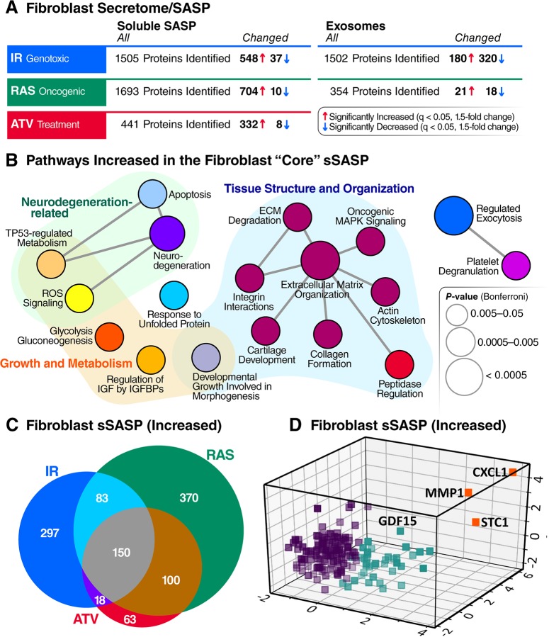
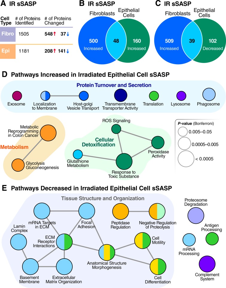
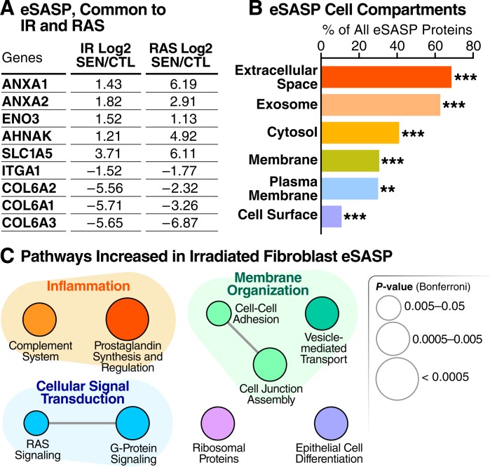
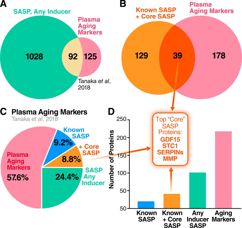
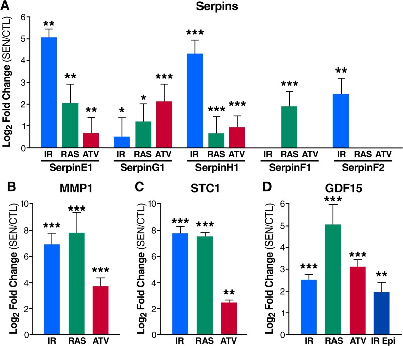
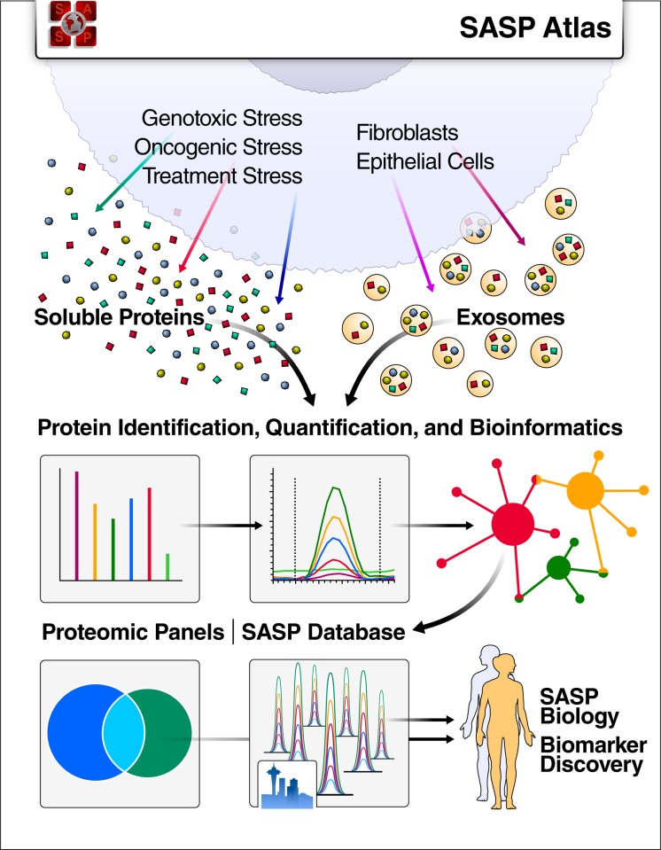
The senescence-associated secretory phenotype (SASP) has recently emerged as a driver of and promising therapeutic target for multiple age-related conditions, ranging from neurodegeneration to cancer. The complexity of the SASP, typically assessed by a few dozen secreted proteins, has been greatly underestimated, and a small set of factors cannot explain the diverse phenotypes it produces in vivo. Here, we present the "SASP Atlas," a comprehensive proteomic database of soluble proteins and exosomal cargo SASP factors originating from multiple senescence inducers and cell types. Each profile consists of hundreds of largely distinct proteins but also includes a subset of proteins elevated in all SASPs. Our analyses identify several candidate biomarkers of cellular senescence that overlap with aging markers in human plasma, including Growth/differentiation factor 15 (GDF15), stanniocalcin 1 (STC1), and serine protease inhibitors (SERPINs), which significantly correlated with age in plasma from a human cohort, the Baltimore Longitudinal Study of Aging (BLSA). Our findings will facilitate the identification of proteins characteristic of senescence-associated phenotypes and catalog potential senescence biomarkers to assess the burden, originating stimulus, and tissue of origin of senescent cells in vivo.


Conflict of interest statement

I have read the journal’s policy and the authors of this manuscript have the following competing interests: JC is a founder and shareholder of Unity Biotechnology, which develops senolytic drugs. All other authors have declared no competing interests.

Figures

Fig 1
Fig 1. Proteomic workflow for isolation and analysis of secreted proteins and exosomes/EVs.
Senescence was induced in cultured primary human lung fibroblasts by either IR, RAS, or ATV. Quiescent control cells were either mock irradiated or vehicle treated. Soluble proteins and exosomes/EVs were then isolated from conditioned media. Samples were digested and subjected to mass spectrometric analysis (DIA), followed by protein identification and quantification using Spectronaut Pulsar [32] and by bioinformatic, pathway, and network analyses in R and Cytoscape [33,34]. ATV, atazanavir treatment; CTL, Control; DIA, data-independent acquisition; EV, extracellular vesicle; IR, X-irradiation; RAS, inducible RAS overexpression; SEN, senescent.
Fig 2
Fig 2. Core sSASP proteins, networks and pathways.
(A) Summary of proteins with significantly altered (q-value <0.05 and >1.5-fold change) secretion by senescent compared with quiescent cells following genotoxic, oncogenic, or ATV treatment stress in senescent human lung fibroblasts. (B) ClueGO [33] pathway enrichment and network analyses of overlapping sSASPs resulting from each senescence inducer. Pathways of the same color have ≥50% similarity. Connecting lines represent Kappa connectivity scores >40%. (C) Venn diagram of proteins showing significantly increased secretion in senescent versus non-senescent fibroblasts following induction of senescence by IR, RAS, or ATV. (D) Unsupervised K-means clustering of proteins significantly increased in the sSASPs of all inducers based on the magnitude of the protein changes (log2-fold change) in senescent versus control groups and partitioned into three clusters. ATV, atazanavir treatment; CXCL1, chemokine C-X-C motif ligand 1; ECM, extracellular matrix; GDF15, growth/differentiation factor 15; IGF, insulin-like growth factor; IGFBP, IGF binding protein; IR, X-irradiation; MMP1, matrix metalloproteinase-1; RAS, RAS oncogene overexpression; ROS, reactive oxygen species; sSASP, soluble senescence-associated secretory phenotype; STC1, stanniocalcin 1; TP53, tumor protein p53.
Fig 3
Fig 3. Inducer-specific RNA and protein signatures of the sSASP.
Tables of top 10 inducer-specific sSASP proteins in (A) IR-induced senescence and (B) RAS-induced senescence. (B) Workflow for generating inducer-specific RNA and protein signatures of senescent cells. Transcriptome analysis of IR- and RAS-induced senescent fibroblasts were obtained from published studies [24,44] and combined. Transcriptome data were filtered for changes that were inducer specific (genes changing exclusively in one inducer but not the other) and were consistent in both studies. Inducer-specific transcriptomes were then compared with inducer-specific secretome changes in the sSASP (from the current study) to produce a combined inducer-specific RNA and protein signature. (D) Log2-fold changes of the top five RAS-specific genes in the sSASP secretome and in two published transcriptome datasets [24,44]. CTL, quiescent control; IR, X-irradiation; RAS, inducible RAS overexpression; SEN, senescent; sSASP, soluble senescence-associated secretory phenotype.
Fig 4
Fig 4. Epithelial cells and fibroblasts exhibit distinct sSASPs.
(A) Number of proteins identified and significantly altered in the sSASP of irradiated fibroblasts and epithelial cells. (B) Venn diagram comparing proteins significantly increased in the sSASPs of senescent fibroblasts and epithelial cells, both induced by IR (q < 0.05). (C) Venn diagram comparing protein increases in the fibroblast sSASP versus decreases in the epithelial sSASP. (D) Pathway and network analysis of secreted proteins significantly increased in epithelial cell sSASP. (E) Pathway and network analysis of proteins significantly decreased in the epithelial cell sSASP. ECM, extracellular matrix; IR, X-irradiation; ROS, reactive oxygen species; sSASP, soluble senescence-associated secretory phenotype.
Fig 5
Fig 5. Cellular senescence alters exosome/EV features and composition.
(A) Table showing overlapping significant protein changes in exosomes/EVs secreted by senescent cells induced by IR versus RAS (q < 0.05). (B) Enrichment analysis of gene-ontology/cellular compartments overrepresented among protein contents of exosomes/EVs released by senescent cells. (C) Network analysis of pathways and functions unique to the eSASP. CTL, quiescent control; eSASP, extracellular vesicle senescence-associated secretory phenotype; EV, extracellular vesicle; IR, X-irradiation; RAS, inducible RAS overexpression; SEN, senescent.
Fig 6
Fig 6. Human plasma aging markers are enriched for sSASP proteins.
(A) Venn diagram comparing sSASP factors secreted by at least one of IR-, RAS-, or ATV- induced senescent cells with markers of aging identified in human plasma [48]. (B) Overlap between the core sSASP (proteins secreted following all senescence-inducing stimuli) and plasma aging markers. (C) Pie chart showing the proportion of known sSASP factors, newly identified core sSASP factors, and sSASP factors found among plasma markers of aging in humans. (D) Number of proteins contained in the originally identified sSASP, core sSASP, noncore sSASP, and markers of aging in human plasma [48] (p < 0.00005). Top core sSASP factors GDF15, STC1, SERPINs, and MMP1 are among the plasma aging markers. ATV, atazanavir treatment; GDF15, growth/differentiation factor 15; IR, X-irradiation; MMP, matrix metalloproteinase; RAS, inducible RAS overexpression; SASP, senescence-associated secretory phenotype; SERPIN, serine protease inhibitors; sSASP, soluble senescence-associated secretory phenotype; STC1, stanniocalcin 1.
Fig 7
Fig 7. The sSASP contains aging and disease biomarkers.
(A) Serpins are secreted at high levels by senescent fibroblasts induced by IR, RAS, or ATV. (B) MMP1 and (C) STC1 are among the most highly secreted proteins by senescent fibroblasts. (D) The plasma aging biomarker GDF15 is increased in the sSASPs of fibroblasts induced to senesce by IR, RAS, and ATV and epithelial cells induced by IR. *q < 0.05, **q < 0.01, ***q < 0.001. ATV, atazanavir treatment; CTL, quiescent control; Epi, renal epithelial cell; GDF15, growth/differentiation factor 15; IR, X-irradiation; MMP1, matrix metalloproteinase-1; RAS, inducible RAS overexpression; SEN, senescent; sSASP, soluble senescence-associated secretory phenotype; STC1, stanniocalcin 1.
Fig 8
Fig 8. SASP Atlas: A comprehensive resource for SASPs.
SASP Atlas (www.SASPAtlas.com) is a curated and freely available database of the secretomes of senescent cells, including both the soluble and exosome SASP, that can be used to identify SASP components or biomarker candidates for senescence burden, aging, and related diseases. SASP, senescence-associated secretory phenotype.

Similar articles

Cited by

References

    1. Acosta JC, O’Loghlen A, Banito A, Guijarro MV, Augert A, Raguz S, et al. Chemokine signaling via the CXCR2 receptor reinforces senescence. Cell. 2008;133: 1006–1018. 10.1016/j.cell.2008.03.038 - DOI - PubMed
    1. Coppé J-P, Patil CK, Rodier F, Sun Y, Muñoz DP, Goldstein J, et al. Senescence-Associated Secretory Phenotypes Reveal Cell-Nonautonomous Functions of Oncogenic RAS and the p53 Tumor Suppressor. PLoS Biol. 2008;6: e301 10.1371/journal.pbio.0060301 - DOI - PMC - PubMed
    1. Coppé J-P, Patil CK, Rodier F, Krtolica A, Beauséjour CM, Parrinello S, et al. A Human-Like Senescence-Associated Secretory Phenotype Is Conserved in Mouse Cells Dependent on Physiological Oxygen. PLoS ONE. 2010;5: e9188 10.1371/journal.pone.0009188 - DOI - PMC - PubMed
    1. Kuilman T, Michaloglou C, Vredeveld LCW, Douma S, van Doorn R, Desmet CJ, et al. Oncogene-induced senescence relayed by an interleukin-dependent inflammatory network. Cell. 2008;133: 1019–1031. 10.1016/j.cell.2008.03.039 - DOI - PubMed
    1. Neves J, Demaria M, Campisi J, Jasper H. Of flies, mice, and men: evolutionarily conserved tissue damage responses and aging. Dev Cell. 2015;32: 9–18. 10.1016/j.devcel.2014.11.028 - DOI - PMC - PubMed

Publication types