. 2020 Jan 16;15(1):e0227887.
doi: 10.1371/journal.pone.0227887. eCollection 2020.

Astrocyte senescence promotes glutamate toxicity in cortical neurons

Affiliations

Astrocyte senescence promotes glutamate toxicity in cortical neurons

Chandani Limbad et al. PLoS One. .

Abstract

Neurodegeneration is a major age-related pathology. Cognitive decline is characteristic of patients with Alzheimer's and related dementias and cancer patients after chemo- or radio-therapies. A recently emerged driver of these and other age-related pathologies is cellular senescence, a cell fate that entails a permanent cell cycle arrest and pro-inflammatory senescence-associated secretory phenotype (SASP). Although there is a link between inflammation and neurodegenerative diseases, there are many open questions regarding how cellular senescence affects neurodegenerative pathologies. Among the various cell types in the brain, astrocytes are the most abundant. Astrocytes have proliferative capacity and are essential for neuron survival. Here, we investigated the phenotype of primary human astrocytes made senescent by X-irradiation, and identified genes encoding glutamate and potassium transporters as specifically downregulated upon senescence. This down regulation led to neuronal cell death in co-culture assays. Unbiased RNA sequencing of transcripts expressed by non-senescent and senescent astrocytes confirmed that glutamate homeostasis pathway declines upon senescence. Our results suggest a key role for cellular senescence, particularly in astrocytes, in excitotoxicity, which may lead to neurodegeneration including Alzheimer's disease and related dementias.


Conflict of interest statement

The authors have declared that no competing interests exist.

Figures

Fig 1
Fig 1. Human primary astrocytes undergo senescence and express a SASP upon X-irradiation.
(a) Immunofluorescence staining for the astrocyte marker GFAP was performed on primary human astrocytes and IMR-90 fibroblasts. (b) Cellular senescence (SEN) was induced in astrocytes by X-irradiation (IR). Fourteen days after IR, SA-β-gal staining was performed on non-senescent (NS) control cells and SEN cells. Quantification is shown in the right panel. (c-d) Real-time PCR was performed on RNA samples from NS and SEN cells. (c) shows the expression of p16INK4a (left) and LMNB1 (right). (d) shows the expression of three SASP factors, IL-6, IL-8 and CXCL-1. (e) High mobility group box 1 (HMGB1) immunofluorescence staining was performed on NS and SEN astrocytes. Quantification is shown in the right panel (n = 3, where n = experimental replicates). (f-g) ELISAs and AlphaLISAs were performed using conditioned media (CM) collected from NS and SEN cells; (f) shows HMGB1 ELISA results (n = 2, where n = experimental replicates), and (g) shows IL-6 AlphaLISA results (n = 3, where n = experimental replicates). For (b-d) (n = 2), shown are representative results from 3 independent experiments. For (b-g): *p<0.05, **p<0.02, ***p<0.001, ****p<0.0001 (unpaired t test).
Fig 2
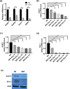
Fig 2. Senescence in astrocytes downregulates genes that modulate excitotoxicity.
(a) Real-time PCR was performed for EAAT1, EAAT2, Kir4.1, and AQP4 mRNA using RNA samples from NS and SEN astrocytes. (b-d) Time course experiments of astrocytes after IR. Samples were collected days 7, 10, 14, 21 after treatment. Real-time PCR was performed on NS and SEN samples at the indicated times; (b) corresponds to EAAT1 gene expression, (c) corresponds to EAAT2 gene expression, and (d) corresponds to Kir4.1 gene expression. (e) Western blotting was performed using protein extracts from NS and SEN 14 days after X-irradiation. Antibodies against EAAT1 and Kir4.1 were used. For (a-e) (n = 2), shown are the representative results from 2 independent experiments. For (a): *p<0.05, **p<0.02, ***p<0.001 (unpaired t test). For (b-d): **p<0.02, ***p<0.001, ****p<0.0001 (ordinary one-way ANOVA).
Fig 3
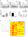
Fig 3. RNA-seq data reveal genes regulated in astrocytes upon senescence induction.
(a) RNA extracted from SEN astrocytes corresponding to 6 different human cell strains was compared to RNA extracted from NS samples. p16INK4a and SASP factors (IL-6 and IL-8) were analyzed by real-time PCR. (b) RNA-seq results (presented as Fragments Per Kilobase Million or FPKM) for the expression of various genes, i.e., p16INK4a (left panel), LMNB1 (center panel) and IL-6, IL-8, CXCL-1 (right panel) were compared between NS and SEN samples. (c) Hierarchical clustering using the Jensen-Shannon distance metric was used to determine the similarity within the 6 NS samples and within the 6 SEN samples. (d) A heatmap based on FPKM of Transporter-associated Astrocyte Enriched Genes (TAEG) in all the NS and SEN samples is shown. For (a), n = 2s. For (b), n = 3. For (a, b): *p<0.05, **p<0.02, ***p<0.001, ****p<0.0001 (unpaired t test).
Fig 4
Fig 4. Increased neuronal death in co-cultures with SEN astrocytes dependent on glutamate.
(a-b) Neurons (only DAPI-stained cells) were co-cultured with NS or SEN astrocytes (CMPTX-red + DAPI-stained cells) in neuronal media. The co-cultures were treated for 24 h with control media without glutamate. In (a) we show the fluorescent images, and in (b) the cell quantification. (c-d) Neurons were also co-cultured with NS or SEN astrocytes in neuronal media. However, here co-cultures were treated for 24 h with media containing 10 mM glutamate. In (c) we show the fluorescent images, and in (d) the cell quantification. Lane 1: surviving neurons (NS astrocytes + neurons co-cultures). Lane 2: surviving neurons (SEN astrocytes + neurons co-cultures). Lane 3: surviving astrocytes (NS astrocytes + neurons co-cultures). Lane 4: surviving astrocytes (SEN astrocytes + neurons co-cultures). For (b, d) (n = 4, where n = experimental replicates): ns = not significant, *p<0.05, **p<0.02 (unpaired t test).

References

    1. Lecot P, Alimirah F, Desprez PY, Campisi J, Wiley C. Context-dependent effects of cellular senescence in cancer development. Br J Cancer. 2016;114(11):1180–4. Epub 2016/05/04. bjc2016115 [pii] 10.1038/bjc.2016.115 - DOI - PMC - PubMed
    1. Rodier F, Campisi J. Four faces of cellular senescence. J Cell Biol. 2011;192(4):547–56. Epub 2011/02/16. jcb.201009094 [pii] 10.1083/jcb.201009094 - DOI - PMC - PubMed
    1. Minamino T, Miyauchi H, Yoshida T, Ishida Y, Yoshida H, Komuro I. Endothelial cell senescence in human atherosclerosis: role of telomere in endothelial dysfunction. Circulation. 2002;105(13):1541–4. Epub 2002/04/03. 10.1161/01.cir.0000013836.85741.17 . - DOI - PubMed
    1. Schnabl B, Purbeck CA, Choi YH, Hagedorn CH, Brenner D. Replicative senescence of activated human hepatic stellate cells is accompanied by a pronounced inflammatory but less fibrogenic phenotype. Hepatology. 2003;37(3):653–64. Epub 2003/02/26. 10.1053/jhep.2003.50097 S027091390214208X [pii]. . - DOI - PubMed
    1. Minamino T, Yoshida T, Tateno K, Miyauchi H, Zou Y, Toko H, et al. Ras induces vascular smooth muscle cell senescence and inflammation in human atherosclerosis. Circulation. 2003;108(18):2264–9. Epub 2003/10/15. 10.1161/01.CIR.0000093274.82929.22 CIR.0000093274.82929.22 [pii]. . - DOI - PubMed

Publication types

MeSH terms

Substances