

A research-driven approach to the identification of novel natural killer cell deficiencies affecting cytotoxic function
- PMID: 31945148
- PMCID: PMC7046607
- DOI: 10.1182/blood.2019000925
A research-driven approach to the identification of novel natural killer cell deficiencies affecting cytotoxic function
Abstract
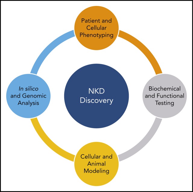
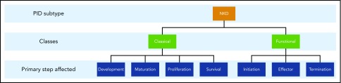
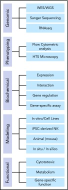
Natural killer cell deficiencies (NKDs) are an emerging phenotypic subtype of primary immune deficiency. NK cells provide a defense against virally infected cells using a variety of cytotoxic mechanisms, and patients who have defective NK cell development or function can present with atypical, recurrent, or severe herpesviral infections. The current pipeline for investigating NKDs involves the acquisition and clinical assessment of patients with a suspected NKD followed by subsequent in silico, in vitro, and in vivo laboratory research. Evaluation involves initially quantifying NK cells and measuring NK cell cytotoxicity and expression of certain NK cell receptors involved in NK cell development and function. Subsequent studies using genomic methods to identify the potential causative variant are conducted along with variant impact testing to make genotype-phenotype connections. Identification of novel genes contributing to the NKD phenotype can also be facilitated by applying the expanding knowledge of NK cell biology. In this review, we discuss how NKDs that affect NK cell cytotoxicity can be approached in the clinic and laboratory for the discovery of novel gene variants.
© 2020 by The American Society of Hematology.
Conflict of interest statement
Conflict-of-interest disclosure: J.S.O. declares royalties from Wolters Kluwer Publishing (UpToDate) for chapters on NK cell deficiency. The remaining authors declare no competing financial interests.
Figures
Similar articles
-
Clinical, immunologic, and genetic characteristics of 148 patients with natural killer cell deficiency.J Allergy Clin Immunol. 2025 May;155(5):1623-1634. doi: 10.1016/j.jaci.2025.01.030. Epub 2025 Feb 4. J Allergy Clin Immunol. 2025. PMID: 39914554
-
How I Manage Natural Killer Cell Deficiency.J Clin Immunol. 2020 Jan;40(1):13-23. doi: 10.1007/s10875-019-00711-7. Epub 2019 Nov 22. J Clin Immunol. 2020. PMID: 31754930 Free PMC article.
-
Emerging insights into human health and NK cell biology from the study of NK cell deficiencies.Immunol Rev. 2019 Jan;287(1):202-225. doi: 10.1111/imr.12725. Immunol Rev. 2019. PMID: 30565241 Free PMC article. Review.
-
Natural killer cell biology illuminated by primary immunodeficiency syndromes in humans.Clin Immunol. 2017 Apr;177:29-42. doi: 10.1016/j.clim.2015.11.004. Epub 2015 Dec 1. Clin Immunol. 2017. PMID: 26592356 Review.
-
Natural killer cell deficiency.J Allergy Clin Immunol. 2013 Sep;132(3):515-525. doi: 10.1016/j.jaci.2013.07.020. J Allergy Clin Immunol. 2013. PMID: 23993353 Free PMC article. Review.
Cited by
-
Clinical, immunologic, and genetic characteristics of 148 patients with natural killer cell deficiency.J Allergy Clin Immunol. 2025 May;155(5):1623-1634. doi: 10.1016/j.jaci.2025.01.030. Epub 2025 Feb 4. J Allergy Clin Immunol. 2025. PMID: 39914554
-
Complete CD16A Deficiency and Defective NK Cell Function in a Man Living with HIV.J Clin Immunol. 2025 May 24;45(1):98. doi: 10.1007/s10875-025-01886-y. J Clin Immunol. 2025. PMID: 40411624 Free PMC article.
-
Diagnostic guidelines for familial hemophagocytic lymphohistiocytosis revisited.Blood. 2024 Nov 28;144(22):2308-2318. doi: 10.1182/blood.2024025077. Blood. 2024. PMID: 39046779 Free PMC article.
References
-
- Prager I, Watzl C. Mechanisms of natural killer cell‐mediated cellular cytotoxicity. J Leukoc Biol. 2019;105(6):1319-1329. - PubMed
Publication types
MeSH terms
Grants and funding
LinkOut - more resources
Full Text Sources
