. 2020 May;71(5):1787-1801.
doi: 10.1002/hep.31116.

c-Jun NH2 -Terminal Protein Kinase Phosphorylates the Nrf2-ECH Homology 6 Domain of Nuclear Factor Erythroid 2-Related Factor 2 and Downregulates Cytoprotective Genes in Acetaminophen-Induced Liver Injury in Mice

Affiliations

c-Jun NH2 -Terminal Protein Kinase Phosphorylates the Nrf2-ECH Homology 6 Domain of Nuclear Factor Erythroid 2-Related Factor 2 and Downregulates Cytoprotective Genes in Acetaminophen-Induced Liver Injury in Mice

Yiping Chen et al. Hepatology. 2020 May.

Abstract

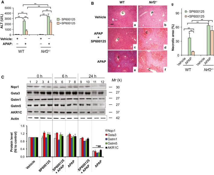
Background and aims: Acetaminophen (APAP) overdose induces severe liver injury and hepatic failure. While the activation of c-Jun NH2 -terminal kinase (JNK) has been implicated as a mechanism in APAP-induced liver injury, the hepatic defense system controlled by nuclear factor erythroid 2-related factor 2 (Nrf2) plays a central role in the mitigation of APAP toxicity. However, the link between the two signaling pathways in APAP-induced liver injury (AILI) remains unclear.

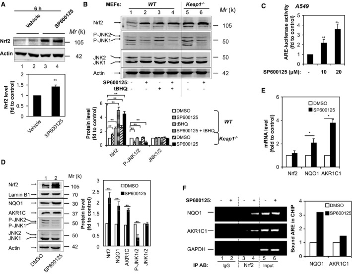
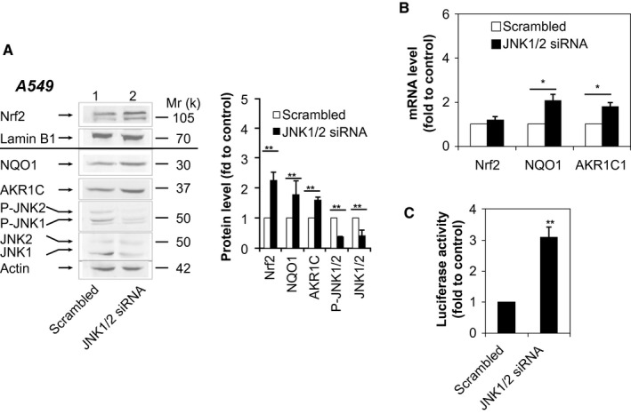
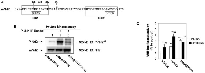
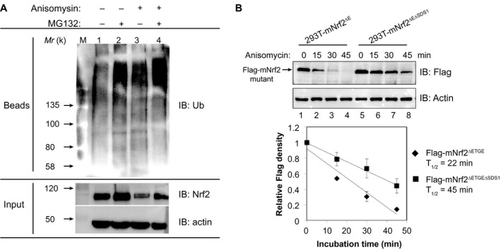
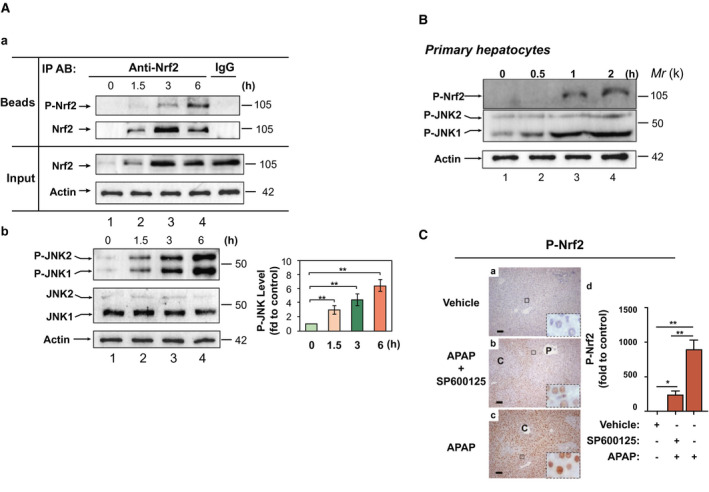
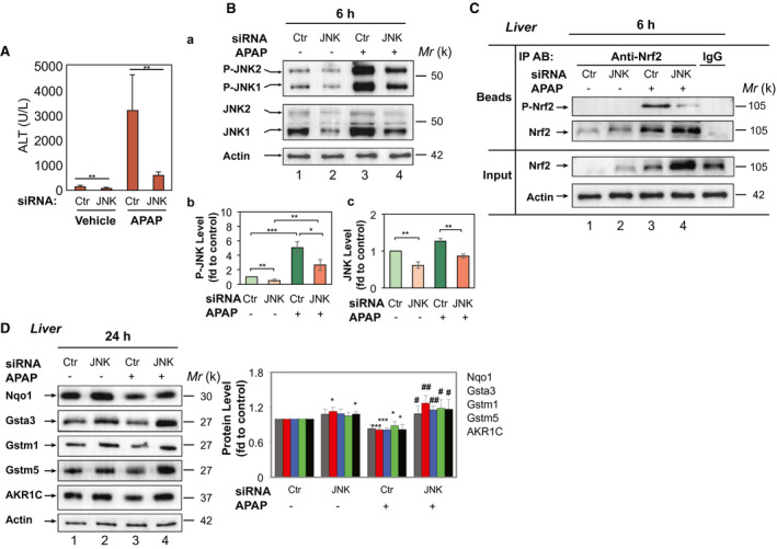
Approach and results: In this study, we demonstrated that the activation of JNK in mouse liver following exposure to APAP was correlated with the phosphorylation of Nrf2 and down-regulation of the antioxidant response element (ARE)-driven genes, NAD(P)H:quinone dehydrogenase 1, glutathione S-transferase α3, glutathione S-transferase M1, glutathione S-transferase M5, and aldo-keto reductase 1C. The JNK inhibitor, SP600125, or knockdown of JNK by infection of adenovirus expressing JNK small interfering RNA, ameliorated the APAP induced liver toxicity, and inhibited the phosphorylation of Nrf2 and down-regulation of detoxifying enzymes by stabilizing the transcription factor. Mechanistically, JNK antagonized Nrf2- and ARE-driven gene expression in a Kelch-like ECH-associated protein 1-independent manner. Biochemical analysis revealed that phosphorylated JNK (P-JNK) directly interacted with the Nrf2-ECH homology (Neh) 1 domain of Nrf2 and phosphorylated the serine-aspartate-serine motif 1 (SDS1) region in the Neh6 domain of Nrf2.

Conclusions: Mass spectrometric analysis identified serine 335 in the SDS1 region of mNrf2 as the major phosphorylation site for modulation of Nrf2 ubiquitylation by P-JNK. This study demonstrates that Nrf2 is a target of P-JNK in AILI. Our finding may provide a strategy for the treatment of AILI.


Figures

Figure 1
Figure 1
The JNK inhibitor, SP600125, blocks down‐regulation of ARE‐driven genes induced by APAP in mouse liver. SP600125 (10 mg/kg IP) was given to WT and Nrf2−/− mice 1 hour before injection of APAP (300 mg/kg IP). Livers were harvested 6 and 24 hours after administration of APAP. (A) Serum ALT levels at 6 hours (n = 3‐6). (B) Hematoxylin and eosin staining of liver sections at 6 hours (original magnification, ×200; scale bars, 50 μm; P, portal venules; C, central venules). g, the percentage of necrotic area by semiquantification (mean ± SD; n = 3‐5). (C) SP600125 blocked the decrease of Nqo1, AKR1C, Gstα3, Gstm1, and Gstm5 expression in APAP‐treated liver. Protein extracts were prepared from livers of WT mice harvested at 6 and 24 hours after administration of APAP. Western immunoblottings were probed with the indicated antibodies. Each lane contains a sample from a single mouse. Lower panel, semiquantitative results of blottings. The value from the WT group treated with vehicle was set at 1. Values are mean ± SD (n = 3). *P < 0.05; **P < 0.01 versus WT vehicle. ## P < 0.01 versus WT treated with SP600125 and APAP at 24 hours.
Figure 2
Figure 2
The JNK inhibitor, SP600125, up‐regulates expression of Nrf2‐ and ARE‐driven genes. (A) SP600125 increases expression of Nrf2 in liver. Mice were given SP600125 (10 mg/kg IP). Livers were harvested 6 hours after administration. Western immunoblottings of protein extracts from livers probed with anti‐Nrf2 or anti‐actin. Each lane contains a sample from a single mouse. Lower panel, semiquantitative results of the blottings. The value from the WT group treated with vehicle was set at 1. Values are mean ± SD (n = 3). (B) SP600125‐induced Nrf2 expression is independent of Keap1. WT and Keap1 −/− MEFs were treated with SP600125 (10 μM) in the presence or absence of tBHQ (20 μM) for 16 hours. Cell lysates were probed by immunoblotting with anti‐Nrf2, anti‐P‐JNK, anti‐JNK, or anti‐actin. Lower panel, semiquantitative results of the blottings. The value for DMSO treatment was set at 1. Values are mean ± SD (n = 3). (C) SP600125 activates ARE‐luciferase activity dose dependently in A549 cells. A549 cells were transfected with pGL‐GSTA2‐41bp‐ARE in combination with pRL‐TK. Twenty‐four hours later, cells were treated with 10 or 20 μM of SP600125 for 16 hours and the dual luciferase activity was determined. The value for DMSO treatment was set at 1 (control). Values are mean ± SD (n = 3). (D,E) SP600125 increases the expression of Nrf2‐ and ARE‐driven genes in A549 cells. Immunoblotting with anti‐Nrf2 and anti‐lamin B1 of nuclear extracts from A549 cells subjected to serum depletion for 16 hours before treatment with 10 μM of SP600125 for 16 hours. Whole‐cell extracts were probed by immunoblotting with anti‐NQO1, anti‐AKR1C, anti‐P‐JNK, and anti‐JNK. Right panel, semiquantitative results of the blottings. Relative levels of Nrf2 normalized to lamin B1. Relative levels of NQO1, AKR1C, P‐JNK, and JNK normalized to actin. The value for DMSO treatment was set at 1. Values are mean ± SD (n = 3). (E) Statistics for mRNA levels of NQO1 and AKR1C as determined by RT‐PCR. 18S rRNA was used as an internal control. The value for DMSO treatment was set at 1. Data are presented as the mean ± SD of triplicate experiments. (F) SP600125 increases Nrf2 binding to ARE sites in the promoters of NQO1 and AKR1C1. A549 cells were treated with 10 μM of SP600125 for 6 hours and used for ChIP analysis. GAPDH served as a negative control. PCR reactions were not saturated. Results are representative of three separate experiments. The relative value of NRF2 binding to ARE sites was determined as described in Materials and Methods. *P < 0.05; **P < 0.01. Abbreviations: DMSO, dimethyl sulfoxide; fd, fold; GAPDH, glyceraldehyde 3‐phosphate dehydrogenase; IgG, immunoglobulin G; IP AB, immunoprecipitation antibody; rRNA, ribosomal RNA; tBHQ, tert‐butylhydroquinone.
Figure 3
Figure 3
Knockdown of JNK increases expression of Nrf2‐ and ARE‐driven genes. (A,B) A549 cells were transfected with JNK1/2 siRNA or scrambled siRNA. Forty‐eight hours later, nuclear extracts were probed by immunoblotting with anti‐Nrf2 and anti‐lamin B1, and whole‐cell lysates were probed by immunoblotting with anti‐NQO1, anti‐AKR1C, anti‐P‐JNK, anti‐JNK, and anti‐actin (A). Right panel (A), semiquantitative result of blotting. Relative levels of Nrf2 normalized to lamin B1. Relative levels of NQO1, AKR1C, P‐JNK, and JNK normalized to actin. The value for scrambled siRNA was set at 1. Values are mean ± SD (n = 3). mRNA levels of NQO1 and AKR1C1 were determined by RT‐PCR (B). The level of 18S rRNA was used as an internal control. The value for scrambled siRNA was set at 1. (C) Luciferase activity 48 hours after transfection of A549 cells with JNK1/2 siRNA or scrambled siRNA plus pGL‐GSTA2.41bp‐ARE reporter vector and pRL‐TK. Data are presented as the mean ± SD of triplicate experiments. *P < 0.05; **P < 0.01. Abbreviation: rRNA, ribosomal RNA.
Figure 4
Figure 4
Identification of the site at which Nrf2 is phosphorylated by P‐JNK. (A) Amino‐acid sequences of mNrf2 between residues 329 and 379. (B) Mutation of Ser‐335 to Ala in the Neh6 domain of Nrf2 abolishes its phosphorylation by P‐JNK. HEK293 cells were treated with 5 μg/mL of anisomysin for 30 minutes. The cell lysate was immunoprecipitated with anti‐P‐JNK. Immunoprecipitates were subjected to in vitro kinase assays with purified recombinant His‐mNrf2ΔETGE or His‐mNrf2ΔETGE,S335A proteins, followed by western immunoblotting. Upper blottings, immunodetection of P‐Nrf2 with anti‐P‐Nrf23S. Lower blottings, immunodetection of Nrf2 mutant proteins to ensure similar quantity per reaction. Lane 1, kinase assay in the absence of P‐JNK immunoprecipitates. The blottings presented are typical examples of at least three independent experiments. (C) Mutation of Ser‐335 to Ala in the Neh6 domain of Nrf2 abolishes SP600125‐enhanced ARE‐luciferase activity. A549 cells were transfected with pcDNA3.1/V5/His, pcDNA3.1/V5‐mNrf2, or pcDNA3.1/V5‐mNrf2S335A plus pGL‐GSTA2.41bp‐ARE reporter vector and pRL‐TK. Cells were treated with 10 μM of SP600125 for 16 hours before dual luciferase activity was analyzed. The value for cells transfected with pcDNA3.1/V5, pGL‐GSTA2.41bp‐ARE reporter vector, and pRL‐TK and treated with DMSO was set at 1. Data are presented as the mean ± SD of triplicate experiments. *Significantly different from cells transfected with pcDNA3.1/V5/His, pGL‐GSTA2.41bp‐ARE reporter vector, and pRL‐TK and treated with DMSO. #Significantly different from cells transfected with the same plasmids and treated with DMSO. **P < 0.01; ## P < 0.01. Abbreviations: Ala, alanine; DMSO, dimethyl sulfoxide; fd, fold; IB, immunoblotting; IP, immunoprecipitation; Ser‐335, serine 335; V5‐His, V5 peptide with polyhistidine tag.
Figure 5
Figure 5
P‐JNK regulates Nrf2 stability. (A) Activated JNK increases the polyubiquitination of endogenous Nrf2 in A549 cells. A549 cells were pretreated with 10 μM of MG132 for 3 hours, before 1‐hour treatment with 5 μg/mL of anisomysin in the continued presence or absence of 10 μM of MG132. Whole‐cell lysates were subjected to immunoprecipitation with anti‐Nrf2. Immunoprecipitates were analyzed by immunoblotting using antibody against Ub. Input, 10% of the cell lysate used for immunoprecipitation. The results presented are typical examples from at least three independent experiments. (B) Deletion of SDS1 in the Neh6 domain blocks degradation of Nrf2 induced by anisomycin. 293T‐mNrf2ΔE and 293T‐mNrf2ΔEΔSDS1 cells expressing Flag‐tagged mNrf2ΔETGE and Flag‐tagged mNrf2ΔETGEΔSDS1, respectively, were treated with 5 μg/mL of anisomysin for 15‐45 minutes. Whole‐cell lysates were immunoblotted with anti‐Flag or anti‐actin. Lower panel, relative levels of Flag‐mNrf2 mutants normalized to actin. The value at time 0 for the same protein was set at 1. Results are from three separate experiments. Values are mean ± SD. Abbreviations: IB, immunoblotting; Ub, ubiquitin.
Figure 6
Figure 6
APAP induces phosphorylation of Nrf2 by P‐JNK in mouse liver. (A) WT mice were given APAP (300 mg/kg BW IP). Livers were harvested 1.5, 3, or 6 hours later. (a) Soluble extracts (100 μg) from livers in 3 randomly selected mice each group were subjected to immunoprecipitation with anti‐Nrf2. After washing, each half of the immunoprecipitates was then subjected to immunoblotting with anti‐Nrf2 or anti‐P‐Nrf23S. IgG with extracts from livers in the 6‐hour APAP group was used as a negative control. Input, 10% of the lysate used for immunoprecipitation. The blottings shown represent results from three independent experiments. (b) Western blotting of P‐JNK and JNK in 3 randomly selected mice each group. Right panel, semiquantitative results of the blottings. The control (vehicle) was set at 1. Values are mean ± SD (n = 3). **P < 0.01 versus vehicle. (B) APAP induces phosphorylation of Nrf2 in mouse primary hepatocytes. Freshly isolated mouse hepatocytes were exposed to 10 mM of APAP for 30 minutes, 1 hour, and 2 hours. Cell lysates were subjected to immunoprecipitation with anti‐Nrf2. After washing, each of the immunoprecipitates was then subjected to western blotting with anti‐P‐Nrf23S; the flowthroughes blotting with anti‐P‐JNK, and anti‐actin, respectively. Blottings are representative of two independent experiments. (C) SP600125 inhibits APAP‐induced phosphorylation of Nrf2 in mouse liver. SP600125 (10 mg/kg IP) was given to mice 1 hour before injection of APAP (300 mg/kg IP). Livers were harvested 6 hours after APAP. (a‐c) Liver sections were probed with anti‐P‐Nrf23S (original magnification, ×200; scale bars, 50 μm; insets, original magnification, ×1000; P, portal venules; C, central venules). (d) Statistics from experiments as in (a–c). The control (vehicle at 6 h) was set at 1. Values are mean ± SD (n = 3). *P < 0.05; **P < 0.01. Abbreviations: fd, fold; IgG, immunoglobulin G; IP AB, immunoprecipitation antibody.
Figure 7
Figure 7
Knockdown of JNK in mouse liver attenuates phosphorylation of Nrf2 and down‐regulation of Nqo1, AKR1C, Gstα3, Gstm1, and Gstm5 induced by APAP. WT mice were treated with an adenovirus‐expressing control or JNK1/2 siRNA (5 × 10⁹ PFU per mouse IV) for 10 days, then by APAP (300 mg/kg IP) for 6 or 24 hours. (A) Serum ALT levels at 24 hours post‐APAP (n = 5 each, means ± SD). (B) Western blotting of P‐JNK1/2 and JNK1/2 in 3 randomly selected mice each group at 6 hours post‐APAP. Lower panel, semiquantitative results of the blottings. (C) Soluble extracts (100 μg) from livers in 3 randomly selected mice each group at 6 hours post‐APAP were first subjected to immunoprecipitation with anti‐Nrf2. After washing, each half of the immunoprecipitates was then subjected to immunoblotting with anti‐Nrf2 or anti‐P‐Nrf23S antibodies. IgG with extracts from livers in the Ctr siRNA‐APAP group was used as a negative control. Input, 10% of the lysate used for immunoprecipitation. Blottings shown represent results from three independent experiments. (D) Western blotting of Nqo1, AKR1C, Gstα3, Gstm1, and Gstm5 in 3 randomly selected mice each group at 24 hours post‐APAP. Right panel, semiquantitative results of blottings. (B‐D) Each lane represents a different mouse. The control (Ctr siRNA and treated with PBS at 6 hours) was set at 1. Values are mean ± SD (n = 3). *P < 0.05; **P < 0.01 versus Ctr siRNA + vehicle; # P < 0.05; ## P < 0.01 versus Ctr siRNA + APAP (300 mg/kg). Abbreviations: Ctr, control; fd, fold; IgG, immunoglobulin G; IP AB, immunoprecipitation antibody.

Comment in

References

    1. Wallace CI, Dargan PI, Jones AL. Paracetamol overdose: an evidence based flowchart to guide management. Emerg Med J 2002;19:202‐205. - PMC - PubMed
    1. Dahlin DC, Miwa GT, Lu AY, Nelson SD. N‐acetyl‐p‐benzoquinone imine: a cytochrome P‐450‐mediated oxidation product of acetaminophen. Proc Natl Acad Sci U S A 1984;81:1327‐1331. - PMC - PubMed
    1. Kaplowitz N. Idiosyncratic drug hepatotoxicity. Nat Rev Drug Discov 2005;4:489‐499. - PubMed
    1. Pumford NR, Halmes NC. Protein targets of xenobiotic reactive intermediates. Annu Rev Pharmacol Toxicol 1997;37:91‐117. - PubMed
    1. Johnson GL, Nakamura K. The c‐jun kinase/stress‐activated pathway: regulation, function and role in human disease. Biochim Biophys Acta 2007;1773:1341‐1318. - PMC - PubMed

Publication types

MeSH terms