Review
. 2020 Jan 14;9(1):218.
doi: 10.3390/jcm9010218.

Presbycusis: An Update on Cochlear Mechanisms and Therapies

Affiliations
Review

Presbycusis: An Update on Cochlear Mechanisms and Therapies

Jing Wang et al. J Clin Med. .

Abstract

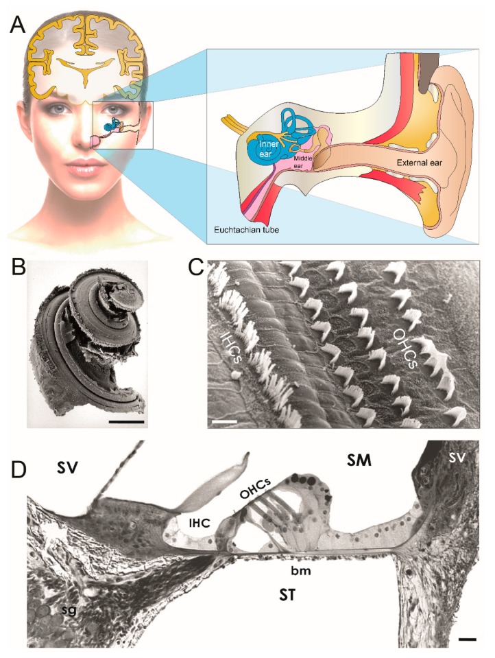
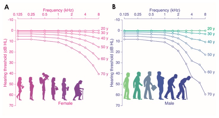
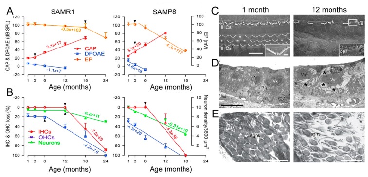
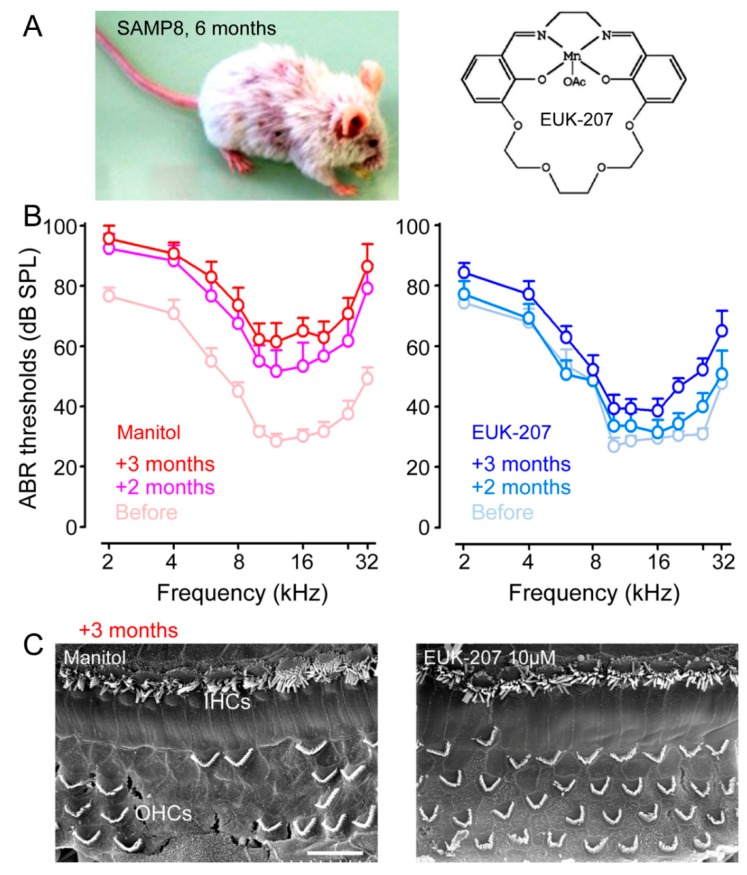
Age-related hearing impairment (ARHI), also referred to as presbycusis, is the most common sensory impairment seen in the elderly. As our cochlea, the peripheral organ of hearing, ages, we tend to experience a decline in hearing and are at greater risk of cochlear sensory-neural cell degeneration and exacerbated age-related hearing impairments, e.g., gradual hearing loss, deterioration in speech comprehension (especially in noisy environments), difficulty in the localization sound sources, and ringing sensations in the ears. However, the aging process does not affect people uniformly; nor, in fact, does the aging process appear to be uniform even within an individual. Here, we outline recent research into chronological cochlear age in healthy people, and exacerbated hearing impairments during aging due to both extrinsic factors including noise and ototoxic medication, and intrinsic factors such as genetic predisposition, epigenetic factors, and aging. We review our current understanding of molecular pathways mediating ARHL and discuss recent discoveries in experimental hearing restoration and future prospects.

Keywords: age-related hearing loss; causal factors; mechanisms; presbycusis; therapies.


Conflict of interest statement

The authors declare no conflict of interest.

Figures

Figure 1
Figure 1
Inner-ear anatomy. (A) Schematic representation of ear anatomy. The ear is divided into three parts (insert): the external and middle ear transfer the sound waves to the inner ear where they are transduced into neural activity. The external ear is closed off from the middle ear by the eardrum. In the middle ear, the eardrum is mechanically linked, by a chain of three tiny bones (the ossicles), to the oval-window membrane which closes the inner ear. Embedded in the temporal bone, the inner ear comprises the balance organ or vestibule, and the hearing organ or cochlea. (B) Scanning electron micrograph of the organ of Corti. The cochlea is a coiled organ that forms a spiral. Scanning electron micrographs show a narrow, linear shape of IHC stereocilial bundles and a V-shape of OHC stereocilia. (C) Transverse section of the basal cochlear turn under light microscopy. The cochlea is made up of three canals wrapped around a bony axis, the modiolus. These canals are the scala tympani (ST), the scala vestibuli (SV), and the scala media (SM). The ST and SV are filled with perilymph. The SM is filled with endolymph. The organ of Corti is situated on the basilar membrane (bm). (B) = 2 mm, (C) = 10 µM, (D) = 50 µm. IHCs: inner hair cells; OHCs: outer hair cells ((BD) micrographs courtesy of Marc Lenoir, Inserm U1051, France).
Figure 2
Figure 2
Age-related hearing loss according to the International Organization for Standardization (ISO) 7029 standard. Shown are audiograms for females (A) and males (B). The x-axis displays the pure tone frequency (Hz) and the y-axis the hearing thresholds (dB HL). Each individual graph is representative of the median audiogram at a particular age (ranging from 20 to 70 years old, with increments of 10 years).
Figure 3
Figure 3
Imbalance between anti-aging and pro-aging mechanisms with age. The scheme drawing numerates several anti-aging and pro-aging mechanisms identified in the cochlear aging process. Anti-aging mechanisms include estrogen, autophagic damage clearance, and mitochondrial dynamic. Pro-aging mechanisms include oxidative stress, DNA damage, mitochondrial dysfunction, senescence-like phenotype, and senescence-associated inflammation. During the aging process, decreased activity of anti-aging molecules and increased activity of pro-aging properties might lead to accumulation of mutations in mitochondrial DNA, increased lysosomal pH with a resulting accumulation of lipofuscin and aggregates, and nuclear DNA damage, leading to cochlear cell degeneration and age-related hearing loss.
Figure 4
Figure 4
Functional and morphological assessments in SAMP8 and in SAMR1 mice. (A) Functional assessment. The compound action potential (CAP) threshold (red line) and distortion product otoacoustic emissions (DPOAE) amplitude (blue line) evoked by 20 kHz tone bursts, and endocochlear potential (EP) recordings (orange line, right axis)) in SAMR1 and SAMP8 mice. Fifty SAMP8 mice (n = 10 per age: 1, 3, 6, 12, 18 months) and 60 SAMR1 (n = 10 per age: 1, 3, 6, 12, 18, 24 months) mice were used for functional assessment. The lifespan of SAMR1 and SAMP8 was approximately 30 and 20 months, respectively. Note the earlier and faster increase in CAP threshold and decrease in DPOAE amplitude and EP value (arrowheads indicated broken-stick nonlinearities) in the SAMP8. In contrast to SAMR1, no CAP threshold nor DPOAEs could be recorded in 12-month-old SAMP8 mice, respectively. (B) Morphological assessment. Age-related loss of inner hair cells (IHCs, red line), outer hair cells (OHCs, blue line), and spiral ganglion neurons (SGNs, green line, right axis). At the end of the functional assessment period, the cochleae were removed and prepared for hair cell counting using SEM (n = 5 per age per strain) and SNG using light microscopy (n = 5 per age per strain). (C) Scanning electron microscopy in one and 12 months SAMP8 mice. Few OHCs are lacking (asterisks) among the three rows, but all IHCs are present at 1 month. The higher magnification insert shows an OHC stereociliary bundle with missing sterocilia (arrow). In a 12-month-old mouse, all OHCs and numerous IHCs (asterisks) have disappeared. The white box indicates a damaged IHC stereociliary bundle. In the insert, enlargement of the same IHC stereociliary bundle shows fused stereocilia. Scale bar = 10 µm; Insert in (A) = 1 µm. (D) Electron transmission microscopy of the stria vascularis. At one month, the three layers of strial cells, marginal (Mc), intermediate (Ic), basal (Bc) cells, and the blood vessels (Vx) appear normal. At 12 months, enlarged intercellular spaces and perivascular edema (asterisks) are seen. Scale bar = 10 µm. (E) Light microscopical evaluation of spiral ganglion loss. Shown is the normal aspect and density of neurons at 1 month, and a reduced number of spiral ganglion neurons at 12 months. Scale bar = 50 µm. (Adapted from Ménardo et al., [59]).
Figure 5
Figure 5
Pharmacological mitigation of ROS prevents loss of hearing and hair cells in SAMP8 mice (A) SAMP8 mice and EUK-207. Shown are a SAMP8 mouse aged six months and the synthetic superoxide dismutase/catalase mimetic EUK-207. (B) Physiological assessment. The auditory brainstem response (ABR) thresholds recorded before (pale red plot) and after two months (pink plot) and three months (red plot) of Manitol treatments, or before (pale blue plot) and after two months (azure plot) and three months (blue plot) of EUK-207 (10 µM) treatments. (C) Morphological assessment. Representative scanning electron micrographs showing the basal regions of cochleae from Manitol-treated (left panel) and EUK-207-treated (right panel) SAMP8 mice after three months. Scale bar = 15 µm (Adapted from Benkafadar et al. [70]).

References

    1. Franceschi C., Garagnani P., Morsiani C., Conte M., Santoro A., Grignolio A., Monti D., Capri M., Salvioli S. The Continuum of Aging and Age-Related Diseases: Common Mechanisms but Different Rates. Front. Med. 2018;5:61. doi: 10.3389/fmed.2018.00061. - DOI - PMC - PubMed
    1. Addressing the Rising Prevalence of Hearing Loss. World Health Organization; Geneva, Switzerland: 2018.
    1. Woodcock K., Pole J.D. Educational attainment, labour force status and injury: A comparison of Canadians with and without deafness and hearing loss. Int. J. Rehabil. Res. 2008;31:297–304. doi: 10.1097/MRR.0b013e3282fb7d4d. - DOI - PubMed
    1. Moscicki E.K., Elkins E.F., Baum H.M., McNamara P.M. Hearing loss in the elderly: An epidemiologic study of the Framingham Heart Study Cohort. Ear Hear. 1985;6:184–190. doi: 10.1097/00003446-198507000-00003. - DOI - PubMed
    1. Cruickshanks K.J., Wiley T.L., Tweed T.S., Klein B.E., Klein R., Mares-Perlman J.A., Nondahl D.M. Prevalence of hearing loss in older adults in Beaver Dam, Wisconsin. The Epidemiology of Hearing Loss Study. Am. J. Epidemiol. 1998;148:879–886. doi: 10.1093/oxfordjournals.aje.a009713. - DOI - PubMed