. 2020 Jan 14;9(1):210.
doi: 10.3390/cells9010210.

Hippocampal Neurogenesis Is Enhanced in Adult Tau Deficient Mice

Affiliations

Hippocampal Neurogenesis Is Enhanced in Adult Tau Deficient Mice

Marangelie Criado-Marrero et al. Cells. .

Abstract

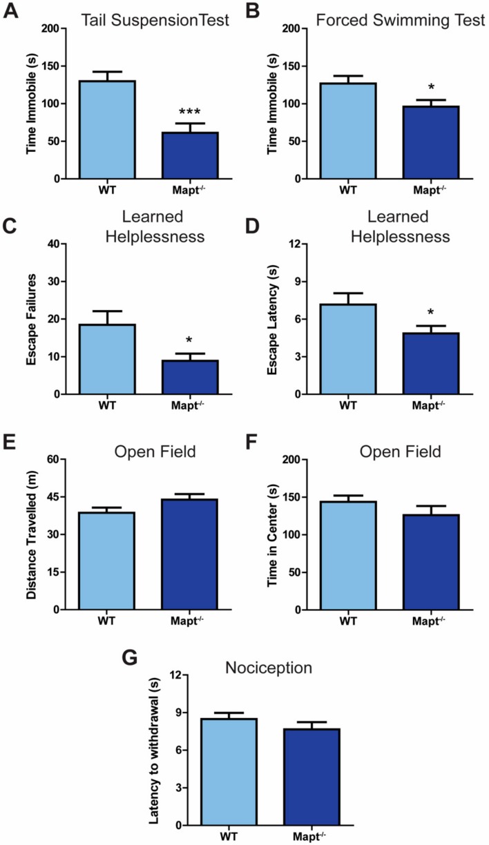
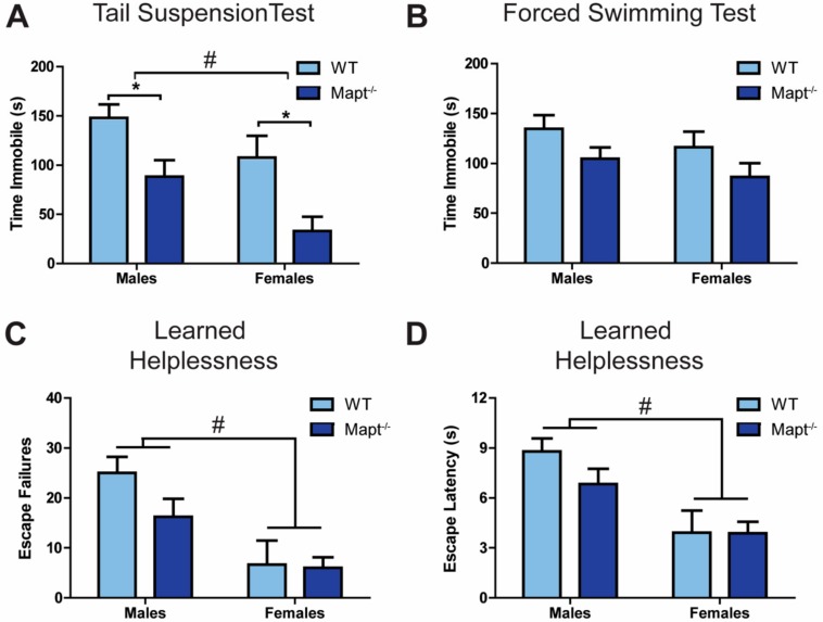
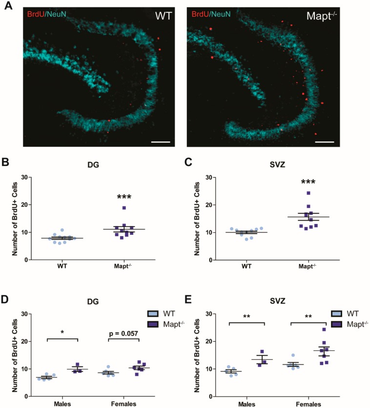
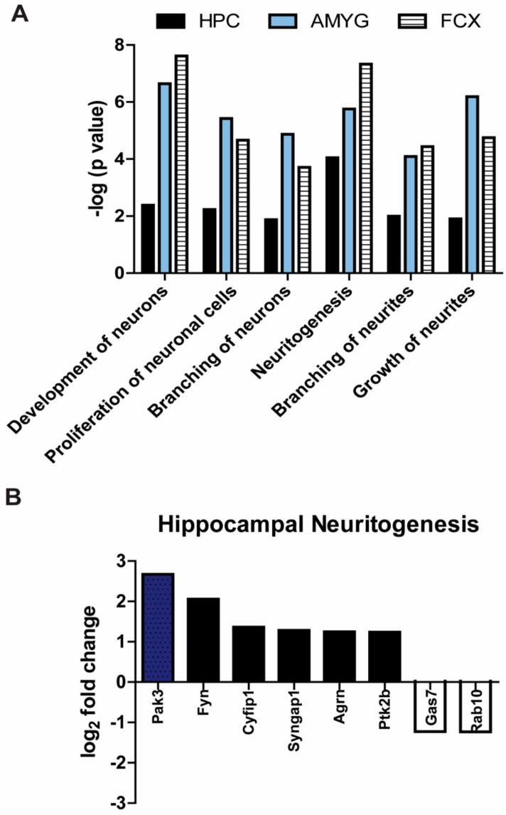
Tau dysfunction is common in several neurodegenerative diseases including Alzheimer's disease (AD) and frontotemporal dementia (FTD). Affective symptoms have often been associated with aberrant tau pathology and are commonly comorbid in patients with tauopathies, indicating a connection between tau functioning and mechanisms of depression. The current study investigated depression-like behavior in Mapt-/- mice, which contain a targeted deletion of the gene coding for tau. We show that 6-month Mapt-/- mice are resistant to depressive behaviors, as evidenced by decreased immobility time in the forced swim and tail suspension tests, as well as increased escape behavior in a learned helplessness task. Since depression has also been linked to deficient adult neurogenesis, we measured neurogenesis in the hippocampal dentate gyrus and subventricular zone using 5-bromo-2-deoxyuridine (BrdU) labeling. We found that neurogenesis is increased in the dentate gyrus of 14-month-old Mapt-/- brains compared to wild type, providing a potential mechanism for their behavioral phenotypes. In addition to the hippocampus, an upregulation of proteins involved in neurogenesis was observed in the frontal cortex and amygdala of the Mapt-/- mice using proteomic mass spectrometry. All together, these findings suggest that tau may have a role in the depressive symptoms observed in many neurodegenerative diseases and identify tau as a potential molecular target for treating depression.

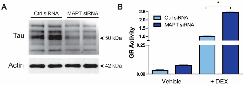
Keywords: Alzheimer’s disease; depression; glucocorticoid receptor; hippocampal neurogenesis; stress; tauopathies.


Conflict of interest statement

The authors declare no conflict of interest. The funders had no role in the design of the study; in the collection, analyses, or interpretation of data; in the writing of the manuscript, or in the decision to publish the results.

Figures

Figure 1
Figure 1
Tau ablation increased resiliency to depressive-like symptoms. Immobility time was measured for Mapt−/− and WT mice using the (A) tail suspension and (B) forced swim tasks. In the learned helplessness paradigm, (C) escape failures and (D) escape times were measured. (E) Total distance traveled and (F) time spent in the center in the open field task was measured for Mapt−/− and WT mice. (G) A hot-plate was used to measure nociception in Mapt−/− and WT mice. Data are represented as standard error of the mean (SEM) and analyzed by Student’s t-test. WT, wild type. A total number of 15–20 animals were used per genotype. Significant results were considered when * p < 0.05, *** p < 0.001.
Figure 2
Figure 2
Depressive-like behavior is more pronounced in female mice. Data from Figure 1 broken down by sex for Mapt−/− and WT mice in the (A) tail suspension and (B) forced swim tasks, and (C,D) the learned helplessness paradigm. Data are represented as standard error of the mean (SEM) and analyzed by two-way ANOVA followed by Bonferroni posthoc test. WT, wild type. Significant results by genotype * p < 0.05 or by sex # p < 0.05.
Figure 3
Figure 3
Tau knockdown increased GR activity in vitro. (A) M17 cells were incubated with either control or MAPT siRNAs for 72 hrs. Following transfection, cells lysates were collected and reduction of tau levels was confirmed by Western blot. (B) After MAPT and control siRNA transfection, M17 cells were cotransfected with a GRE-luciferase reporter and a GR cDNA plasmid for 2 days prior to treatment with vehicle (DMSO) (n =3) or 50 nM DEX (n =3) for 4 h. Experiment was run in triplicate. GR activity was measured using a GRE reporter luciferase assay. Data are represented as standard error of the mean (SEM) and analyzed by Student’s t-test. Significant results were considered when * p < 0.05. DEX, dexamethasone; GR, glucocorticoid receptor; MAPT siRNA, short interfering RNA targeting MAPT.
Figure 4
Figure 4
HPA axis function was unaltered in tau knockout mice. (A) Serum CORT levels were measured by ELISA in Mapt−/− and WT mice in unstressed and stressed conditions (n = 10 per group). (B) CORT induction following dexamethasone injection was measured in serum from Mapt−/− and WT mice (WT = 10, Mapt−/− = 12). Data are represented as standard error of the mean (SEM) and analyzed by two-way ANOVA (non-stressed vs. stressed) or Student’s t-test (dexamethasone suppression test). CORT, corticosterone; Dex, dexamethasone; WT, wild type.
Figure 5
Figure 5
Upregulation of neurogenesis occurs in the dentate gyrus and subventricular zone of Mapt−/− mice. Brains from 14- months-old WT and Mapt−/− mice were collected and stained with anti-BrdU (proliferating cell marker) and anti-NeuN (neuronal nuclei marker). (A) A representative 10× image of BrdU+/NeuN staining in Mapt−/− and WT tissue is shown. Scale bar represents 100 µm. Quantification of BrdU+ staining was performed in the (B) DG and (C) SVZ. Quantification of BrdU+ staining by sex is also shown in the (D) DG and (E) SVZ brain areas. Data are represented as standard error of the mean (SEM) and analyzed by two-way ANOVA (neurogenesis by sex) and Student’s t-test (WT vs. Mapt−/−). Significant results are shown as * p < 0.05, ** p < 0.01, *** p < 0.001. DG, dentate gyrus; SVZ, subventricular zone; WT, wild type; BrdU+, 5-bromo-2-deoxyuridine positive cells.
Figure 6
Figure 6
Processes involved in neurogenesis are significantly upregulated in adult Mapt−/− mice. (A) Neural tissue homogenates from three different brain regions Hippocampus (HPC); amygdala (AMYG); frontal cortex (FCX) of 14-month-old Mapt−/− and WT mice were analyzed by proteomic mass spectrometry and then by Ingenuity Pathway Analysis software. We examined top processes involved in neurogenesis. (B) Our proteomics data showed that neuritogenesis was greatly increased in the hippocampus, so we selected the top proteins altered in this process. Fold change of proteins in the frontal cortex and amygdala can be found in Table S1. Data are displayed as difference in Mapt−/− mice compared to WT (n = 3 per genotype).

References

    1. Braak H., Braak E. Staging of Alzheimer’s Disease-Related Neurofibrillary Changes. Neurobiol. Aging. 1995;16:271–278. doi: 10.1016/0197-4580(95)00021-6. - DOI - PubMed
    1. Pozueta J., Lefort R., Shelanski M.L. Synaptic Changes in Alzheimer’s Disease and Its Models. Neuroscience. 2013;251:51–65. doi: 10.1016/j.neuroscience.2012.05.050. - DOI - PubMed
    1. Mufson E.J., Ward S., Binder L. Prefibrillar Tau Oligomers in Mild Cognitive Impairment and Alzheimer’s Disease. Neurodegener. Dis. 2014;13:151–153. doi: 10.1159/000353687. - DOI - PMC - PubMed
    1. Bierer L.M., Haroutunian V., Gabriel S., Knott P.J., Carlin L.S., Purohit D.P., Perl D.P., Schmeidler J., Kanof P., Davis K.L. Neurochemical Correlates of Dementia Severity in Alzheimer’s Disease: Relative Importance of the Cholinergic Deficits. J. Neurochem. 2002;64:749–760. doi: 10.1046/j.1471-4159.1995.64020749.x. - DOI - PubMed
    1. Gómez-Isla T., Hollister R., West H., Mui S., Growdon J.H., Petersen R.C., Parisi J.E., Hyman B.T. Neuronal Loss Correlates with but Exceeds Neurofibrillary Tangles in Alzheimer’s Disease. Ann. Neurol. 1997;41:17–24. doi: 10.1002/ana.410410106. - DOI - PubMed

Publication types

LinkOut - more resources