. 2020 Jan 3;21(1):326.
doi: 10.3390/ijms21010326.

Age Associated Decrease of MT-1 Melatonin Receptor in Human Dermal Skin Fibroblasts Impairs Protection Against UV-Induced DNA Damage

Affiliations

Age Associated Decrease of MT-1 Melatonin Receptor in Human Dermal Skin Fibroblasts Impairs Protection Against UV-Induced DNA Damage

Kelly Dong et al. Int J Mol Sci. .

Abstract

The human body follows a physiological rhythm in response to the day/night cycle which is synchronized with the circadian rhythm through internal clocks. Most cells in the human body, including skin cells, express autonomous clocks and the genes responsible for running those clocks. Melatonin, a ubiquitous small molecular weight hormone, is critical in regulating the sleep cycle and other functions in the body. Melatonin is present in the skin and, in this study, we showed that it has the ability to dose-dependently stimulate PER1 clock gene expression in normal human dermal fibroblasts and normal human epidermal keratinocytes. Then we further evaluated the role of MT-1 melatonin receptor in mediating melatonin actions on human skin using fibroblasts derived from young and old subjects. Using immunocytochemistry, Western blotting and RT-PCR, we confirmed the expression of MT-1 receptor in human skin fibroblasts and demonstrated a dramatic age-dependent decrease in its level in mature fibroblasts. We used siRNA technology to transiently knockdown MT-1 receptor in fibroblasts. In these MT-1 knockdown cells, UV-dependent oxidative stress (H2O2 production) was enhanced and DNA damage was also increased, suggesting a critical role of MT-1 receptor in protecting skin cells from UV-induced DNA damage. These studies demonstrate that the melatonin pathway plays a pivotal role in skin aging and damage. Moreover, its correlation with skin circadian rhythm may offer new approaches for decelerating skin aging by modulating the expression of melatonin receptors in human skin.

Keywords: aging; circadian rhythm; melatonin; skin fibroblasts; ultraviolet (UV) irradiation.


Conflict of interest statement

The authors declare no conflict of interest.

Figures
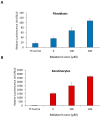
Figure 1
Figure 1
PER1 expression increases in response to higher concentration of melatonin. (A) Normal Human Dermal Fibroblasts (NHDF) and (B) Normal Human Epidermal Keratinocytes (NHEK) were incubated with different concentration of melatonin for 24 h, and the level of expression of PER1 was evaluated using a reporter gene assay. Tf Control, transfection control. Error bars are SEM. N = 5.
Figure 2
Figure 2
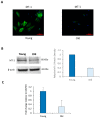
Expression of melatonin MT-1 receptor in adult NHDF declines with age. (A) MT-1 localization was visualized using specific antibodies as described in Methods. (B) Protein analysis by Western blot and (C) mRNA analysis by RT-PCR of fibroblast extracts derived from young and old subjects. Error bars are SEM.
Figure 3
Figure 3
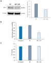
Knockdown of MT-1 receptor. (A) Young NHDF were electroporated with MT-1 receptor specific siRNA. Protein levels were measured by Western blot 72 h post-electroporation and (B) mRNA levels by RT-PCR 48 h post-electroporation. NHDF were electroporated with non-targeting (NT) siRNA for comparison. (C) The viability of electroporated NHDF was compared with the untreated cells and assessed by Alamar Blue staining. Error bars are SEM. N = 4.
Figure 4
Figure 4
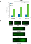
DNA damage is elevated in MT-1 knockdown NHDF exposed to UVA (5 J/cm2) and UVB (40 mJ/cm2). The cells were then assayed for DNA damage by subjecting the cells embedded in agarose to electrophoresis and then staining for DNA with SYBR Gold. An increased length of DNA staining in the immunocytochemistry figures (“comet”) indicates increased damage. Panel (A) shows the graphical representation of the “comet tail” and, panel (B) shows the actual immunocytochemistry photos (*** p < 0.0001, ** p < 0.01). Error bars are SEM. N = 18.
Figure 5
Figure 5
H2O2 generation is elevated in MT-1 knockdown NHDF exposed to UV. NHDF were treated with non-targeted siRNA (NT), targeted siRNA (MT-1 KD) or untreated, and exposed to UV (5 J/cm2 of UVA + 50 mJ/cm2 of UVB). The amount of H2O2 generated were quantitated as described in Methods and normalized to the total cellular protein. Error bars are SEM. N = 4.

References

    1. Bickers D.R., Athar M. Oxidative Stress in the Pathogenesis of Skin Disease. J. Investig. Dermatol. 2006;126:2565–2575. doi: 10.1038/sj.jid.5700340. - DOI - PubMed
    1. Thiele J.J., Schroeter C., Hsieh S.N., Podda M., Packer L. Oxidants and Antioxidants in Cutaneous Biology. Volume 29. Karger Publishers; Basel, Switzerland: 2001. The antioxidant network of the stratum corneum; pp. 26–42. - PubMed
    1. Briganti S., Picardo M. Antioxidant activity, lipid peroxidation and skin diseases. What’s new. J. Eur. Acad. Dermatol. Venerol. 2003;17:663–669. doi: 10.1046/j.1468-3083.2003.00751.x. - DOI - PubMed
    1. Oresajo C., Pillai S., Yatskayer M., Puccetti G., McDaniel D. Antioxidants and Skin Aging: A Review. Cosmet. Dermatol. 2009;22:563–570.
    1. Slominski A., Tobin D.J., Zmijewski M.A., Wortsman J., Paus R. Melatonin in the skin: Synthesis, metabolism and functions. Trends Endocrinol. Metab. 2007;19:17–24. doi: 10.1016/j.tem.2007.10.007. - DOI - PubMed

MeSH terms

Substances

LinkOut - more resources