. 2020 Jan 3;10(1):78.
doi: 10.3390/biom10010078.

Voltage Sensing in Bacterial Protein Translocation

Affiliations

Voltage Sensing in Bacterial Protein Translocation

Denis G Knyazev et al. Biomolecules. .

Abstract

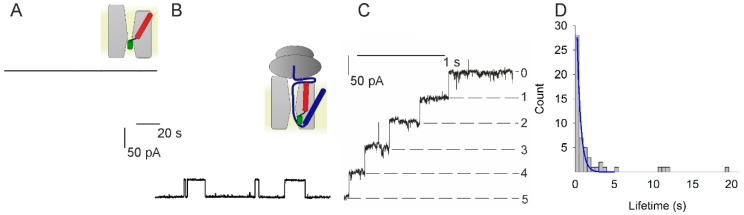
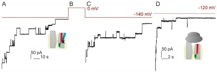
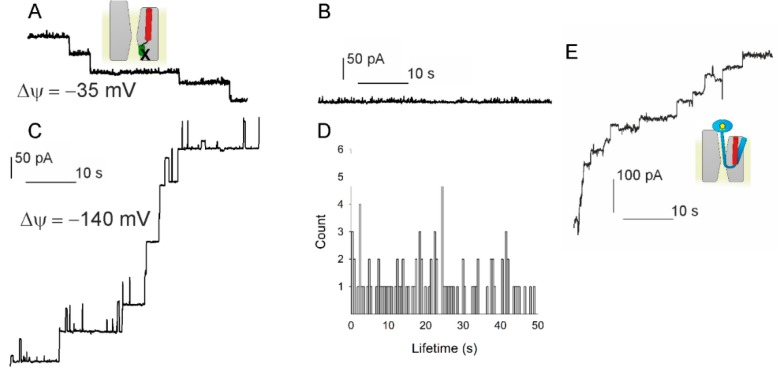
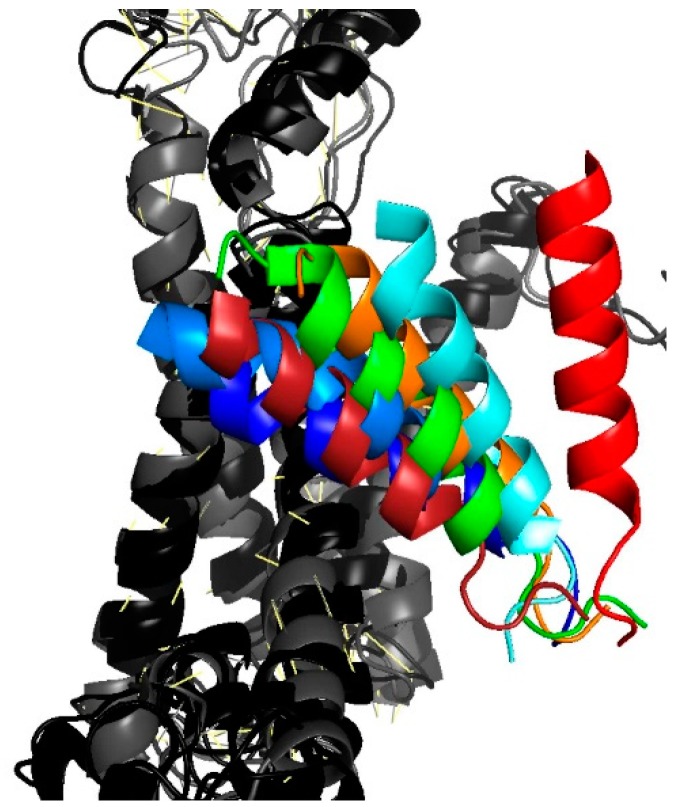
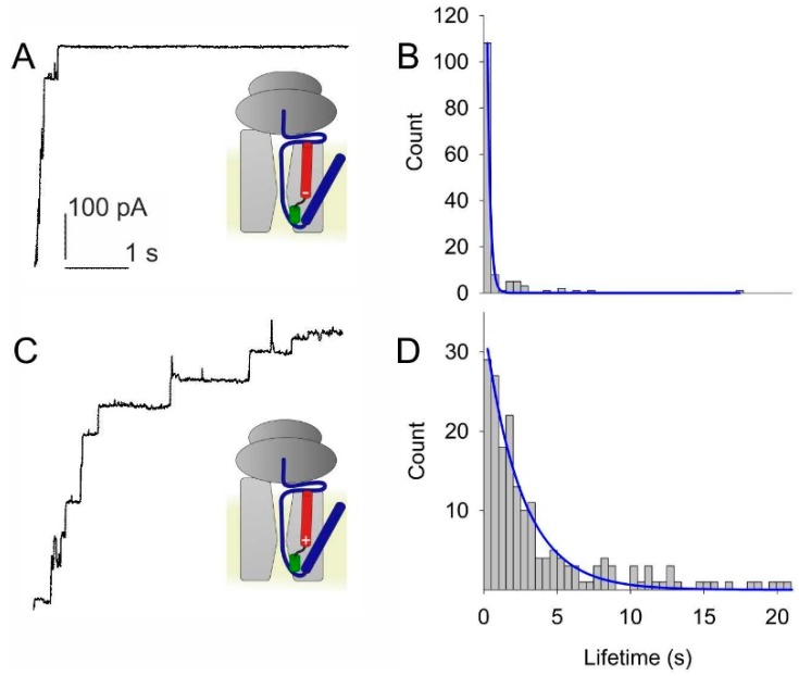
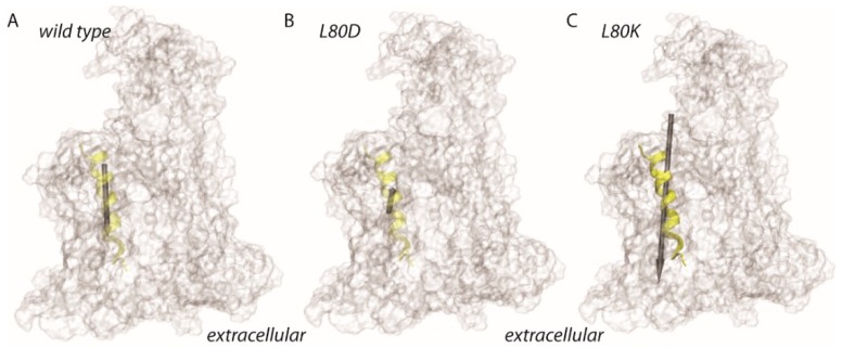
The bacterial channel SecYEG efficiently translocates both hydrophobic and hydrophilic proteins across the plasma membrane. Translocating polypeptide chains may dislodge the plug, a half helix that blocks the permeation of small molecules, from its position in the middle of the aqueous translocation channel. Instead of the plug, six isoleucines in the middle of the membrane supposedly seal the channel, by forming a gasket around the translocating polypeptide. However, this hypothesis does not explain how the tightness of the gasket may depend on membrane potential. Here, we demonstrate voltage-dependent closings of the purified and reconstituted channel in the presence of ligands, suggesting that voltage sensitivity may be conferred by motor protein SecA, ribosomes, signal peptides, and/or translocating peptides. Yet, the presence of a voltage sensor intrinsic to SecYEG was indicated by voltage driven closure of pores that were forced-open either by crosslinking the plug to SecE or by plug deletion. We tested the involvement of SecY's half-helix 2b (TM2b) in voltage sensing, since clearly identifiable gating charges are missing. The mutation L80D accelerated voltage driven closings by reversing TM2b's dipolar orientation. In contrast, the L80K mutation decelerated voltage induced closings by increasing TM2b's dipole moment. The observations suggest that TM2b is part of a larger voltage sensor. By partly aligning the combined dipole of this sensor with the orientation of the membrane-spanning electric field, voltage may drive channel closure.

Keywords: Sec61; SecY; gating; translocon.


Conflict of interest statement

The authors declare no conflict of interest.

Figures

Figure 1
Figure 1
Channel activity of the SecYEG-RNC (ribosome-nascent chain) complex. (A) The planar bilayer did not show channel activity when only SecYEG-containing vesicles were added to the cis compartment (φ = −85 mV). (B) However, the subsequent addition of RNCs resulted in channel activity (φ = −85 mV). (C) Augmenting the membrane potential to φ = −140 mV resulted in channel closure. The negative potential induces the current to flow in the negative direction of the y-axis. Thus, the apparent movement upstairs indicates stepwise channel closure. The number of open channels is indicated on the right side. It decreases from five open channels at the moment of switching the potential to −140 mV, to zero open channels after little more than one second has elapsed. (D) Channel lifetime histogram for φ = −140 mV. From the single-exponential fit (blue) we extracted a channel lifetime of about 0.5 s. The colored schemes show SecYEG (light gray), the ribosome (dark grey), the nascent chain (blue), SecYEG’s helix 2b (red), and SecYEG’s plug domain (green).
Figure 2
Figure 2
Voltage sensitivity is not conferred to the translocon by the signal peptide. (AC) Closings of SecY(S329C)EG crosslinked to the signal peptide of OmpA (21C) at φ = −140 mV. The voltage protocol is shown in red above. (D) SecYEG in complex with empty ribosome also closes in response to voltage. Note the shorter time scale here as compared to (A,C). The small schemes obey the color code from Figure 1: with the signal peptide in light blue, nascent chain in blue and the cross-link depicted as X.
Figure 3
Figure 3
Testing the hypothesis that voltage driven plug movement confers voltage sensitivity to SecYEG (scheme in the upper left panel). (A) Crosslinking the plug to SecE by 1 mM potassium tetrathionate (KTT) in the SecY(F67C)E(S120C)G mutant forces reconstituted translocons to open. Single channels were recorded at φ = −35 mV. (B) At small φ values the channels virtually do not close. (C) φ = −140 mV elicits a conformational change that closes the channel. Scale bars for A–C are the same. (D) The channel lifetime histogram shows a wide scatter for −140 mV. (E) The plug deletion mutant SecYEG (Δ60–74) retains voltage sensitivity. Channel activity was observed when fusing the mutant containing proteoliposomes in the presence of SecA, ATP, Methotrexate (MTX), and proOmpA-DHFR. MTX binding to DHFR maintains the globular structure of the latter and thus prevents full translocation. φ = −85 mV closes the channels. The schematic representations use the same colour code as in Figure 1: proOmpA in light blue, MTX in complex with DHFR as a yellow pentagon inside a blue ellipse and the cross-link depicted as X. The experiments showed that plug immobilization or removal does not abolish SecYEG’s voltage sensitivity.
Figure 4
Figure 4
Movement of TM2b as gathered from an overlay of different translocon structures. The position of the colored TM2b is shown relative to the second half of the protein that consists of helixes 6 through 10 (in shades of gray). Closed conformations are represented by the PDB entries 4CG7 in dark blue and 4CG5 in light blue [9], 3J45 in dark red [28]. Partially or fully open conformations: 3J7R in green [29] (Sec61-RNC), 5EUL in orange [30] (SecYEG-pOA-SecA), 4CG6 in cyan [9] (RNC-SecYEG), 3J46 in red [28] (RNC-SecYEG).
Figure 5
Figure 5
Test of the hypothesis that the partial charges at the poles of helix 2b act as voltage sensors. (A) Adding a negatively charged residue to the positive pole of the helix by site directed mutagenesis (L80D) accelerate voltage induced channel closings. (B) A single exponential fit (blue line) to the lifetime histogram indicates a channel lifetime of about 0.2 s at φ = −140 mV. (C) In contrast, closings at φ = −140 mV of SecYEG-RNC complex are slower with the addition of a positive residue to the tip of helix 2b (L80K). (D) The single exponential fit to the lifetime histogram shows a lifetime of about 2.5 s. The color code of the small schemes is the same as in Figure 1.
Figure 6
Figure 6
Schematic representation of the dipole moment estimates for TM2b of E. coli SecY. Dipole moments were estimated for the amino acid residues A79-P100, which are depicted as a transparent yellow ribbon. The protein is shown as a transparent surface. (AC) Dipole moment estimates for helix 2 of wild type SecY (62.4D, panel A), L80D (25.7D, panel B), and for L80K (153.1D, panel C). The size of the gray arrow indicates the relative size of the dipole moments.

References

    1. Spiess M., Junne T., Janoschke M. Membrane protein integration and topogenesis at the ER. Protein J. 2019;38:306–316. doi: 10.1007/s10930-019-09827-6. - DOI - PubMed
    1. Seinen A.B., Driessen A.J.M. Single-molecule studies on the protein translocon. In: Dill K.A., editor. Annual Review of Biophysics, 48. Annual Reviews; Palo Alto, CA, USA: 2019. pp. 185–207. - PubMed
    1. Saparov S.M., Erlandson K., Cannon K., Schaletzky J., Schulman S., Rapoport T.A., Pohl P. Determining the conductance of the SecY protein translocation channel for small molecules. Mol. Cell. 2007;26:501–509. doi: 10.1016/j.molcel.2007.03.022. - DOI - PubMed
    1. van den Berg B., Clemons W.M., Jr., Collinson I., Modis Y., Hartmann E., Harrison S.C., Rapoport T.A. X-ray structure of a protein-conducting channel. Nature. 2004;427:36–44. doi: 10.1038/nature02218. - DOI - PubMed
    1. Simon S.M., Blobel G. Signal peptides open protein-conducting channels in e. Coli. Cell. 1992;69:677–684. doi: 10.1016/0092-8674(92)90231-Z. - DOI - PubMed

Publication types

MeSH terms

LinkOut - more resources