. 2020 Mar 3;141(9):751-767.
doi: 10.1161/CIRCULATIONAHA.119.042559. Epub 2020 Jan 17.

Natural Compound Library Screening Identifies New Molecules for the Treatment of Cardiac Fibrosis and Diastolic Dysfunction

Affiliations

Natural Compound Library Screening Identifies New Molecules for the Treatment of Cardiac Fibrosis and Diastolic Dysfunction

Katharina Schimmel et al. Circulation. .

Abstract

Background: Myocardial fibrosis is a hallmark of cardiac remodeling and functionally involved in heart failure development, a leading cause of deaths worldwide. Clinically, no therapeutic strategy is available that specifically attenuates maladaptive responses of cardiac fibroblasts, the effector cells of fibrosis in the heart. Therefore, our aim was to develop novel antifibrotic therapeutics based on naturally derived substance library screens for the treatment of cardiac fibrosis.

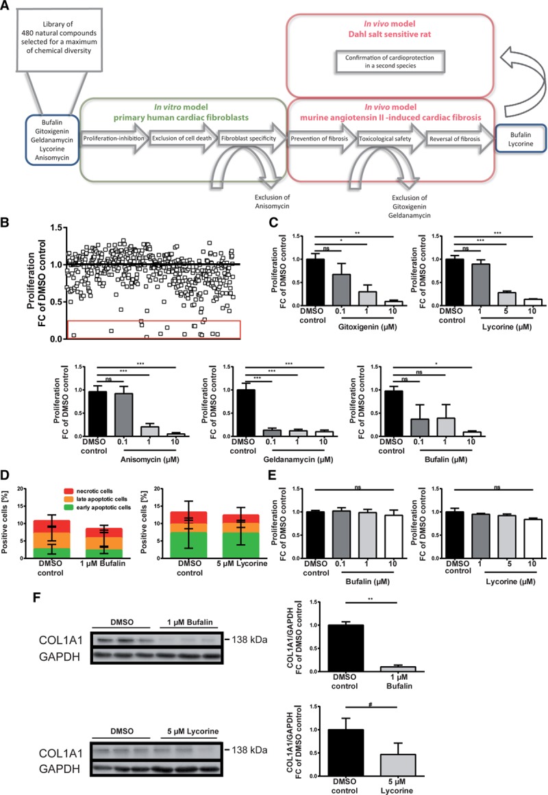
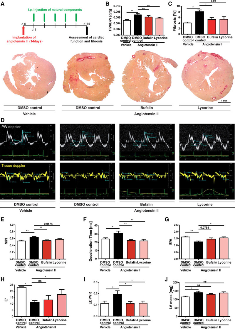
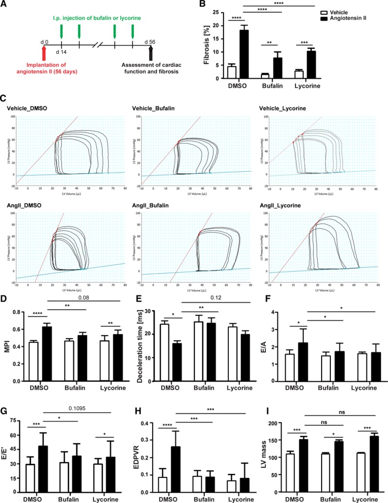
Methods: Antifibrotic drug candidates were identified by functional screening of 480 chemically diverse natural compounds in primary human cardiac fibroblasts, subsequent validation, and mechanistic in vitro and in vivo studies. Hits were analyzed for dose-dependent inhibition of proliferation of human cardiac fibroblasts, modulation of apoptosis, and extracellular matrix expression. In vitro findings were confirmed in vivo with an angiotensin II-mediated murine model of cardiac fibrosis in both preventive and therapeutic settings, as well as in the Dahl salt-sensitive rat model. To investigate the mechanism underlying the antifibrotic potential of the lead compounds, treatment-dependent changes in the noncoding RNAome in primary human cardiac fibroblasts were analyzed by RNA deep sequencing.

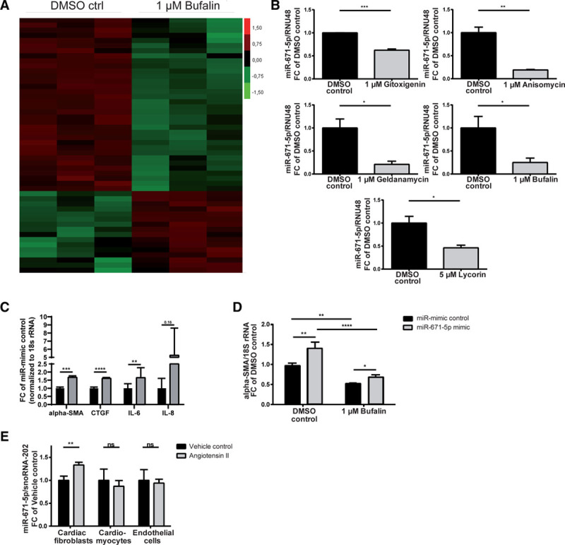
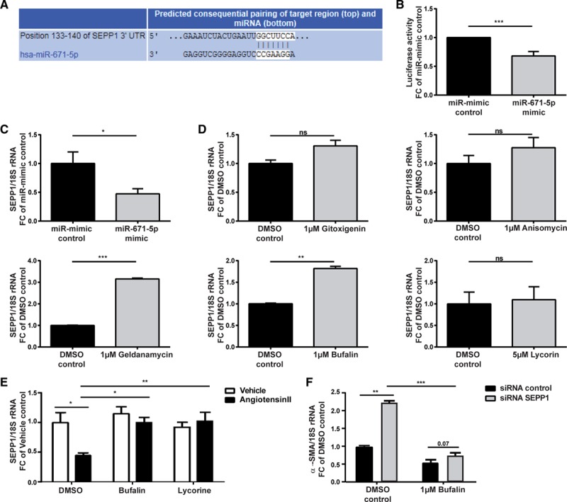
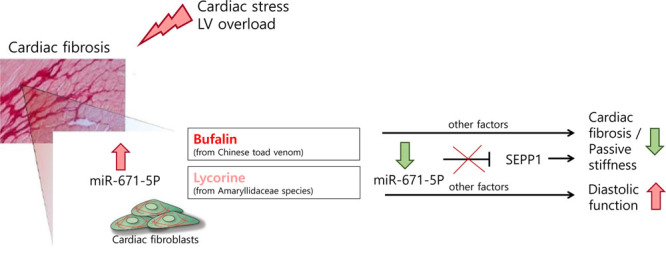
Results: High-throughput natural compound library screening identified 15 substances with antiproliferative effects in human cardiac fibroblasts. Using multiple in vitro fibrosis assays and stringent selection algorithms, we identified the steroid bufalin (from Chinese toad venom) and the alkaloid lycorine (from Amaryllidaceae species) to be effective antifibrotic molecules both in vitro and in vivo, leading to improvement in diastolic function in 2 hypertension-dependent rodent models of cardiac fibrosis. Administration at effective doses did not change plasma damage markers or the morphology of kidney and liver, providing the first toxicological safety data. Using next-generation sequencing, we identified the conserved microRNA 671-5p and downstream the antifibrotic selenoprotein P1 as common effectors of the antifibrotic compounds.

Conclusions: We identified the molecules bufalin and lycorine as drug candidates for therapeutic applications in cardiac fibrosis and diastolic dysfunction.

Keywords: diastole; fibrosis; hypertension; microRNAs.


Figures

Figure 1.
Figure 1.
Bufalin and lycorine potently and specifically repress fibrotic responses in human cardiac fibroblasts (HCFs). A, Filtering strategy, followed by in vitro and in vivo pipeline of analysis, uncovers antifibrotic lead compounds bufalin and lycorine. B, Functional screen of 480 naturally derived substances in vitro yielding natural compounds inhibiting the proliferation of HCFs. The cells were incubated for 24 hours as indicated, and proliferation of HCFs was measured by BrdU-ELISA. Fifteen candidates inhibiting the fibroblast proliferation fold change (FC) ≥75% are highlighted by the red rectangle. C, Dose-dependent inhibitory effects of bufalin, gitoxigenin, lycorine, anisomycin, and geldanamycin on proliferation of HCFs. The cells were incubated for 24 hours as indicated, and the proliferation of HCFs was measured by BrdU-ELISA (1-way ANOVA, Dunnett multiple-comparisons test, n=4-6). D, Positively validated hits do not induce cell death in HCFs. Cells were treated with respective compounds for 24 hours as indicated and subjected to fluorescence-activated cell sorter analysis after annexin-7AAD staining (unpaired t test, n=3). E, Dose-dependent inhibitory effects of bufalin and lycorine on proliferation of HCFs are fibroblast specific, as evidenced by no impact of the same concentrations of respective substances on the proliferation of the cardiomyocyte cell line HL-1. Cells were treated with the compounds for 24 hours as indicated, and proliferation of HL-1 cells was measured by BrdU-ELISA (1-way ANOVA, Dunnett multiple-comparisons test, n=3). F, Bufalin and, to a lesser extent, lycorine decrease expression levels of collagen type I, α1 (COL1A1) in HCFs shown in a representative Western blot. Cells were treated with respective substances for 24 hours as indicated, lysed, and analyzed for COL1A1 protein levels (normalized to GAPDH; unpaired t test). All values from C through F are presented as mean±SEM. DMSO indicates dimethyl sulfoxide; and ns, not significant. *P<0.05, **P<0.01, ***P<0.001, #P=0.265.
Figure 2.
Figure 2.
Bufalin and lycorine protect from angiotensin II (Ang II)–induced fibrotic cardiac disease in mice. A, Schematic representation of the preventive in vivo study using a murine model of hypertensive heart disease. Diastolic dysfunction was induced in C57BL/6 mice via implantation of Ang II–filled minipumps, and bufalin or lycorine was injected intraperitoneally every other day during 2 consecutive weeks starting 1 day after the operation. Fourteen days after the operation, hearts were explanted for biochemical and histological analysis. B, Reduced heart-to-body weight ratios (HW/BW) on treatment as indicated compared with control animals (1-way ANOVA, Tukey multiple-comparisons test, n=14/17/17/14). C, Prevention of collagen deposition shown in representative images of cardiac histological sections of the whole heart and quantification of Picrosirius red–stained areas on administration of bufalin and lycorine (1-way ANOVA, Tukey multiple-comparisons test, n=10/11/10/11), scale bar=1 mm. D, Isovolumic contraction time, isovolumic relaxation time, aortic ejection time, and E and A waves are measured by pulsed-wave (PW) Doppler from the apical 4-chamber view from the lateral mitral valve (top). Early (E’) and atrial (A’) peak velocities are measured from tissue Doppler signal at the mitral annulus (bottom). Preservation of myocardial performance index (MPI; E; 1-way ANOVA, Tukey multiple-comparisons test, n=11/21/16/12), deceleration time (F; 1-way ANOVA, Tukey multiple-comparisons test, n=11/21/16/12), improvement of peak E/A ratio (G; 1-way ANOVA, Tukey multiple-comparisons test, n=8/16/11/7), and improved E’ peak value (H; 1-way ANOVA, Tukey multiple-comparisons test, n=8/16/11/7) after treatment with compounds as indicated. I, Preservation of left ventricular (LV) compliance by bufalin or lycorine, assessed by end-diastolic pressure-volume relationship (EDPVR) obtained by linear fits of the EDPVR slope, resulting from the shift of pressure-volume loops after transient vena cava occlusions (1-way ANOVA, Tukey multiple-comparisons test, n=8/9/6/8). J, Significant increase in LV mass by Ang II infusion in animals treated with the solvent only (controls) but not in mice treated with bufalin or lycorine (1-way ANOVA, Tukey multiple-comparisons test, n=8/11/9/9). All values from B through J are presented as mean±SEM. DMSO indicates dimethyl sulfoxide. *P<0.05. **P<0.01.
Figure 3.
Figure 3.
Bufalin and lycorine reverse established fibrosis and improve heart function in hypertensive mice. A, Therapeutic in vivo study using a murine model of systemic hypertension. Bufalin, lycorine, or dimethyl sulfoxide (DMSO) was injected intraperitoneally every other day for 6 consecutive weeks starting 2 weeks after the implantation of minipumps filled with angiotensin II (Ang II). B, Significant amelioration of cardiac fibrosis in established diastolic heart failure on treatment with bufalin and lycorine by 50% (2-way ANOVA, Tukey multiple-comparisons test, n=19/8/7/17/18/16). C, Representative images of hemodynamic pressure-volume loops. Both compounds were able to partly reverse diastolic dysfunction; bufalin recovered hemodynamic parameters (dP/dtmin, tau, and end-diastolic pressure-volume relationship [EDPVR]) compared with the Ang II group. D through F, Strong trend of reduction of myocardial performance index (MPI; 1-way ANOVA, n=19/8/7/17/18/16) and prolongation of deceleration time (2-way ANOVA, Tukey multiple-comparisons test, n=19/8/7/17/18/16) by the lead compounds and decreased peak E/A (2-way ANOVA, Tukey multiple-comparisons test, n=18/8/7/14/15/15) on treatment with compounds. G,Strong trend in reduction of peak E/E’ on treatment with both bufalin and lycorine (2-way ANOVA, Tukey multiple-comparisons test, n=14/8/7/14/14/15). H, Significant improvement of load-independent EDPVR by bufalin and lycorine on systemic hypertension (2-way ANOVA, Tukey multiple-comparisons test, n=14/8/7/14/14/15). I, Significant increase in left ventricular (LV) mass by Ang II infusion in animals treated with the solvent only (controls) but not in mice treated with bufalin or lycorine (2-way ANOVA, Tukey multiple-comparisons test, n=19/8/7/16/17/16). All values from B through I are presented as mean±SEM. ESPVR indicates end-systolic pressure-volume relationship; and ns, not significant. *P<0.05, **P<0.01, ***P<0.001, ****P<0.0001.
Figure 4.
Figure 4.
Lycorine and bufalin diminish fibrosis, diastolic dysfunction, and pulmonary congestion in the Dahls salt-sensitive rat. A, In vivo study using a model of hypertension-induced diastolic dysfunction. Lycorine, bufalin, or dimethyl sulfoxide (DMSO) was injected intraperitoneally every other day for 5 consecutive weeks starting 2 weeks after induction of hypertension by a high-salt diet. B through D, Effective and significant reduction of collagen volume fraction (CVF; 1-way ANOVA, Tukey multiple-comparisons test, n=17/16/10/13), collagen cross-linking (CCL; 1-way ANOVA, Tukey multiple comparisons test, n=17/16/10/13), and collagen accumulation in the myocardium as shown in representative images of Picrosirius red staining; scale bar=1 mm. E, No significant impact on cardiomyocyte area shown in representative images and corresponding quantification of heart sections (1-way ANOVA, Tukey multiple-comparisons test, n=17/16/10/13); scale bar=200 μm. F through H, Significantly ameliorated myocardial performance index (MPI) and isovolumic relaxation time/aortic ejection time (IVRT/AET) in compound-treated rats compared with rats fed a high-salt diet (1-way ANOVA, Tukey multiple-comparisons test, n=10/9/10/13), as well as significant reduction of peak E/A (1-way ANOVA, Tukey multiple-comparisons test, n=10/9/10/13) on treatment with lycorine but not with bufalin. I, Representative pulsed-wave (PW) Doppler and tissue Doppler images from each group. All values from B, C, and E through H are presented as mean±SEM. *P<0.05, **P<0.01, ***P<0.001.
Figure 5.
Figure 5.
Antifibrotic substances converge on profibrotic microRNA (miR) 671-5p. A, Representative heat map of differentially regulated miRNAs (unpaired t test P<0.05) and (B) validation of significantly reduced miR-671-5p expression in primary human cardiac fibroblasts (HCFs) on treatment as indicated compared with control (unpaired ttest, n=3). C, Activation of fibrosis markers α-smooth muscle actin (α-SMA), connective tissue growth factor (CTGF), and proinflammatory cytokines interleukin (IL)-6 and IL-8 in primary HCFs after overexpression of miR-671-5p (unpaired t test, n=5, miR-mimic control vs miR671-5p mimic). D, Restoration of diminished α-SMA expression of primary HCFs after treatment with bufalin by miR-671-5p (2-way ANOVA, Tukey multiple-comparisons test, dimethyl sulfoxide [DMSO] control vs 1 μmol/L bufalin; unpaired t test, miR-mimic control vs miR-671-5p mimic; n=5). E, MiR-671-5p expression in murine heart cell fractions after infusion with angiotensin II for 2 weeks (unpaired t test, n=6/10). All values from B through E are presented as mean±SEM. RNU48 indicates small-nucleolar RNA48. *P<0.05, **P<0.01, ***P<0.001, ****P<0.0001.
Figure 6.
Figure 6.
MicroRNA (miR) 671-5p drives fibrosis via repression of selenoprotein P1 (SEPP1). A, Predicted base pairing between the seed sequence of human miR-671-5p and target sequence in the 3′ untranslated region (UTR) of SEPP1 (TargetScanHuman, version 7.1). B, Luciferase activity levels on transfection of a luciferase construct containing the 3′ UTR of SEPP1 with miR-mimic control or miR-671-5p mimic (1-sample ttest, n=27 replicates). C, Significant reduction of SEPP1 mRNA levels in primary human cardiac fibroblasts (HCFs) after overexpression of miR-671-5p (unpaired t test, n=3). D, SEPP1 levels follow the opposite pattern compared with miR-671-5p in primary HCFs on treatment with the lead antifibrotic substances (unpaired t test, n=2-3). E, Decrease of SEPP1 levels in cardiac tissue after 8 weeks of angiotensin II infusion (therapeutic mouse study) is significantly counteracted by bufalin and lycorine (2-way ANOVA, Tukey multiple-comparison test, n=18/8/7/17/18/16). F, Restoration of diminished α-SMA expression in primary HCFs after treatment with bufalin by siRNA-mediated silencing of SEPP1 (2-way ANOVA, Sidak multiple-comparisons test, dimethyl sulfoxide [DMSO] control vs 1 μmol/L bufalin; unpaired t test, siRNA control vs siRNA SEPP1; n=4). All values from C through F are presented as mean±SEM. *P<0.05, **P<0.01, ***P<0.001.
Figure 7.
Figure 7.
Mechanistic overview. Proposed therapeutic mode of action for the antifibrotic compounds bufalin and lycorine in angiotensin II–mediated diastolic dysfunction. Treatment with the lead natural compound results in a decline in microRNA (miR) 671-5p levels in cardiac fibroblasts, which, in turn, leads to derepression of its target selenoprotein P1 (SEPP1). LV indicates left ventricular.

References

    1. Jessup M, Brozena S. Heart failure. N Engl J Med. 2003;348:2007–2018. doi: 10.1056/NEJMra021498. - PubMed
    1. Kong P, Christia P, Frangogiannis NG. The pathogenesis of cardiac fibrosis. Cell Mol Life Sci. 2014;71:549–574. doi: 10.1007/s00018-013-1349-6. - PMC - PubMed
    1. Braunwald E. Heart failure. JACC Heart Fail. 2013;1:1–20. doi: 10.1016/j.jchf.2012.10.002. - PubMed
    1. Heymans S, Gonzalez A, Pizard A, Papageorgiou AP, Lopez-Andres N, Jaisser F, Thum T, Zannad F, Diez J. Searching for new mechanisms of myocardial fibrosis with diagnostic and/or therapeutic potential. Eur J Heart Fail. 2015;17:764–771. doi: 10.1002/ejhf.312. - PubMed
    1. Cao Z, Yu D, Fu S, Zhang G, Pan Y, Bao M, Tu J, Shang B, Guo P, Yang P, et al. Lycorine hydrochloride selectively inhibits human ovarian cancer cell proliferation and tumor neovascularization with very low toxicity. Toxicol Lett. 2013;218:174–185. doi: 10.1016/j.toxlet.2013.01.018. - PubMed

Publication types

MeSH terms