Review
. 2020 Jan 17;27(1):3.
doi: 10.1186/s12929-019-0594-x.

Photoacoustic imaging of cells in a three-dimensional microenvironment

Affiliations
Review

Photoacoustic imaging of cells in a three-dimensional microenvironment

Wei-Wen Liu et al. J Biomed Sci. .

Abstract

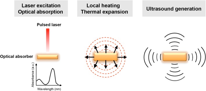
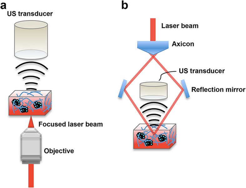
Imaging live cells in a three-dimensional (3D) culture system yields more accurate information and spatial visualization of the interplay of cells and the surrounding matrix components compared to using a two-dimensional (2D) cell culture system. However, the thickness of 3D cultures results in a high degree of scattering that makes it difficult for the light to penetrate deeply to allow clear optical imaging. Photoacoustic (PA) imaging is a powerful imaging modality that relies on a PA effect generated when light is absorbed by exogenous contrast agents or endogenous molecules in a medium. It combines a high optical contrast with a high acoustic spatiotemporal resolution, allowing the noninvasive visualization of 3D cellular scaffolds at considerable depths with a high resolution and no image distortion. Moreover, advances in targeted contrast agents have also made PA imaging capable of molecular and cellular characterization for use in preclinical personalized diagnostics or PA imaging-guided therapeutics. Here we review the applications and challenges of PA imaging in a 3D cellular microenvironment. Potential future developments of PA imaging in preclinical applications are also discussed.

Keywords: Biomedical imaging; Photoacoustic imaging; Three-dimensional cell culture; Tumor microenvironment.


Conflict of interest statement

The author declares that he has no competing interests.

Figures

Fig. 1
Fig. 1
Illustration of PA signal generation. Optical energy excited from a short-pulse laser is absorbed by optical absorbers, which causes an increase in the local temperature. An US pressure wave, the so-called PA signal, is then generated by the thermal expansion of the absorber
Fig. 2
Fig. 2
Schematics of two types of PAM system: (a) OR-PAM and (b) AR-PAM. In this setup, 3D tumor spheres labeled with contrast agents are cultured in a cuboidal matrix hydrogel for PA imaging. Note that the laser light is focused in OR-PAM but unfocused in AR-PAM, respectively. Once the laser energy is delivered into the 3D cell culture and absorbed by endogenous or exogenous contrast agents, the absorbed energy is converted into heat, leading to thermal expansion. Ultrasound signals are then generated and detected by the transducer located at the top of the samples
Fig. 3
Fig. 3
Scalability of PAM among multiscale biological systems. The blue circles denote lateral resolution, and green circles denote axial resolution. Solid lines denote OR-PAMs, and dotted lines denote AR-PAMs. LA-PACT, linear-array PA computed tomography [37]; PAMac, PA macroscopy [38]; AR-PAM, acoustic resolution PAM [39]; OR-PAM, optical resolution PAM [40]; 125-MHz-PAM, PAM using a 125-MHz ultrasound detector [41]; SW-PAM, subwavelength resolution PAM [42]; PI-PAM, photoimprint PAM [43]. Figure adapted from [44]

References

    1. Duval K, Grover H, Han LH, Mou Y, Pegoraro AF, Fredberg J, et al. Modeling physiological events in 2D vs. 3D cell culture. Physiology (Bethesda) 2017;32:266–277. - PMC - PubMed
    1. Gillet JP, Calcagno AM, Varma S, Marino M, Green LJ, Vora MI, et al. Redefining the relevance of established cancer cell lines to the study of mechanisms of clinical anti-cancer drug resistance. Proc Natl Acad Sci U S A. 2011;108:18708–18713. doi: 10.1073/pnas.1111840108. - DOI - PMC - PubMed
    1. Katt ME, Placone AL, Wong AD, Xu ZS, Searson PC. In vitro tumor models: advantages, disadvantages, variables, and selecting the right platform. Front Bioeng Biotechnol. 2016;4:12. doi: 10.3389/fbioe.2016.00012. - DOI - PMC - PubMed
    1. Liu Y, Chen YG. 2D- and 3D-based intestinal stem cell cultures for personalized medicine. Cells. 2018;7:225. doi: 10.3390/cells7120225. - DOI - PMC - PubMed
    1. Yamada KM, Cukierman E. Modeling tissue morphogenesis and cancer in 3D. Cell. 2007;130:601–610. doi: 10.1016/j.cell.2007.08.006. - DOI - PubMed

MeSH terms