. 2020 Jan 16;11(1):28.
doi: 10.1038/s41419-019-2214-9.

Targeting alkaline ceramidase 3 alleviates the severity of nonalcoholic steatohepatitis by reducing oxidative stress

Affiliations

Targeting alkaline ceramidase 3 alleviates the severity of nonalcoholic steatohepatitis by reducing oxidative stress

Kai Wang et al. Cell Death Dis. .

Erratum in

Abstract

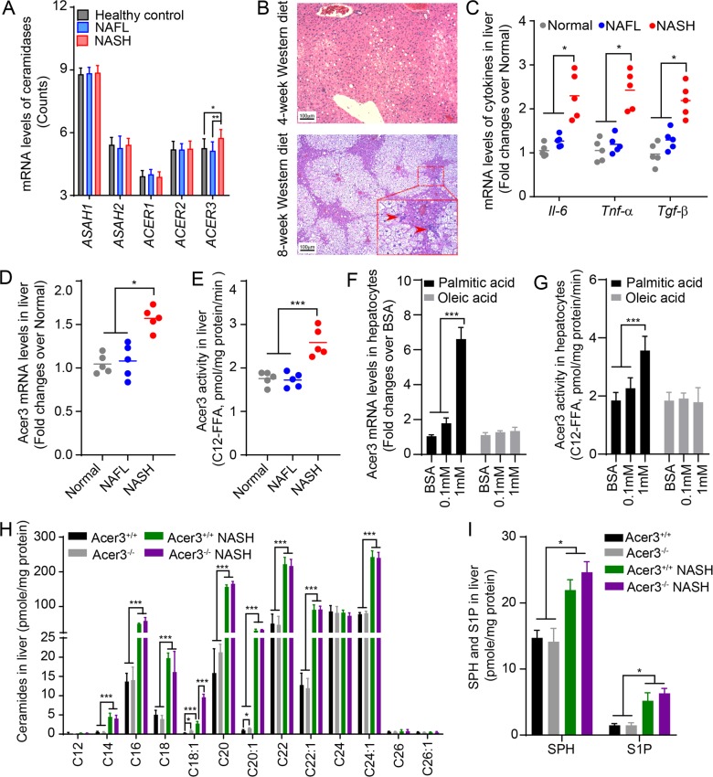
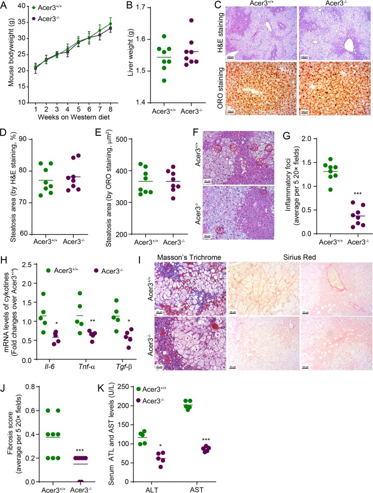
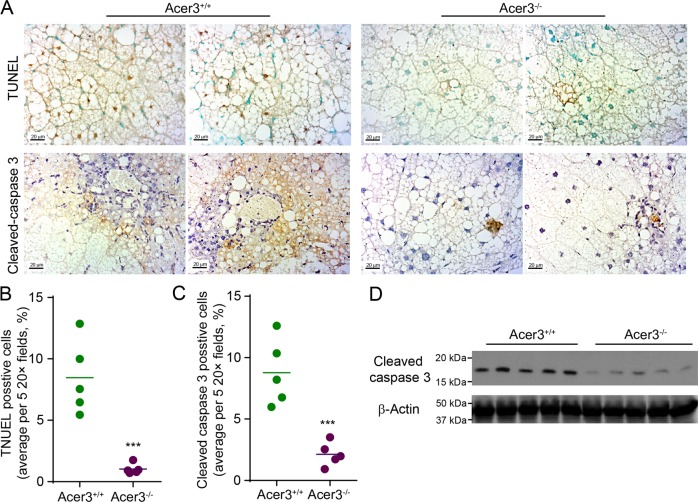
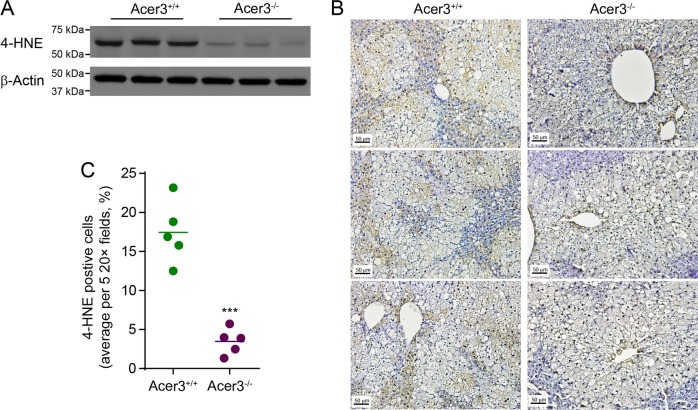
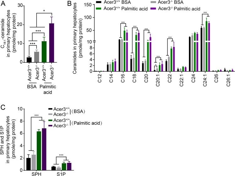
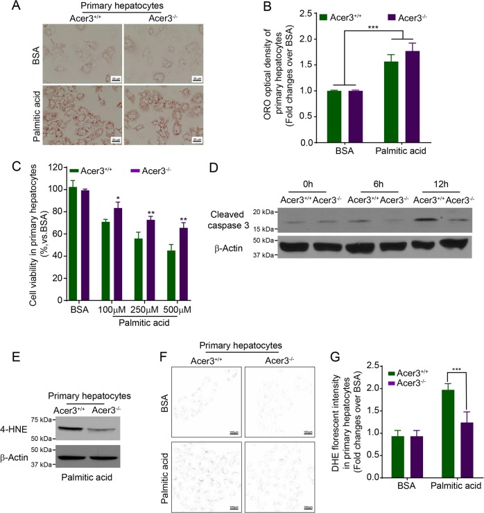
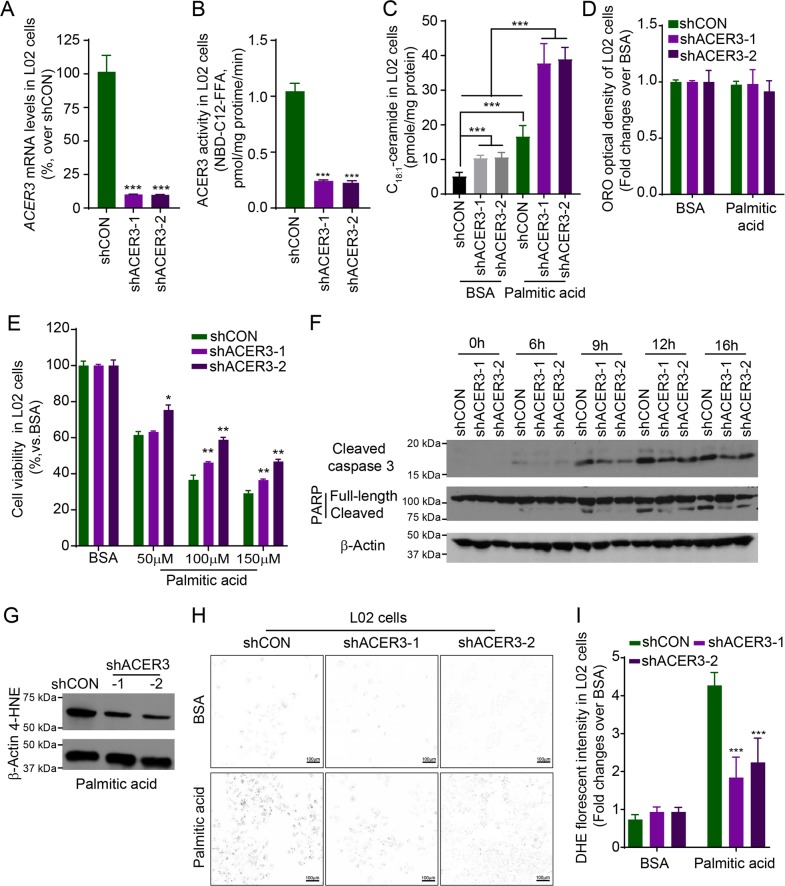
Overload of palmitic acids is linked to the dysregulation of ceramide metabolism in nonalcoholic steatohepatitis (NASH), and ceramides are important bioactive lipids mediating the lipotoxicity of palmitic acid in NASH. However, much remains unclear about the role of ceramidases that catalyze the hydrolysis of ceramides in NASH. By analyzing the National Center for Biotechnology Information (NCBI) Gene Expression Omnibus (GEO) database, we found that alkaline ceramidase 3 (ACER3) is upregulated in livers of patients with NASH. Consistently, we found that Acer3 mRNA levels and its enzymatic activity were also upregulated in mouse livers with NASH induced by a palmitate-enriched Western diet (PEWD). Moreover, we demonstrated that palmitate treatment also elevated Acer3 mRNA levels and its enzymatic activity in mouse primary hepatocytes. In order to investigate the function of Acer3 in NASH, Acer3 null mice and their wild-type littermates were fed a PEWD to induce NASH. Knocking out Acer3 was found to augment PEWD-induced elevation of C18:1-ceramide and alleviate early inflammation and fibrosis but not steatosis in mouse livers with NASH. In addition, Acer3 deficiency attenuated hepatocyte apoptosis in livers with NASH. These protective effects of Acer3 deficiency were found to be associated with suppression of hepatocellular oxidative stress in NASH liver. In vitro studies further revealed that loss of ACER3/Acer3 increased C18:1-ceramide and inhibited apoptosis and oxidative stress in mouse primary hepatocytes and immortalized human hepatocytes induced by palmitic-acid treatment. These results suggest that ACER3 plays an important pathological role in NASH by mediating palmitic-acid-induced oxidative stress.


Conflict of interest statement

The authors declare that they have no conflict of interest.

Figures

Fig. 1
Fig. 1. ACER3 regulates the catabolism of hepatic C18:1-ceramide in the context of NASH.
a The NCBI GEO database, GSE48452, was analyzed for mRNA levels of genes encoding sphingolipid-metabolizing enzymes in liver tissues from 14 healthy individuals, 14 NAFL patients, and 18 NASH patients. The mRNA levels of ceramidases, including ASAH1, ASAH2, ACER1, ACER2, and ACER3, were reported. b and c 6-week-old C57BL/6J mice were fed standard chow (Normal) or palmitic-acid-enriched WD for 4 (NAFL) or 8 weeks (NASH) before mouse liver tissues were dissected. Livers were subjected to histological analyses for steatosis and inflammatory infiltration b or to qPCR analyses for the mRNA levels of Il-6, Tnf-α, Tgf-β, and β-actin as a reference gene c. Inflammatory foci were marked by arrowheads. d and e Mice were fed standard chow (Normal) or PEWD for 4 (NAFL) or 8 (NASH) weeks before livers were subjected to qPCR analysis for Acer3 mRNA levels d or ACER3 enzymatic activity assays e. f and g Primary hepatocytes were isolated from wild-type mice fed standard chow then treated with fatty acid-free bovine serum albumin (BSA), BSA–palmitate or BSA–oleic acid complex at indicated concentrations. At 6 h post treatment, total RNA and membranes were isolated from hepatocytes and assayed for Acer3 mRNA levels f and enzymatic activity g, respectively. h and i Liver tissues were collected from Acer3+/+ and Acer3−/− mice fed on standard chow or PEWD for 8 weeks and the hepatic levels of ceramides h, SPH, and S1P i were determined by LC–MS/MS. Images in b represent results from one of five pairs of mice. Data in dg represent mean ± SD of three independent experiments. Data in h and i represent mean ± SD, n = 3. *P < 0.05, **P < 0.01, ***P < 0.001.
Fig. 2
Fig. 2. Acer3 deficiency attenuates early inflammation and fibrosis in the liver of mice with NASH.
a and b Acer3+/+ and Acer3−/− mice were fed PEWD for 8 weeks and their body weights were recorded weekly a. At week 8, mice were euthanized and their liver weights were measured b. The liver tissues were subjected to the following assays. ce Liver tissues were sectioned for H&E and ORO staining c. Liver sections stained with H&E d or ORO e were analyzed for steatosis areas as described in “Materials and methods” section. f and g Liver sections were stained with H&E f and hepatic inflammatory foci that marked by red cycle were counted to evaluate inflammatory infiltration g. h Total RNA was extracted from liver tissues and subjected to qPCR analyses for the mRNA levels of Il-6, Tnf-α, Tgf-β, and β-Actin as a reference gene. i and j Liver sections were stained with Mason’s trichrome and Sirius Red i and fibrosis was scored as described in “Materials and methods” section j. k Whole blood was collected from Acer3+/+ and Acer3−/− mice fed PEWD for 8 weeks and serum levels of ALT and AST were determined to evaluate hepatocellular injury. Data in a represent mean ± SD, n = 8. Images in c, f, and i represent results from one of eight pairs of mice. *P < 0.05, **P < 0.01, ***P < 0.001.
Fig. 3
Fig. 3. Acer3 deficiency alleviates apoptosis in hepatocytes in mice with NASH.
a-c Liver sections from Acer3+/+ and Acer3−/− mice fed PEWD for 8 weeks were stained with TUNEL or a cleaved caspase 3 antibody a. Cells stained positive for TUNEL b and cleaved caspase 3 c were counted to evaluate hepatocellular apoptosis induced by NASH. d Liver tissues from the above mice were subjected to Western blot analyses with antibodies against cleaved caspase 3 or β-Actin (a protein loading control). Images in a represent results from five pairs of mice. ***P < 0.001.
Fig. 4
Fig. 4. Acer3 deficiency inhibits oxidative stress in hepatocytes in mice with NASH.
a Liver tissues from Acer3+/+ and Acer3−/− mice fed PEWD for 8 weeks were subjected to western blot analyses with the antibody against 4-HNE or β-Actin (a protein loading control). b and c Liver tissue sections from the above mice were stained with 4-HNE antibody to evaluate oxidative stress b and 4-HNE-positive hepatocytes were numerated c. Images in b represent results from five pairs of mice. ***P < 0.001.
Fig. 5
Fig. 5. Acer3 deficiency augments an increase in the levels of C18:1-ceramide in mouse primary hepatocytes in response to overload of palmitate.
Following 6-h treatment with BSA or BSA–palmitate complex (500 μM), LC–MS/MS was performed to analyze the levels of ceramides a and b, SPH c, and S1P c in primary hepatocytes from Acer3+/+ and Acer3−/− mice. Data represent mean ± SD of three independent experiments. *P < 0.05, ***P < 0.001.
Fig. 6
Fig. 6. Acer3 deficiency inhibits apoptosis and oxidative stress in mouse hepatocytes in response to overload of palmitate.
a and b Mouse primary hepatocytes treated with BSA or BSA–palmitate complex (500 μM) for 24 h were stained with ORO a and the optical density of ORO staining was measured as described in “Materials and methods” section b. c Hepatocytes were treated with BSA or BSA–palmitate complex at indicated concentrations for 24 h before cell viability was determined by MTT assays as described in “Materials and methods” section. d hepatocytes treated with BSA or BSA–palmitate complex (500 μM) for indicated time durations were subjected to Western blot analyses with an antibody against cleaved capase3 or β-Actin (a protein-loading control). e Hepatocytes treated with BSA or BSA–palmitate complex (500 μM) for 12 h were subjected to Western blot analyses with the antibody against 4-HNE or β-Actin (a protein-loading control). f and g DHE staining was performed to measure the production of reactive oxidation species in hepatocytes treated with BSA or BSA–palmitate complex (500 μM) for 12 h. Hepatocytes stained with DHE were imaged by microscopy g with background in white and the staining in black, and the florescent intensity was quantified as described in “Materials and methods” section h. Data in b, c, and g represent mean ± SD of three independent experiments. Images in a, d, e, and f represent results from three independent experiments. *P < 0.05, **P < 0.01, ***P < 0.001.
Fig. 7
Fig. 7. Knocking down ACER3 elevates C18:1-ceramide and protects human hepatocytes from apoptosis and oxidative stress in response to overload of palmitate.
a and b Human L02 hepatocytes were transfected with each of two short hairpin RNAs (shRNA) specific for the ACER3 gene (shACER3-1 and shACER3-2) or a control shRNA (shCON), ACER3 mRNA a and enzymatic activity b were measured by qPCR and in vitro activity assays, respectively. c L02 cells transfected with shACER3-1, shACER3-2, or shCON were treated with BSA or BSA–palmitate complex (100 μM) for 6 h before the levels of ceramides were determined by LC–MS/MS. d and e L02 cells transfected with shACER3-1, shACER3-2, or shCON were treated with BSA or BSA–palmitate complex (100 μM) for 24 h before ORO staining d or MTT assays were performed e. The optical density of ORO staining was measured as described in “Materials and methods” section. f L02 cells transfected with shACER3-1, shACER3-2, or shCON were treated with BSA or BSA–palmitate complex (100 μM) for indicated time durations before western blot analyses were performed with an antibody against PARP, cleaved caspase 3, or β-Actin (a protein-loading control). g L02 cells transfected with shACER3-1, shACER3-2, or shCON were treated with BSA or BSA–palmitate complex (100 μM) for 12 h before Western blot analyses were performed with the antibody against 4-HNE or β-actin (a protein-loading control). h and i L02 cells transfected with shACER3-1, shACER3-2, or shCON were treated with BSA or BSA–palmitate complex (100 μM) for 12 h before DHE staining was imaged h with background in white and the staining in black, and the florescent intensity was detected as described in “Materials and methods” section i. Data in ae and i represent results mean ± SD of three independent experiments. Images in fh represent results from three independent experiments. *P < 0.05, **P < 0.01, ***P < 0.001.

References

    1. Younossi Z, et al. Global burden of NAFLD and NASH: trends, predictions, risk factors and prevention. Nat. Rev. Gastroenterol. Hepatol. 2018;15:11. doi: 10.1038/nrgastro.2017.109. - DOI - PubMed
    1. Tiniakos DG, Vos MB, Brunt EM. Nonalcoholic fatty liver disease: pathology and pathogenesis. Annu. Rev. Pathol. 2010;5:145. doi: 10.1146/annurev-pathol-121808-102132. - DOI - PubMed
    1. Ambrosini GL, et al. Adolescent dietary patterns are associated with lifestyle and family psycho-social factors. Public Health Nutr. 2009;12:1807. doi: 10.1017/S1368980008004618. - DOI - PubMed
    1. Oddy WH, et al. The Western dietary pattern is prospectively associated with nonalcoholic fatty liver disease in adolescence. Am. J. Gastroenterol. 2013;108:778. doi: 10.1038/ajg.2013.95. - DOI - PubMed
    1. Gambino R, et al. Different serum free fatty acid profiles in NAFLD subjects and healthy controls after oral fat load. Int. J. Mol. Sci. 2016;17:479. doi: 10.3390/ijms17040479. - DOI - PMC - PubMed

Publication types

MeSH terms