. 2020 Jan 16;11(1):303.
doi: 10.1038/s41467-019-14220-z.

Crystal structures of fukutin-related protein (FKRP), a ribitol-phosphate transferase related to muscular dystrophy

Affiliations

Crystal structures of fukutin-related protein (FKRP), a ribitol-phosphate transferase related to muscular dystrophy

Naoyuki Kuwabara et al. Nat Commun. .

Abstract

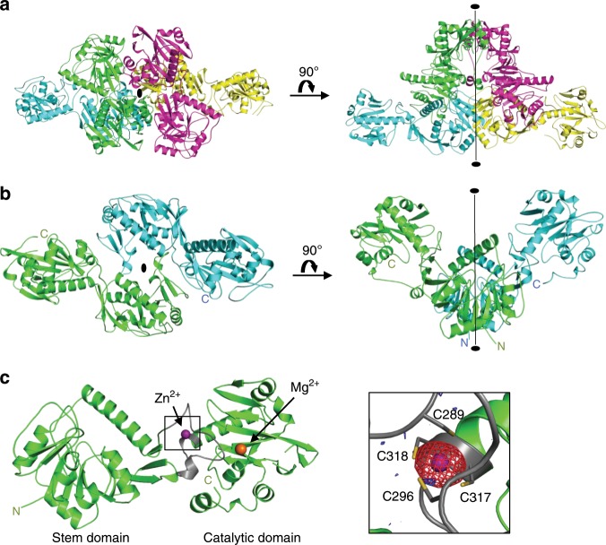
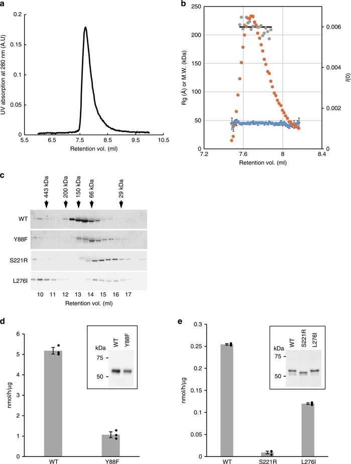
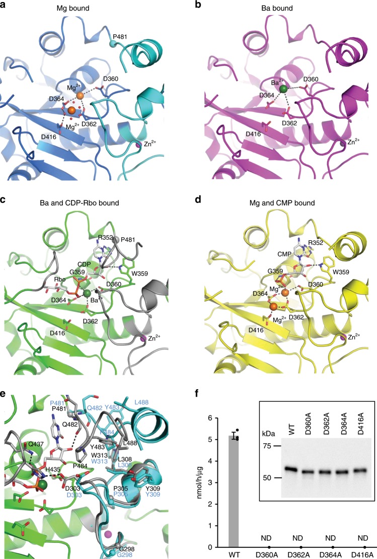
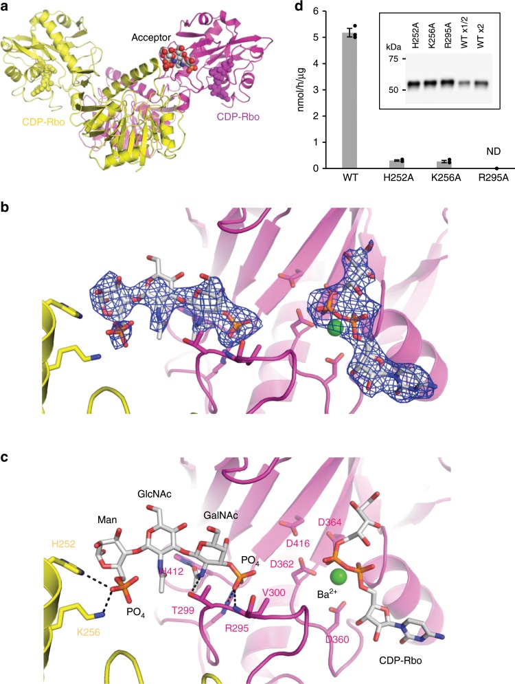
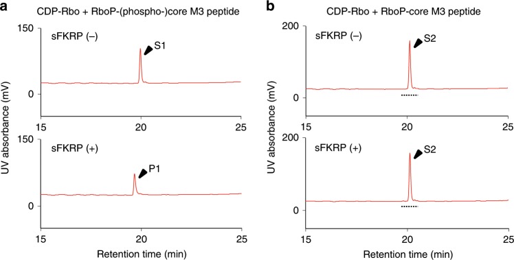
α-Dystroglycan (α-DG) is a highly-glycosylated surface membrane protein. Defects in the O-mannosyl glycan of α-DG cause dystroglycanopathy, a group of congenital muscular dystrophies. The core M3 O-mannosyl glycan contains tandem ribitol-phosphate (RboP), a characteristic feature first found in mammals. Fukutin and fukutin-related protein (FKRP), whose mutated genes underlie dystroglycanopathy, sequentially transfer RboP from cytidine diphosphate-ribitol (CDP-Rbo) to form a tandem RboP unit in the core M3 glycan. Here, we report a series of crystal structures of FKRP with and without donor (CDP-Rbo) and/or acceptor [RboP-(phospho-)core M3 peptide] substrates. FKRP has N-terminal stem and C-terminal catalytic domains, and forms a tetramer both in crystal and in solution. In the acceptor complex, the phosphate group of RboP is recognized by the catalytic domain of one subunit, and a phosphate group on O-mannose is recognized by the stem domain of another subunit. Structure-based functional studies confirmed that the dimeric structure is essential for FKRP enzymatic activity.


Conflict of interest statement

The authors declare no competing interests.

Figures

Fig. 1
Fig. 1. Crystal structure of the sFKRP.
All models were prepared using an Mg2+ bound structure. a Crystal structure of sFKRP showing four subunits in the asymmetrical unit. The subunits are colored green, blue, red, and yellow, respectively. The two-fold axis of the tetramer is shown as a black ellipse and a line. b The protomeric dimer of sFKRP. The local two-fold axis of the protomeric dimer is shown as a black ellipse and a line. c Monomer structure of sFKRP. Zn2+ and Mg2+ are shown in purple and orange, respectively. The zinc finger loop (G288 to C318) is shown in gray. The anomalous difference Fourier maps around the zinc finger for the peak data set (red mesh) and the low remote data set (blue mesh) at a resolution of 2.41 Å are shown in the inset. The contour levels of the peak and the low remote are 5.0 and 3.5 σ, respectively. Labels N and C indicate the N- and C-terminus of sFKRP, respectively.
Fig. 2
Fig. 2. Oligomerization analysis.
Oligomerization states in solution and enzymatic activities of wild-type and disease-related mutants of sFKRP were studied. SEC-SAXS profiles of wild-type sFKRP are shown in (a) (UV absorption) and (b). In (b), the SAXS intensity I(0) (orange), radii of gyration (Rg) (blue), and estimated molecular weight from Porod volume (gray) are plotted. a Black line indicates the average of estimated molecular weight from the Porod volumes. c Expressed sFKRP proteins were analyzed by usual SEC and detected by SDS-PAGE (5–20% acrylamide; ATTO) followed by Western blotting. Arrows indicate fractions at which protein markers were eluted. d, e Enzymatic activities of sFKRP with CDP-Rbo and the RboP-(phospho-)core M3 peptide. Insets: immunoblot analyses of sFKRP proteins to normalize input sFKRP. d sFKRP (WT and Y88F) immunoprecipitated from the culture supernatant were used as the enzyme sources. e Cell lysates expressing sFKRP (WT, S221R, and L276I) were used as the enzyme sources. Average values ± SE of three independent experiments are shown. Each dot represents one data point. Source data are provided as a Source Data file.
Fig. 3
Fig. 3. Detailed structures around the active site.
The main chain of sFKRP is shown in the cartoon model. Black dotted lines represent hydrogen bonds or ionic interactions. Each metal ion is shown by colored spheres: Mg2+ by orange, Zn2+ by purple, and Ba2+ by green. a Mg2+ bound structure. Water molecules which coordinate Mg2+ at site II are shown as red dots. b Ba2+ bound structure. c CDP-Rbo (stick model) and Ba2+ complex structure. The regions involved in the interaction with CDP-Rbo are shown in gray. d CMP (stick model) and Mg2+ complex structure. e Comparison with CDP-Rbo bound (corresponds to (c), green and gray) and substrate-free (corresponds to (a), sky blue) structures. The start point (P481) of conformational change induced by ligand binding is marked with a small sphere. f Enzymatic activities of sFKRP (WT, D360A, D362A, D364A, and D416A) with CDP-Rbo and the RboP-(phospho-)core M3 peptide. ND, not detected. Average values ± SE of three independent experiments are shown. Each dot represents one data point. Inset: immunoblot analysis of sFKRP proteins immunoprecipitated from the culture supernatant to normalize input sFKRP. Source data are provided as a Source Data file.
Fig. 4
Fig. 4. Structure of sFKRP in complex with the acceptor glycopeptide.
a Two sFKRP subunits are shown in yellow and purple, respectively. CDP-Rbo (yellow and purple) and the phospho-(phospho-)core M3 moiety (CPK color) are shown by sphere models. b Fo-Fc omit map around phospho-(phospho-)core M3 (left) and CDP-Rbo (right). The contour level of the map is 3.0 σ. c Interaction between the protomeric dimer and phospho-(phospho-)core M3 moiety. The trisaccharide is shown as a stick model and interactions between sFKRP are depicted as dotted black lines. d Enzymatic activities of sFKRP (WT, H252A, K256A, and R295A) with CDP-Rbo and the RboP-(phospho-)core M3 peptide. ND, not detected. Average values ± SE of three independent experiments are shown. Each dot represents one data point. Inset: immunoblot analysis of sFKRP proteins immunoprecipitated from the culture supernatant to normalize input sFKRP. Source data are provided as a Source Data file.
Fig. 5
Fig. 5. Requirement of the phosphate residue at the 6th-position of O-Man.
a Enzymatic activity of sFKRP with CDP-Rbo and the RboP-(phospho-)core M3 peptide. The products were analyzed by HPLC. Upper, without sFKRP; lower, with sFKRP. S1, acceptor substrate [RboP-(phospho-)core M3 peptide]; P1, product of the reaction of sFKRP with CDP-Rbo. b No enzymatic activity was observed for sFKRP with CDP-Rbo and the RboP-core M3 peptide. The products were analyzed by HPLC. Upper, without sFKRP; lower, with sFKRP. S2, acceptor substrate (RboP-core M3 peptide). Eluates with the retention times indicated by dotted lines were subjected to MS analysis (Supplementary Fig. 6).

References

    1. Manya H, Endo T. Glycosylation with ribitol-phosphate in mammals: New insights into the O-mannosyl glycan. BBA - Gen. Subj. 2017;1861:1–41. doi: 10.1016/j.bbagen.2017.06.024. - DOI - PubMed
    1. Yoshida-Moriguchi T, Campbell KP. Matriglycan: a novel polysaccharide that links dystroglycan to the basement membrane. Glycobiology. 2015;25:702–713. doi: 10.1093/glycob/cwv021. - DOI - PMC - PubMed
    1. Endo T. Glycobiology of α-dystroglycan and muscular dystrophy. J. Biochem. 2015;157:1–12. doi: 10.1093/jb/mvu066. - DOI - PubMed
    1. Michele DE, Campbell KP. Dystrophin-glycoprotein complex: post-translational processing and dystroglycan function. J. Biol. Chem. 2003;278:15457–15460. doi: 10.1074/jbc.R200031200. - DOI - PubMed
    1. Yoshida-Moriguchi T, et al. SGK196 is a glycosylation-specific O-mannose kinase required for dystroglycan function. Science. 2013;341:896–899. doi: 10.1126/science.1239951. - DOI - PMC - PubMed

Publication types