. 2020 Jan 16;10(1):437.
doi: 10.1038/s41598-019-57398-4.

Fam151b, the mouse homologue of C.elegans menorin gene, is essential for retinal function

Affiliations

Fam151b, the mouse homologue of C.elegans menorin gene, is essential for retinal function

Amy S Findlay et al. Sci Rep. .

Abstract

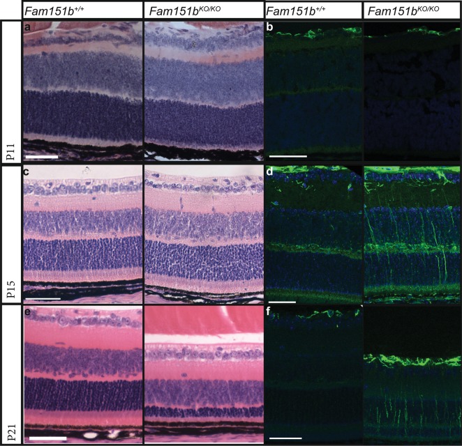
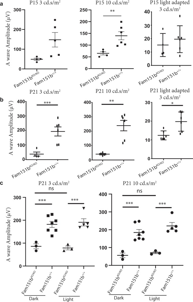
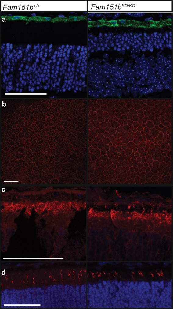
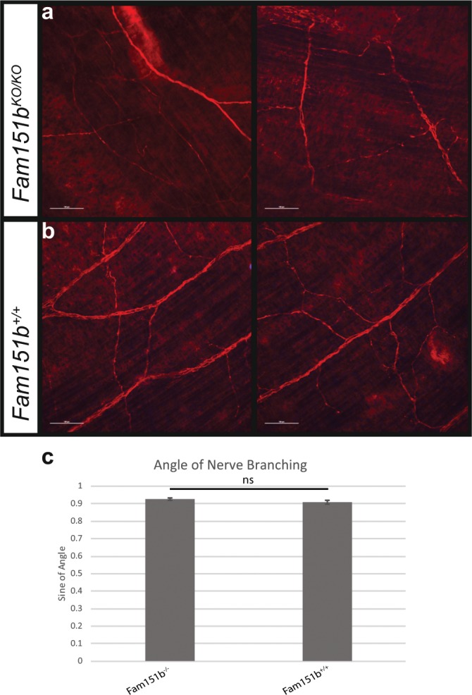
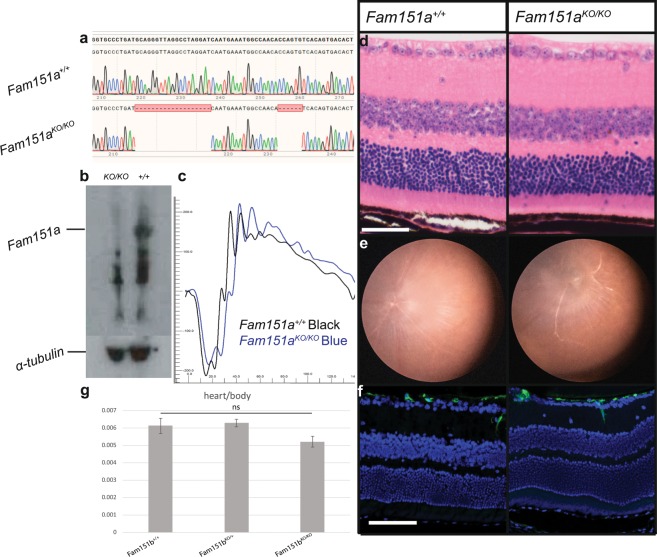
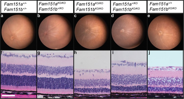
Fam151b is a mammalian homologue of the C. elegans menorin gene, which is involved in neuronal branching. The International Mouse Phenotyping Consortium (IMPC) aims to knock out every gene in the mouse and comprehensively phenotype the mutant animals. This project identified Fam151b homozygous knock-out mice as having retinal degeneration. We show they have no photoreceptor function from eye opening, as demonstrated by a lack of electroretinograph (ERG) response. Histological analysis shows that during development of the eye the correct number of cells are produced and that the layers of the retina differentiate normally. However, after eye opening at P14, Fam151b mutant eyes exhibit signs of retinal stress and rapidly lose photoreceptor cells. We have mutated the second mammalian menorin homologue, Fam151a, and homozygous mutant mice have no discernible phenotype. Sequence analysis indicates that the FAM151 proteins are members of the PLC-like phosphodiesterase superfamily. However, the substrates and function of the proteins remains unknown.


Conflict of interest statement

The authors declare no competing interests.

Figures

Figure 1
Figure 1
(a) Multiple sequence alignment (MSA) of representative members of FAM151 family and PLC-like phosphodiesterases. This MSA was generated with the program T-Coffee using default parameters and slightly refined manually. The final superfamily alignment was generated using a combination of profile-to-profile comparisons and sequence alignments derived from structural superimpositions using DALI, for those families whose tertiary structure is known, such us: Phospholipase D toxins (PDB-IDs: 4Q6X, 1XX1, and 3RLG), GDPD enzymes (PDB-IDs: 1YDY, 3QVQ, 2OTD, and 1VD6) and PLC (Phosphoinositide phospholipase C) enzyme (PDB-ID:1DJY). Families are indicated by coloured background to the left of the alignment: FAM151, Phospholipase D toxins, GDPD and PLC enzymes are indicated in red, pink, blue and green, respectively. The limits of the protein sequence conserved regions included in the alignment are indicated by flanking residue positions. Secondary structure predictions were performed for the FAM151 family and these are consistent with X-ray determined secondary structures of the PLC-like phosphodiesterases. Alpha-helices and beta-strands are indicated by cylinders and arrows, respectively. The alignment was presented with the program Belvu using a colouring scheme indicating the average BLOSUM62 scores (which are correlated with amino acid conservation) of each alignment column: red (>3), violet (between 3 and 1.5) and light yellow (between 1.5 and 0.5). Sequences are named according to their UniProt identification. (b) Structural superimposition of two members of the PLC-like phosphodiesterases superfamily: GDPD and PLCD1. Active site residues are labelled and coloured according to their reference protein sequences: human FAM151B, GDPD (PDB-ID: 3QVQ) and PLCD1 (PDB-ID:1djy) in red, blue and green, respectively. Structures are presented using Pymol (http://www.pymol.org).
Figure 2
Figure 2
Adult Fam151bKO/KO mice have extensive retinal degeneration. (a) Comparison of 11 week old Fam151bKO/KO mice and their wild-type littermates fundal images shows an uneven appearance on the Fam151bKO/KO mouse retina indicative of abnormal retinal morphology. Histological analysis (b) shows that this was due to a loss of the outer nuclei layer, in which the nuclei of photoreceptor cells are located. The outer segments are shortened. This loss of nuclei is directly related to a loss of photoreceptor cells. (c) Functional analysis of the retina through ERG traces shows a loss of scotopic a-wave, produced from photoreceptor cells response to a light stimulus, Fam151bKO/KO (blue line) compared with wild type littermates (black line). RGC = Retinal Ganglion Cells, INL = Inner Nuclei Layer, ONL = Outer Nuclei Layer, RPE = Retinal Pigment Epithelium. Scale bar = 50 µm.
Figure 3
Figure 3
Histological analysis of disease progression in Fam151bKO/KO mice. Haematoxylin and eosin staining of P11 (a), P15 (c) and P21 (e) Fam151bKO/KO retinal sections compared with Fam151b+/+ littermates shows the loss of photoreceptor nuclei by P21. At P11 and P15 mutant mice sections show comparable numbers of nuclei to wild-type. All other retinal cell layers seem unaffected by the loss of Fam151b expression. GFAP staining in green shows that at P11 (b) Fam151bKO/KO sections have similar staining in the RGC layer as wild-type littermates. However, by P15 (d) and continuing on to P21 (f) mutant sections exhibit a clear upregulation of GFAP indicative of retinal stress. Scale bar = 50 µm.
Figure 4
Figure 4
ERG a-wave amplitudes plotted for comparison. (a,b) Compares P15 (a) and P21 (b) mutant mice with wild-type littermates, using 3, 10 cd.s/m2 light stimuli on dark adapted mice followed by 3 cd.s/m2 light stimuli on light adapted mice (n = 4 (mutant), 6 (wild type)). There is a significant reduction in a-wave amplitude in the Fam1511bKO/KO mice compared to wild-type controls except at P15 for 3 cd.s/m2 light stimulus in both dark and light adapted mice. (c) Comparison of P21 mice kept in either darkened cage conditions or a normal 12 hour light cycle (n = 3 (mutant dark), 7 (wild type dark), 3 (mutant light), 5 (wild type light)). **p < 0.01, ***p < 0.001 by unpaired t-test with Welch’s correction.
Figure 5
Figure 5
RPE health was assessed by examining typical RPE markers. (a) RPE65 showed correct localisation to the RPE in retinal sections of P15 mice and (b) ZO-1 localised to tight junctions when observed on flat mount RPE in both P30 Fam151bKO/KO and wild type. The correct localisation of light sensitive opsins is essential for the proper function of photoreceptors, in P15 Fam151bKO/KO mutant eyes both rhodopsin (c) and m-opsin (d) localise to the outer segments of photoreceptor cells as expected. Scale bar 50 µm.
Figure 6
Figure 6
Nerve branching in the skin was normal in Fam151bKO/KO mice. Neurofilament staining was done on flat mount skin from embryonic day 16.5 (E16.5) Fam151bKO/KO mice (a) and wild type littermates (b) in order to analyse the branching patterns of the nerves. (c)The angle of branching nerves was measured and the sine of the angle was used for analysis. No significant difference was found between Fam151bKO/KO mice and their wild-type counterparts (p = 0.1869, unpaired t-test, n = 6). Scale bar 100 µm.
Figure 7
Figure 7
Fam151aKO/KO mice were aged to one year to observe whether they exhibit late onset retinal degeneration not picked up on at the 15 week fundal examination by IMPC. (a) We used CRISPR Cas-9 to produce the Fam151aKO/KO mice and here we show the two separate deletion caused in exon 1 of the gene (bottom line) compared to wild type sequence (top line). (b) Western blot showing loss of Fam151a protein in kidney samples of Fam151aKO/KO mice compared to Fam151a+/+, band at 66 kDa highlighted. Alpha-tubulin used as a loading control. Functional analysis by ERG (c) shows comparable response to a light stimulus as that seen in wild-type littermates. (d) Histology and (e) fundal imaging both show normal retinal morphology, no decrease in photoreceptor layer thickness was seen and fundal images showed an even and healthy appearance. (f) GFAP staining showed no upregulation, indicative of no retinal stress. (g) Comparison of heart weight in relation to body weight between Fam151bKO/KO mutant mice and wild-type littermates. No significant difference was found by t-test (p = 0.1275). Scale bar = 50 µm.
Figure 8
Figure 8
Loss-of-function of Fam151a does not exacerbate the Fam151bKO/KO mutant eye phenotype. Fam151aKO/KOFam151bKO/KO double mutants were analysed alongside control littermates. Fundal images (ae) and histology (fj) were performed on P21 mice. The retinal morphology and loss of photoreceptor cell nuclei of Fam151bKO/KO mice was unaffected by Fam151a status. Scale bar 50 µm.

References

    1. Phelan JK, Bok D. A brief review of retinitis pigmentosa and the identified retinitis pigmentosa genes. Mol Vis. 2000;6:24. - PubMed
    1. Chang B, et al. Retinal degeneration mutants in the mouse. Vision research. 2002;42:517–525. doi: 10.1016/S0042-6989(01)00146-8. - DOI - PubMed
    1. Brown SD, Moore MW. The International Mouse Phenotyping Consortium: past and future perspectives on mouse phenotyping. Mammalian Genome. 2012;23:632–640. doi: 10.1007/s00335-012-9427-x. - DOI - PMC - PubMed
    1. Meehan TF, et al. Disease model discovery from 3,328 gene knockouts by The International Mouse Phenotyping Consortium. Nature genetics. 2017;49:1231. doi: 10.1038/ng.3901. - DOI - PMC - PubMed
    1. Salzberg Y, et al. Skin-derived cues control arborization of sensory dendrites in Caenorhabditis elegans. Cell. 2013;155:308–320. doi: 10.1016/j.cell.2013.08.058. - DOI - PMC - PubMed

Publication types

Substances