Case Reports
. 2018 Jul 1;11(7):3770-3776.
eCollection 2018.

MiT family translocation renal cell carcinoma after malignant infantile osteopetrosis in childhood: a case report

Affiliations
Case Reports

MiT family translocation renal cell carcinoma after malignant infantile osteopetrosis in childhood: a case report

Jing Wang et al. Int J Clin Exp Pathol. .

Abstract

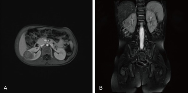
Malignant infantile osteopetrosis (MIOP) is a rare inherited bone metabolism disorder characterized by increased bone mineral density (BMD) and abnormal hematopoiesis. Hematopoietic stem cell transplantation (HSCT) is currently the only curative therapy for MIOP. However, a higher risk of secondary malignancy occurs in children previously exposed to cytotoxic drugs. Here we report a rare case of a 3-year-old female patient with MiT family translocation renal cell carcinoma (MiTF tRCC), who is a survivor of HSCT for MIOP 2 years earlier. The patient had a complete resection of the tumor. Microscopically, we detected diffusely and papillary-like arranged tumor cells whose cytoplasm was bright and clear. Immunohistochemistry showed tumor cells diffusely expressed TFE3, and fluorescence in situ hybridization (FISH) demonstrated disruption of the TFE3 locus, confirming the diagnosis of Xp11 translocation RCC, the subtype of MiTF tRCC. This case supports the view that chemotherapy exposure is a risk factor for MiTF tRCC and indicates the possible association of HSCT with MiTF tRCC.

Keywords: Malignant infantile osteopetrosis; MiT family translocation renal cell carcinoma; Xp11 translocation renal cell carcinoma; chemotherapy; hematopoietic stem cell transplantation.


Conflict of interest statement

None.

Figures

Figure 1
Figure 1
Radiological findings of MIOP. A. MRI showed thin corpus callosum and plump lateral ventricle. B. The pelvis X-ray revealed uneven bone density increased, periosteal reaction could be seen on both sides of the sacrum and femur. C. The left lower extremity X-ray revealed extensively increased BMD and medullary cavity stenosis. D. Whole spine lateral X-ray showed skull bones, vertebrae, pelvis had increased BMD. The vertebral bodies showed a ‘bone-in-bone’ appearance, and the anterior ribs were widened with reduced uneven bone density.
Figure 2
Figure 2
A. MR on fat-suppressed T1WI sequence revealed a 26×29×22 mm solid mass with isointensity signal, low signal and patchy high signal in the middle of the right kidney. B. MR on fat-suppressed T2WI sequence revealed isointensity signal and high signal.
Figure 3
Figure 3
Pathological findings of the tumor. A. Gross appearance demonstrated a well encapsulated tumor measuring 3.0×2.7×2.5 cm. The section was grayish red, yellow and soft, with focal areas of necrosis and calcification. B-D. The microscopic examination revealed papillary-like arranged tumor cells whose cytoplasm was bright and clear, and nucleolus was obvious (HE staining, ×100, ×200, ×400). E. Tumor cells with focal areas of hemorrhage and necrosis (HE staining, ×100). F. The tumor cells broke through the capsule in some areas, invading the kidney tissue (HE staining, ×100).
Figure 4
Figure 4
Immunohistochemical manifestations of the tumor cells. A. TFE3+. B. PAX8+. C. CD10+, membranous. D. PAX5+. E. PAX2+, weak. F. CD68+, sparse. G. Ki67 5%+. H. P53 40%+, weak. I. MelonA-. J. HMB45-. K. WT1-. L. Vimentin+, scarce. (A-G, ×200; H-L, ×100).
Figure 5
Figure 5
Fluorescence in situ hybridization. Red arrow, a break-apart TFE3 probe showed disruption of the TFE3 gene with separation of green and red signals. Green arrow, normal gene: green and red signal merged together.

References

    1. Sobacchi C, Schulz A, Coxon FP, Villa A, Helfrich MH. Osteopetrosis: genetics, treatment and new insights into osteoclast function. Nat Rev Endocrinol. 2013;9:522–536. - PubMed
    1. Eapen M, Davies SM, Ramsay NK, Orchard PJ. Hematopoietic stem cell transplantation for infantile osteopetrosis. Blood. 2015;2:270–276. - PubMed
    1. Srigley JR, Delahunt B, Eble JN, Egevad L, Epstein JI, Grignon D, Hes O, Moch H, Montironi R, Tickoo SK, Zhou M, Argani P. The international society of urological pathology (ISUP) vancouver classification of renal neoplasia. Am J Surg Pathol. 2013;37:1469–1489. - PubMed
    1. Argani P, Zhang L, Reuter VE, Tickoo SK, Antonescu CR. RBM10-TFE3 renal cell carcinoma: a potential diagnostic pitfall due to cryptic intrachromosomal xp11.2 inversion resulting in false-negative TFE3 FISH. Am J Surg Pathol. 2017;41:655–662. - PMC - PubMed
    1. Liu N, Wang Z, Gan W, Xiong L, Miao B, Chen X, Guo H, Li D. Renal cell carcinoma associated with xp11.2 translocation/TFE3 gene fusions: clinical features, treatments and prognosis. PLoS One. 2016;11:e166897. - PMC - PubMed

Publication types

LinkOut - more resources