

The transplantation of induced pluripotent stem cells into the cochleae of mature mice
- PMID: 31949839
- PMCID: PMC6962958
The transplantation of induced pluripotent stem cells into the cochleae of mature mice
Abstract
Objective: Stem cell transplantation is an effective method for treating sensorineural hearing loss (SNHL), but its safety needs further study. This study aimed to reveal the differentiation outcome of induced pluripotent stem cells (iPSCs) after they were transplanted into cochleae.
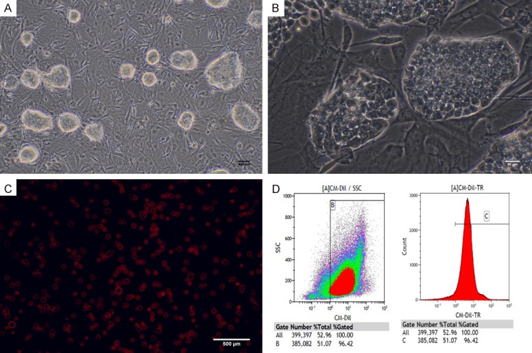
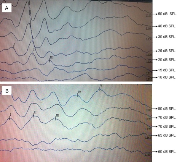
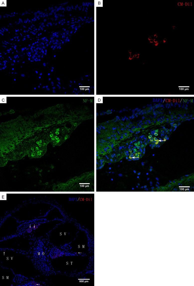
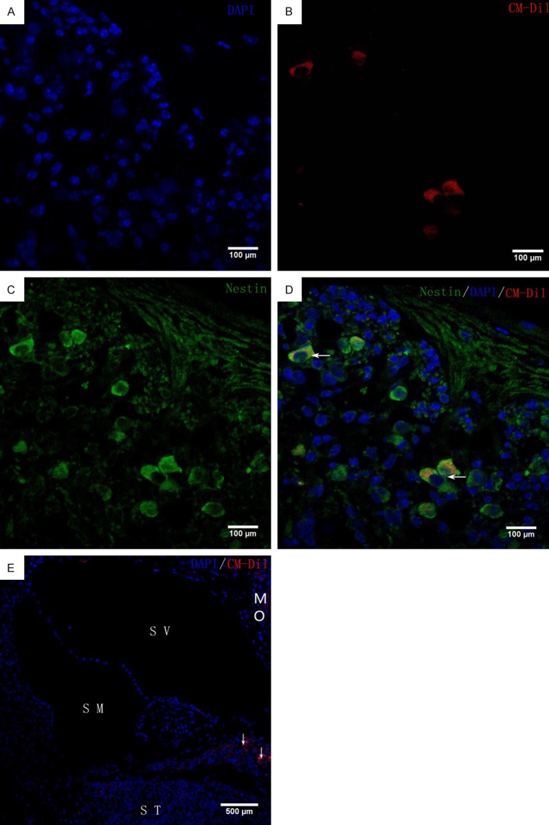
Methods: iPSCs were labelled with CM-Dil and identified by flow cytometry. Twenty 6-8-week-old ICR mice were divided into experimental (A) and control (B) groups. Ten mice were microinjected with CM-Dil-labelled iPSC suspension (group A) or an equal volume DMEM (group B) into the left ear cochlea. The tthresholds of all mice were tested by auditory brainstem response (ABR) at 1 week pre-surgery and 4 weeks post-surgery. Differentiated cells were identified by immunohistochemical staining for neuronal cell markers (nestin, neurofilament-M), and teratoma formation was determined by HE staining.
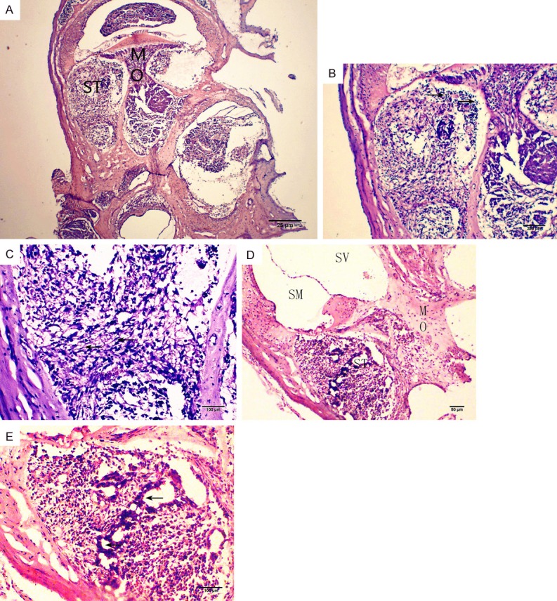
Results: The ABR thresholds in groups A and B at one week pre-surgery (24.50±5.50 vs. 26.00±6.15 dB SPL) and at 4 weeks post-surgery (70.50±4.97 vs. 68.00±5.37 dB SPL) were not significantly different; however, in both groups, the thresholds were lower at pre-surgery than at 4 weeks post-surgery. In group A, CM-Dil-labelled iPSCs were observed in the cochlear perilymph, endolymph, and modiolus, and some red fluorescence-labelled cells expressed neural cell markers. In group B, no fluorescence was observed in the cochleae, but teratomas were observed in some cochleae. A teratoma was observed in each of two cochleae after iPSCs transplantation by HE staining.
Conclusion: Mouse iPSCs can differentiate into cells with neuronal cell markers 4 weeks post-cochlear transplantation, and transplanted undifferentiated iPSCs may form teratomas. However, in the short-term, hearing loss in mice caused by cell transplantation through round window pathways cannot be improved by cochlear iPSC transplantation.
Keywords: Induced pluripotent stem cells (iPSCs); cell differentiation; inner ear transplantation; spiral ganglion neuron; teratoma.
IJCEP Copyright © 2018.
Conflict of interest statement
None.
Figures
References
-
- Matsui JI, Parker MA, Ryals BM, Cotanche DA. Regeneration and replacement in the vertebrate inner ear. Drug Discov Today. 2005;10:1307–12. - PubMed
-
- Wang Z, Jiang H, Yan Y, Wang Y, Shen Y, Li W, Li H. Characterization of proliferating cells from newborn mouse cochleae. Neuroreport. 2006;17:767–71. - PubMed
LinkOut - more resources
Full Text Sources
Miscellaneous