

Adrenal Cavernous Hemangioma: A Rarely Perceived Pathology-Case Illustration and Review of Literature
- PMID: 31949968
- PMCID: PMC6944974
- DOI: 10.1155/2019/8463890
Adrenal Cavernous Hemangioma: A Rarely Perceived Pathology-Case Illustration and Review of Literature
Abstract
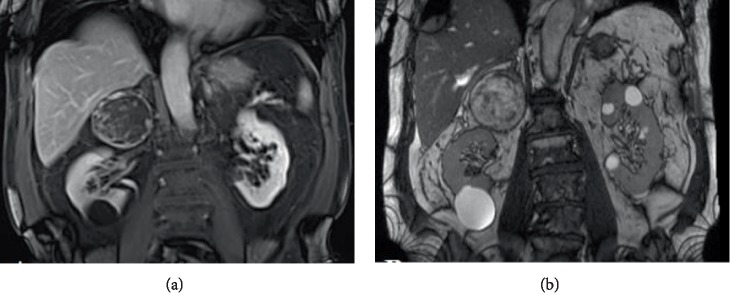
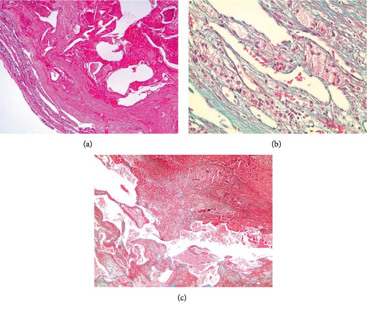
Cavernous hemangiomas are endothelial tumors that rarely affect the adrenal glands. Most of these tumors remain silent and are incidentally found on abdominal imaging. Hardly ever, these tumors are endocrinologically functional. They may present as vague abdominal pain. Surgical resection remains the mainstay for large masses. In this paper, we are presenting a case of adrenal cavernous hemangioma in a 83-year-old male patient who initially presented for workup of vague abdominal and bilateral flank pain. A computed tomography scan of the abdomen showed an 8 cm right adrenal adenoma which was metabolically nonfunctional. The mass was completely resected through an open subcostal incision, with no encountered postoperative complications. A highlight of all published cases of adrenal hemangiomas since 1955 is also presented and reviewed.
Copyright © 2019 Jad A. Degheili et al.
Conflict of interest statement
The authors declare that they have no conflicts of interest.
Figures
Similar articles
-
A Rare Presentation of Lisegang Rings in Adrenal Cavernous Hemangioma : Case Report and Literature Review.Case Rep Med. 2021 Aug 3;2021:9998729. doi: 10.1155/2021/9998729. eCollection 2021. Case Rep Med. 2021. PMID: 34400913 Free PMC article.
-
Rare cavernous hemangioma of adrenal gland: case report.Sao Paulo Med J. 2014;132(4):249-52. doi: 10.1590/1516-3180.2014.1324715. Sao Paulo Med J. 2014. PMID: 25055072 Free PMC article.
-
Adrenal cavernous hemangioma: A rare tumor that mimics adrenal cortical carcinoma.Surg Open Sci. 2019 Apr 27;1(1):7-13. doi: 10.1016/j.sopen.2019.04.001. eCollection 2019 Jul. Surg Open Sci. 2019. PMID: 32754687 Free PMC article.
-
Adrenal cavernous hemangioma: a case report with review of the literature.JOP. 2014 May 27;15(3):254-7. doi: 10.6092/1590-8577/2402. JOP. 2014. PMID: 24865537 Review.
-
Adrenal hemangiomas: two case reports with a review of the literature.Surgery. 1989 May;105(5):674-81. Surgery. 1989. PMID: 2650008 Review.
Cited by
-
A Rare Case of Cavernous Haemangioma of the Adrenal Gland: A Case Report and Review of Literature.Cureus. 2022 Oct 4;14(10):e29917. doi: 10.7759/cureus.29917. eCollection 2022 Oct. Cureus. 2022. PMID: 36348862 Free PMC article.
-
Adrenal cavernous Hemangioma;A rare diagnosis of adrenal incidentaloma:A case report, and literature review.Urol Case Rep. 2020 Oct 31;34:101477. doi: 10.1016/j.eucr.2020.101477. eCollection 2021 Jan. Urol Case Rep. 2020. PMID: 33204642 Free PMC article.
-
Adrenal cavernous hemangioma misdiagnosed as pheochromocytoma: a case report.BMC Surg. 2021 Apr 26;21(1):210. doi: 10.1186/s12893-021-01195-2. BMC Surg. 2021. PMID: 33902538 Free PMC article.
-
A case of adrenal cavernous hemangioma resected due to tumor growth accompanied by intratumoral hemorrhage.IJU Case Rep. 2024 Jul 9;7(5):379-382. doi: 10.1002/iju5.12760. eCollection 2024 Sep. IJU Case Rep. 2024. PMID: 39224674 Free PMC article.
-
[Adrenal cavernous hemangioma: A case report and literature review].Beijing Da Xue Xue Bao Yi Xue Ban. 2021 Aug 18;53(4):808-810. doi: 10.19723/j.issn.1671-167X.2021.04.032. Beijing Da Xue Xue Bao Yi Xue Ban. 2021. PMID: 34393250 Free PMC article. Review. Chinese.
References
Publication types
LinkOut - more resources
Full Text Sources
