

A cadaveric study of ovarian veins: variations, measurements and clinical significance
- PMID: 31949976
- PMCID: PMC6952686
- DOI: 10.5115/acb.19.062
A cadaveric study of ovarian veins: variations, measurements and clinical significance
Abstract
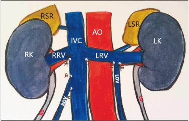
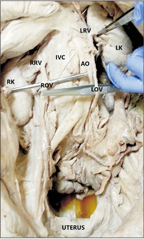
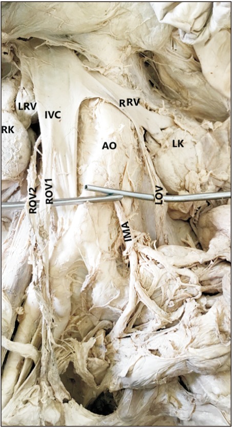
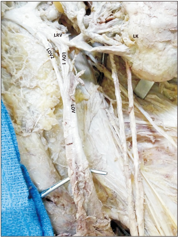
The literature showing information regarding ovarian venous variation, its diameter and termination distance from respective renal venous origin are limited. This information is important in various surgical and clinical procedures including venous embolization, vascular reconstruction during renal transplantation and localizing the source of origin of a pelvic mass. We examined 94 sides of 47 formalin fixed female cadavers and noted the course and termination of ovarian veins. We measured the diameter of ovarian veins at their termination point and the termination distance in respect to the termination point of renal veins at inferior vena cava (IVC) on respective sides. We found two cases of variations related to right ovarian vein -one, right ovarian vein joined the right renal vein; two, right ovarian vein duplicated and joined with IVC at two different points. We found one case of variation related to left ovarian vein-a partially duplicated left ovarian vein. All the variations were unilateral. The mean diameters of right and left ovarian veins were 3.66±1.18 and 4.20±0.96 mm, respectively. The distance of termination of ovarian veins ranged from 19-40 mm and 13-41 mm, respectively from termination points of right and left renal veins at IVC on respective sides. Our study presents a set of data regarding variation of ovarian veins, diameters and termination distances which could be useful for gynecologists, surgeons and radiologists.
Keywords: Clinical significance of ovarian vein; Diameter of ovarian vein; Termination of ovarian vein; Variation of ovarian vein.
Copyright © 2019. Anatomy & Cell Biology.
Conflict of interest statement
Conflicts of Interest: No potential conflict of interest relevant to this article was reported.
Figures
References
-
- Karaosmanoglu D, Karcaaltincaba M, Karcaaltincaba D, Akata D, Ozmen M. MDCT of the ovarian vein: normal anatomy and pathology. AJR Am J Roentgenol. 2009;192:295–299. - PubMed
-
- Jeanneret C, Beier K, von Weymarn A, Traber J. Pelvic congestion syndrome and left renal compression syndrome: clinical features and therapeutic approaches. Vasa. 2016;45:275–282. - PubMed
-
- Abdelsalam H. Clinical outcome of ovarian vein embolization in pelvic congestion syndrome. Alexandria J Med. 2016;53:15–20.
-
- Favorito LA, Costa WS, Sampaio FJ. Applied anatomic study of testicular veins in adult cadavers and in human fetuses. Int Braz J Urol. 2007;33:176–180. - PubMed
-
- Wong VK, Baker R, Patel J, Menon K, Ahmad N. Renal transplantation to the ovarian vein: a case report. Am J Transplant. 2008;8:1064–1066. - PubMed
LinkOut - more resources
Full Text Sources