Review
. 2020 Mar;14(1):97-108.
doi: 10.1007/s12105-019-01086-2. Epub 2020 Jan 16.

Soft Tissue Special Issue: Giant Cell-Rich Lesions of the Head and Neck Region

Affiliations
Review

Soft Tissue Special Issue: Giant Cell-Rich Lesions of the Head and Neck Region

Jen-Chieh Lee et al. Head Neck Pathol. 2020 Mar.

Abstract

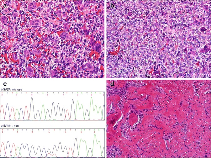
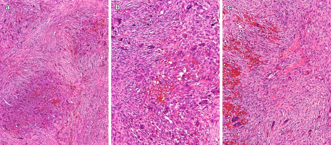
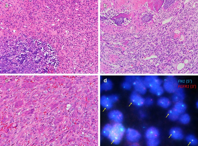
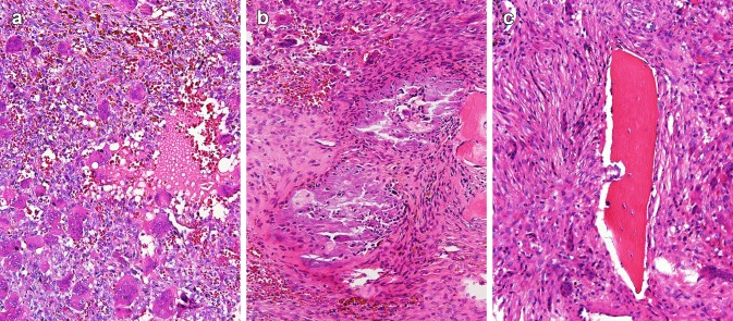
Giant cell-rich lesions represent a heterogeneous group of tumors and non-neoplastic lesions, usually arising in bone, which harbor varying number of reactive osteoclastic-type multinucleate giant cells as a common feature. Among these entities, some are confined to the head and neck region (e.g., central giant cell granuloma and mimicking lesions, i.e., peripheral giant cell granuloma and cherubism) or show a relative predilection for this region (e.g., aneurysmal bone cyst and brown tumor of hyperparathyroidism), while others are rare but associated with distinct underlying disease (e.g., giant cell tumor of bone) or histology (e.g., tenosynovial giant cell tumor of the temporomandibular joint and phosphaturic mesenchymal tumor of the jaws) when occurring in the head and neck. Collectively, these lesions pose great challenge in the pathologic diagnosis, which often requires combinatory assessment from the clinical, histopathologic, and/or molecular aspects. This review provides a summary of pertinent clinical and pathologic features and an update of recent molecular and genetic findings of these entities. The considerations in differential diagnosis as well as the effects of the emerging therapeutic RANKL-antagonizing antibody denosumab will also be addressed.

Keywords: Aneurysmal bone cyst; Brown tumor of hyperparathyroidism; Central giant cell granuloma; Giant cell tumor of bone; Phosphaturic mesenchymal tumor; Tenosynovial giant cell tumor.


Conflict of interest statement

The authors declare no conflict of interest.

Figures

Fig. 1
Fig. 1
Evenly distributed osteoclastic giant cells in most parts of this sinonasal giant cell tumor of bone (a). Note the ovoid to spindle “stromal” cells and a vascular stroma. Areas devoid of giant cells are also noticed in this case, where the tumor bulk is composed mainly of quite uniform ovoid to spindle tumor cells with pale cytoplasm (b). This tumor contains a rare H3F3B p.G34L mutation instead of more common H3F3A mutations (c). A post-denosumab vertebral giant cell tumor exhibits complete depletion of giant cells and prominent woven bone deposition (d). Note the lack of conspicuous atypia and the generally low cellularity. The mitotic activity is also low
Fig. 2
Fig. 2
A solid aneurysmal bone cyst in the occipital bone shows the proliferation of bland spindled cells arranged in fascicular and storiform patterns, in a collagenous to fibromyxoid stroma (a). The picture is somewhat reminiscent of nodular fasciitis. Osteoclast-type giant cells and inflammatory cells are focally present and unevenly distributed. Note a microscopic hemorrhagic cyst (middle lower field). The diagnosis is confirmed with FISH demonstrating break-apart of the USP6 gene (b)
Fig. 3
Fig. 3
Central giant cell granuloma. Nodules of hemorrhagic areas surrounded by fascicles or whorls of bland spindle cells (a). The osteoclastic giant cells tend to aggregate near the hemorrhagic areas (b). Osteoid deposition is sometimes observed (c)
Fig. 4
Fig. 4
A temporomandibular joint tenosynovial giant cell tumor presents scattered giant cells, many small macrophages, and variable number of larger histiocytoid to epithelioid tumor cells often containing cytoplasmic hemosiderin pigment, which tend to be located near the periphery of the cytoplasm (a). Chondroid metaplasia with calcification, a common feature of tenosynovial giant cell tumor in this location (b). FISH reveals CSF1 rearrangement, which is present in a small subset of cells (genuine neoplastic cells) among abundant non-neoplastic macrophages and inflammatory cells (c)
Fig. 5
Fig. 5
Phosphaturic mesenchymal tumors arising in the soft part of nasal cavity (a) and in the mandible bone (b), respectively. Both tumors are composed of bland spindle cells, with grungy calcification, osteoid-like matrix deposition (in b), and a rich vasculature which is somewhat hemangiopericytoma-like. Note the absence of osteoclastic giant cell in these two examples. By contrast, other phosphaturic mesenchymal tumors often harbor variable number of giant cells, sometimes so numerous that they can mimic giant cell tumor of bone (c). The tumor in a contains FN1-FGFR1 fusion (d)
Fig. 6
Fig. 6
Brown tumor of hyperparathyroidism. Note hemorrhage, hemosiderin deposition, and a fibrovascular stroma (a). Harboring numerous osteoclastic giant cells (especially near the hemorrhagic cyst), this example is quite difficult to distinguish from other giant cell-rich lesions. Focal calcification, possibly representing metaplastic calcification in the context of hyperparathyroidism, may also raise the suspicion of phosphaturic mesenchymal tumor (b). Osteoclastic tunneling of the trabecular bone (dissecting osteitis) is a hallmark feature of hyperparathyroidism sometimes identifiable in or adjacent to a brown tumor (c)

References

    1. Athanasou NA, Bansal M, Forsyth R, Reid RP, Sapi Z. Giant cell tumour of bone. In: Fletcher CDM, Bridge JA, Hogendoorn PCW, Mertens F, editors. World Health Organization classification of tumours of soft tissue and bone. Lyon: IARC Press; 2013. pp. 321–324.
    1. Dahlin DC, Cupps RE, Johnson EW., Jr Giant-cell tumor: a study of 195 cases. Cancer. 1970;25(5):1061–1070. - PubMed
    1. Campanacci M, Baldini N, Boriani S, Sudanese A. Giant-cell tumor of bone. J Bone Joint Surg Am. 1987;69(1):106–114. - PubMed
    1. Unni KK, Inwards CY. Dahlin’s bone tumors. 6. Philadelphia: LWW; 2010.
    1. Huvos AG. Bone tumors: diagnosis, treatment, and prognosis. 2. Philadelphia: WB Saunders; 1991.

MeSH terms

LinkOut - more resources