

Exogenous miR-29a Attenuates Muscle Atrophy and Kidney Fibrosis in Unilateral Ureteral Obstruction Mice
- PMID: 31950871
- PMCID: PMC7087404
- DOI: 10.1089/hum.2019.287
Exogenous miR-29a Attenuates Muscle Atrophy and Kidney Fibrosis in Unilateral Ureteral Obstruction Mice
Abstract
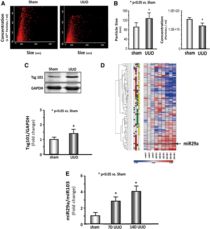
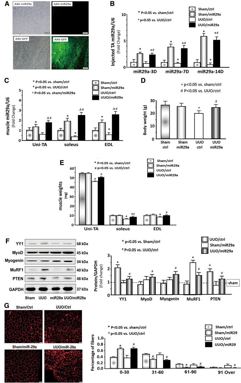
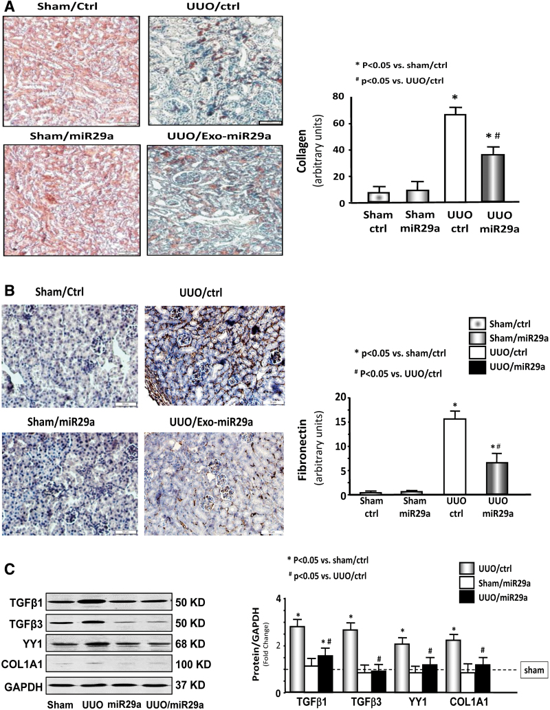
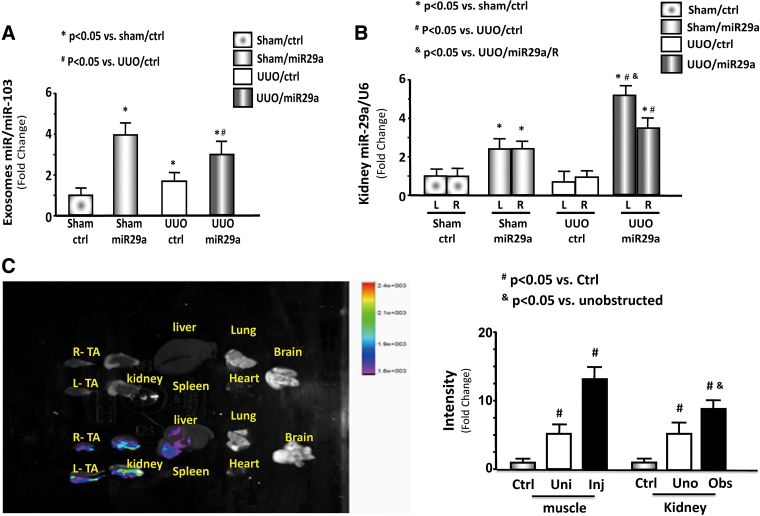
Renal fibrosis leads to end-stage renal disease, but antifibrotic drugs are difficult to develop. Chronic kidney disease often results in muscle wasting, and thereby increases morbidity and mortality. In this work, adeno-associated virus (AAV)-mediated overexpressing miR-29a was hypothesized to counteract renal fibrosis and muscle wasting through muscle-kidney crosstalk in unilateral ureteral obstruction (UUO) mice. miR-29a level was downregulated in the kidney and skeletal muscle of UUO mice. The secretion of exosome-encapsulated miR-29a increased in cultured skeletal muscle satellite cells and HEK293 renal cells after stimulation with serum from UUO mice. This result was confirmed by qPCR and microRNA deep sequencing in the serum exosomes of mice with obstructed ureters. A recombinant AAV-miR-29a was generated to overexpress miR-29a and injected into the tibialis anterior muscle of the mice 2 weeks before UUO surgery. AAV-miR-29a abrogated the UUO-induced upregulation of YY1 and myostatin in skeletal muscles. Renal fibrosis was also partially improved in the UUO mice with intramuscular AAV-miR-29a transduction. AAV-miR-29a overexpression reversed the increase in transforming growth factor β, fibronectin, alpha-smooth muscle actin, and collagen 1A1 and 4A1 levels in the kidney of UUO mice. AAV-green fluorescent protein was applied to trace the AAV route in vivo, and fluorescence was significantly visible in the injected/uninjected muscles and in the kidneys. In conclusion, intramuscular AAV-miR-29a injection attenuates muscle wasting and ameliorates renal fibrosis by downregulating several fibrotic-related proteins in UUO mice.
Keywords: MuRF1; TGF-β1; TGF-β3; YY1; kidney fibrosis; αSMA.
Conflict of interest statement
No competing financial interests exist.
Figures
Similar articles
-
Exosome-Mediated miR-29 Transfer Reduces Muscle Atrophy and Kidney Fibrosis in Mice.Mol Ther. 2019 Mar 6;27(3):571-583. doi: 10.1016/j.ymthe.2019.01.008. Epub 2019 Jan 18. Mol Ther. 2019. PMID: 30711446 Free PMC article.
-
miRNA-23a/27a attenuates muscle atrophy and renal fibrosis through muscle-kidney crosstalk.J Cachexia Sarcopenia Muscle. 2018 Aug;9(4):755-770. doi: 10.1002/jcsm.12296. Epub 2018 Mar 26. J Cachexia Sarcopenia Muscle. 2018. PMID: 29582582 Free PMC article.
-
Exogenous miR-26a suppresses muscle wasting and renal fibrosis in obstructive kidney disease.FASEB J. 2019 Dec;33(12):13590-13601. doi: 10.1096/fj.201900884R. Epub 2019 Oct 8. FASEB J. 2019. PMID: 31593640 Free PMC article.
-
The microRNA-29 family: role in metabolism and metabolic disease.Am J Physiol Cell Physiol. 2022 Aug 1;323(2):C367-C377. doi: 10.1152/ajpcell.00051.2022. Epub 2022 Jun 15. Am J Physiol Cell Physiol. 2022. PMID: 35704699 Review.
-
The long-term renal effects of short periods of unilateral ureteral obstruction.Int J Physiol Pathophysiol Pharmacol. 2022 Apr 15;14(2):60-72. eCollection 2022. Int J Physiol Pathophysiol Pharmacol. 2022. PMID: 35619661 Free PMC article. Review.
Cited by
-
The role of miR-29 family in disease.J Cell Biochem. 2021 Jul;122(7):696-715. doi: 10.1002/jcb.29896. Epub 2021 Feb 2. J Cell Biochem. 2021. PMID: 33529442 Free PMC article. Review.
-
Hochuekkito accelerates recovery from cisplatin induced-muscle atrophy accompanied by slow-twitch fiber-specific microRNA upregulation in mice.Front Pharmacol. 2025 May 15;16:1502563. doi: 10.3389/fphar.2025.1502563. eCollection 2025. Front Pharmacol. 2025. PMID: 40487396 Free PMC article.
-
Using RNA-based therapies to target the kidney in cardiovascular disease.Front Cardiovasc Med. 2023 Oct 6;10:1250073. doi: 10.3389/fcvm.2023.1250073. eCollection 2023. Front Cardiovasc Med. 2023. PMID: 37868774 Free PMC article. Review.
-
Effects of Exosomes Derived from Kidney Tubular Cells on Diabetic Nephropathy in Rats.Cell J. 2022 Jan;24(1):28-35. doi: 10.22074/cellj.2022.7591. Cell J. 2022. PMID: 35182062 Free PMC article.
-
Pathogenesis of Sarcopenia in Chronic Kidney Disease-The Role of Inflammation, Metabolic Dysregulation, Gut Dysbiosis, and microRNA.Int J Mol Sci. 2024 Aug 3;25(15):8474. doi: 10.3390/ijms25158474. Int J Mol Sci. 2024. PMID: 39126043 Free PMC article. Review.
References
Publication types
MeSH terms
Substances
Grants and funding
LinkOut - more resources
Full Text Sources
Medical
