. 2020 Mar 5;135(10):713-723.
doi: 10.1182/blood.2019002779.

Bispecific and split CAR T cells targeting CD13 and TIM3 eradicate acute myeloid leukemia

Affiliations

Bispecific and split CAR T cells targeting CD13 and TIM3 eradicate acute myeloid leukemia

Xin He et al. Blood. .

Abstract

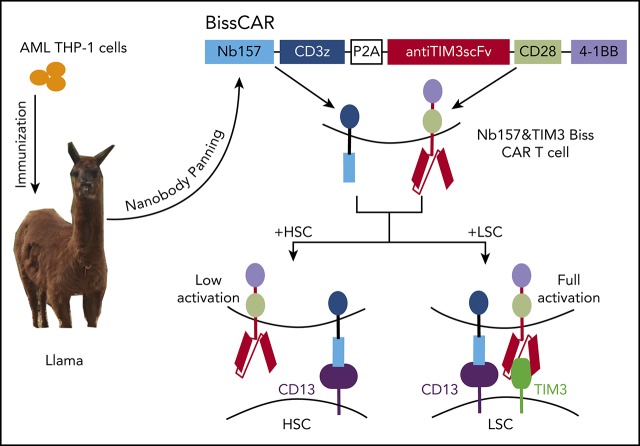
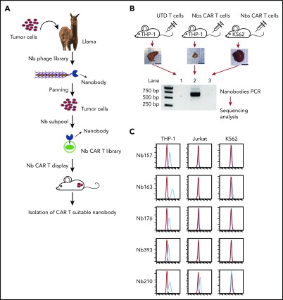
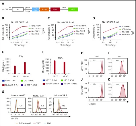
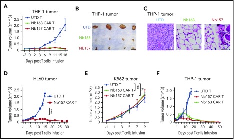
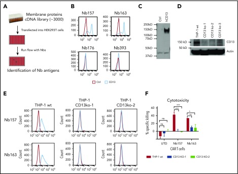
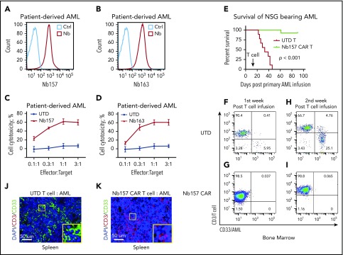
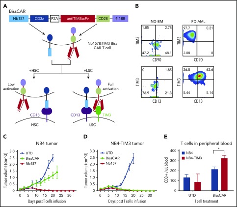
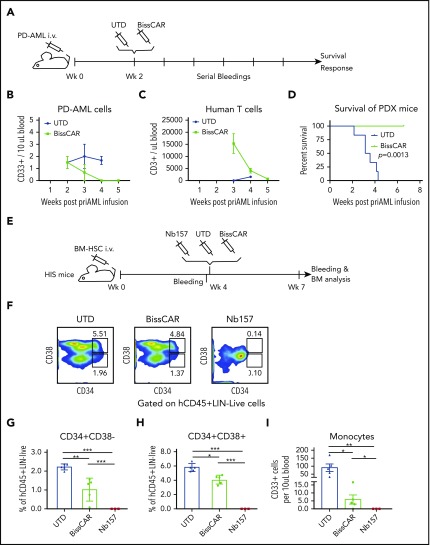
Chimeric antigen receptor (CAR) T cells have radically improved the treatment of B cell-derived malignancies by targeting CD19. The success has not yet expanded to treat acute myeloid leukemia (AML). We developed a Sequentially Tumor-Selected Antibody and Antigen Retrieval (STAR) system to rapidly isolate multiple nanobodies (Nbs) that preferentially bind AML cells and empower CAR T cells with anti-AML efficacy. STAR-isolated Nb157 specifically bound CD13, which is highly expressed in AML cells, and CD13 CAR T cells potently eliminated AML in vitro and in vivo. CAR T cells bispecific for CD13 and TIM3, which are upregulated in AML leukemia stem cells, eradicated patient-derived AML, with much reduced toxicity to human bone marrow stem cells and peripheral myeloid cells in mouse models, highlighting a promising approach for developing effective AML CAR T cell therapy.


Conflict of interest statement

Conflict-of-interest disclosure: The authors declare no competing financial interests.

Figures

None
Graphical abstract
Figure 1.
Figure 1.
Generating Nbs that differentially bind tumor cells and empower CAR T cells to kill the tumor cells. (A) Flowchart of AML-specific CAR-compatible Nbs in vivo screening. A llama was immunized with the AML cell line THP-1. An Nb library was generated from the llama PBMCs by molecular cloning. Two rounds of conventional cell-based phage display were applied, which took the T-acute lymphoblastic leukemia cell line Jurkat and the chronic myelogenous leukemia cell line K562 as negative absorption. Thereafter, 1 round of counter-selection was applied to obtain the nanobodies with high affinity. The resultant THP-1–specific Nbs were inserted into a CAR-expressing lenti-vector to generate the Nb–sub-lib CAR (Nb-CAR) library. Human primary T cells were transduced by the Nb-CAR library and injected into NSG mice with THP-1 or K562 tumors to perform the in vivo selection. Nbs that can redirect T cells to enrich in the tumor were amplified using polymerase chain reaction and sequenced. (B) Ten million THP-1 cells or 5 million K562 cells were transplanted into NSG mice subcutaneously, followed by treatment with UTD T cells or Nb–sub-lib CAR T cells. Two weeks later, Nbs from tumor-infiltrated T cells were isolated and identified using polymerase chain reaction amplification (n = 3). (C) The 5 most frequent Nbs in the THP-1 tumor are shown. The Nb-expressing phage was directly used to test the binding to THP-1 cells, Jurkat cells, or K562 cells using a flow cytometry assay, in which the red line was flow with Nb-expressing phage, and the blue line was isotype control. bp, base pair.
Figure 2.
Figure 2.
All Nbs isolated by the STAR system empower CAR T cells to potently kill AML cells in vitro. (A) Schematic diagram of Nb CAR structure, including signal peptide (SP), IgG4 mutant (IgG4m) hinge, CD8 transmembrane domain (TM), 4-1BB, and CD3z domain. (B-D) Nb CAR T cells showed potent and specific cytotoxicity against THP-1 or HL60 cells, but not K562 or Jurkat cells, in a dose-dependent manner. UTD T cells did not exert obvious killing (n = 3). THP-1 cells stimulated Nb157 or Nb163 CAR T cells, but not UTD T cells, to release cytokines, including IFN-γ (E) and TNF-α (F) (n = 3). (G) Only THP-1 cells induced Nb157 or Nb163 CAR T cells to degranulate (ie, CD107a localization to the cell membrane) after a 4-hour coculture (n = 3). CellTrace Far Red–labeled Nb157 (H-I) or Nb163 (J-K) CAR T cells were coincubated with heat-inactivated THP-1 cells (I-K) or K562 cells (H-J) for 4 days, followed by flow cytometry analysis. CAR T cells were gated on GFP+ signals (n = 3). ***P < .001, 1-way analysis of variance. ns, not significant (P > .05).
Figure 3.
Figure 3.
Nb-redirected CAR T cells potently eradicate AML tumor in vivo. (A-B) Ten million THP-1 cells were transplanted into NSG mice subcutaneously. The tumor reached 150 mm3 after ∼14 days. Three million Nb157 T cells, Nb163 CAR T cells, or UTD T cells were injected IV into the mice separately (n = 4). Tumor engraftment was monitored every other day. Scale bar, 10 mm. (C) Hematoxylin and eosin–stained THP-1 xenografts after treatment with UTD T cells, Nb163 CAR T cells, or Nb157 CAR T cells. Scale bars, 100 μm. (D) Three million Nb157 CAR T cells or UTD T cells were injected IV separately into NSG mice bearing HL60 tumors. Tumor engraftment was monitored every other day (n = 4). (E) Five million K562 cells were transplanted into NSG mice subcutaneously. The tumor reached 150 mm3 after ∼10 days. Three million Nb157 CAR T cells, Nb163 CAR T cells, or UTD T cells were injected into NSG mice separately. Tumor engraftment was monitored every other day (n = 4). (F) A total of 1.5 million Nb157 CAR T cells, Nb163 CAR T cells, or UTD T cells was injected IV separately into NSG mice bearing THP-1 tumor. Tumor engraftment was monitored every other day until the tumors were gone completely (n = 4). ***P < .001, 1-way analysis of variant. ns, not significant.
Figure 4.
Figure 4.
Identification of CD13 as a target to kill AML cells by CAR T cells. (A) Experimental schema. About 3000 cell membrane protein cDNAs were purified and transfected into HEK293T cells separately, followed by flow analysis with Nbs expressing phage and FITC-labeled secondary antibody against phage M13 protein. (B) Flow analysis of Nbs binding to HEK293T cells with CD13 overexpression. (C) Confirmation of CD13 cDNA expression in HEK293T cells by western blot. (D) Western blot was performed to confirm the gRNA/CRISPR-guided CD13-knockout effect in THP-1 cells. Three independent gRNAs were transduced into THP-1 separately, followed by puromycin selection and single individual clone expansion. (E) Flow analysis of Nb157- or Nb163-binding CD13-knockout THP-1 cells. (F) Cytotoxicity assay of CAR/UTD T cells to wild-type (wt) THP-1 or 2 CD13-knockout THP-1 cell lines (CD13 KO) (n = 4). *P < .05, **P <.01, d***P < .001, Student t test. ns. not significant.
Figure 5.
Figure 5.
Nb157 CAR T cells display antitumor activity in patient-derived AML cells in an NSG mouse model. Nb157 (A) and Nb163 (B) recognized PD AML cells by flow analysis. Nb157 (C) and Nb163 (D) CAR T cells specifically killed PD AML cells in vitro in a dose-dependent manner (n = 4). (E) Nb157 CAR T cells efficiently prolonged survival of NSG mice bearing PD AML. In brief, 20 million PD AML cells were injected into NSG mice, followed by treatment with 3 million Nb157 CAR T cells or UTD T cells, and survival of mice was monitored (n = 10, each group). (F-I) PD AML in NSG bone marrow and spleen was monitored after Nb157 CAR T cell treatment by staining with anti-human CD45/CD3/CD33, followed by flow cytometry analysis (n = 3). (J-K) At the end points of each group of experiments, mice spleens were harvested and fixed with paraformaldehyde, followed by immunofluorescence staining of anti-human CD3(red)/CD33(green) and DAPI (blue; nuclear).
Figure 6.
Figure 6.
In vivo, combinatorial bispecific and split CD13 and TIM3 CAR T cells eradicate tumor expressing CD13 and TIM3, but not tumor expressing only CD13. (A) Schematic diagram of combinatorial bispecific and split CD13 and TIM3. Nb157 linked with CD3z recognized CD13 on normal HSCs or LSCs. Anti-TIM3 linked with CD28 and 4-1BB recognized TIM3 only on LSCs. Such Biss CAR T cells can be fully activated only by LSCs but not by HSCs. (B) Flow cytometry showing the expression of TIM3, CD90, CD13 on normal donor bone marrow cells (ND-BM) or PD AML cells, which were gated from CD45+LinCD34+CD38 subsets. Ten million NB4 (C) or NB4-TIM3 (D) cells were transplanted subcutaneously into NSG mice to form 100-mm3 tumors. Three million combinatorial BissCAR T cells, conventional Nb157 CAR T cells, or UTD T cells were injected IV into each NSG mouse with the tumors. The engraftment volume was monitored by measuring the length and width of the tumor every other day (n = 4). (E) Three weeks after mice with NB4 or NB4-TIM3 tumors were treated with BissCAR T cells or UTD T cells, human T-cell (CD3+) numbers in mouse peripheral blood were analyzed by flow cytometry and quantified using CountBright counting beads (n = 3). *P < .05, Student t test.
Figure 7.
Figure 7.
BissCAR T cells targeting CD13 and TIM3 eradicate AML PDXs, but with reduced toxicity to human HSCs in vivo. (A) Schematic diagram of AML PDX mice treated with control or BissCAR T cells. Twenty million PD AML cells were injected into NSG mice, followed by injection of 5 million BissCAR T cells or UTD T cells 2 weeks later. Next, human peripheral blood CD3+ cells were analyzed by serial bleeding weekly. (B-C) PD AML cells or T cells in mouse peripheral blood were monitored weekly by flow staining with anti-human CD33 or anti-human CD3 antibodies. Blood volume was normalized and quantified using CountBright counting beads (n = 3). (D) Mice survival was monitored and recorded (n = 6 per group). (E) Schematic diagram of HIS mice for evaluation of human HSC toxicity. A total of 1.5 million normal donor bone marrow (BM) CD34+ cells was injected into each NSG mouse. Four weeks later, 3 million Nb157 anti-TIM3 BissCAR T cells, conventional Nb157 CAR T cells, or UTD T cells were injected IV, followed by flow cytometry analysis of peripheral blood and bone marrow (n = 5 per group for BissCAR and UTD T cells; n = 3 per group for Nb157 T cells). (F) Bone marrow of HIS mice, which were treated with T cells for 3 weeks, was analyzed by flow cytometry after staining with CD45/Lin/CD34/CD38/7-AAD. Representative fluorescence-activate cell sorting plots were used to identify HSC (CD34+CD38) and myeloid progenitors (CD34+CD38+). (G) HSCs (CD45+LinCD34+CD38) in the bone marrow of HIS mice were analyzed by flow cytometry 3 weeks after the initial treatment. (H) Myeloid progenitors (CD45+LinCD34+CD38) in the bone marrow of HIS mice were analyzed by flow cytometry 3 weeks after the initial treatment. (I) Monocytes (human CD45+CD33+) from peripheral blood of HIS mice were analyzed by flow cytometry 3 weeks after the initial treatment; cell number and blood volume were quantified using CountBright counting beads. In (G-I), n = 5 per group for BissCAR T cells and UTD T cells, n = 3 per group for Nb157 T cells. *P < .05, **P < .01, ***P < .001, Student t test.

Comment in

References

    1. Couzin-Frankel J. Breakthrough of the year 2013. Cancer immunotherapy. Science. 2013;342(6165):1432-1433. - PubMed
    1. Grupp SA, Kalos M, Barrett D, et al. . Chimeric antigen receptor-modified T cells for acute lymphoid leukemia. N Engl J Med. 2013;368(16):1509-1518. - PMC - PubMed
    1. Brudno JN, Kochenderfer JN. Chimeric antigen receptor T-cell therapies for lymphoma. Nat Rev Clin Oncol. 2018;15(1):31-46. - PubMed
    1. June CH, Sadelain M. Chimeric antigen receptor therapy. N Engl J Med. 2018;379(1):64-73. - PMC - PubMed
    1. Sadelain M, Rivière I, Riddell S. Therapeutic T cell engineering. Nature. 2017;545(7655):423-431. - PMC - PubMed

Publication types

Substances