. 2020:25:102154.
doi: 10.1016/j.nicl.2019.102154. Epub 2019 Dec 27.

Altered modulation of beta band oscillations during memory encoding is predictive of lower subsequent recognition performance in post-traumatic stress disorder

Affiliations

Altered modulation of beta band oscillations during memory encoding is predictive of lower subsequent recognition performance in post-traumatic stress disorder

Mihai Popescu et al. Neuroimage Clin. 2020.

Abstract

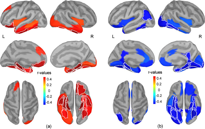
We studied the relationship between electrophysiological markers of memory encoding, subsequent recognition performance, and severity of PTSD symptoms in service members with combat exposure (n = 40, age: 41.2 ± 7.2 years) and various levels of PTSD symptom severity assessed using the PTSD Check List for DSM V version (PCL-5). Brain activity was recorded using magnetoencephalography during a serial presentation of 86 images of outdoor scenes that were studied by participants for an upcoming recognition test. In a second session, the original images were shown intermixed with an equal number of novel images while participants performed the recognition task. Participants recognized 76.0% ± 12.1% of the original images and correctly categorized as novel 89.9% ± 7.0% of the novel images. A negative correlation was present between PCL-5 scores and discrimination performance (Spearman rs = -0.38, p = 0.016). PCL-5 scores were also negatively correlated with the recognition accuracy for original images (rs = -0.37, p = 0.02). Increases in theta and gamma power and decreases in alpha and beta power were observed over distributed brain networks during memory encoding. Higher PCL-5 scores were associated with less suppression of beta band power in bilateral ventral and medial temporal regions and in the left orbitofrontal cortex. These regions also showed positive correlations between the magnitude of suppression of beta power during encoding and subsequent recognition accuracy. These findings indicate that the lower recognition performance in participants with greater PTSD symptom severity may be due in part to ineffective encoding reflected in altered modulation of beta band oscillatory activity.

Keywords: Beta band oscillations; Magnetoencephalography; Memory encoding; Post-traumatic stress disorder.


Conflict of interest statement

Declaration of Competing Interest None.

Figures

Fig. 1
Fig. 1
Results of the t-tests comparing the relative change in power during the temporal window from 0.3 s to 3 s against baseline are shown for theta (a), alpha (b), beta (c), and gamma bands (d). Statistical maps (t-values) show only regions with significant results after controlling the FDR. Maps are shown for each frequency band in lateral (upper row) and medial views (lower row) of the two hemispheres.
Fig. 2
Fig. 2
Temporal variation of the relative change in power is exemplified for the lateral occipital and parahippocampal regions of the left hemisphere. Power was integrated over 250 ms intervals centered at each time sample. The vertical dotted lines mark the temporal interval from 0.3 s to 3 s used to integrate the power for the primary analysis.
Fig. 3
Fig. 3
Maps of correlation coefficients between the regional change in beta band power and PCL-5 scores (a), and between the regional relative change in beta band power and recognition performance for original images (b). The regional relative change in beta band power was estimated during the temporal window from 0.3 s to 3 s. Maps show all regions with correlations significant at p<0.05 uncorrected for multiple comparisons (blue colors indicate negative correlations while red colors indicate positive correlations). The thin white lines mark the borders for brain regions with correlations that are significant after adjusting to control the FDR. Maps are shown in lateral (upper row) and medial views (middle row) of the two hemispheres, as well as in top and bottom views of the brain (lower row).
Fig. 4
Fig. 4
The mean change in beta band power for subgroups of patients with high (n = 21) vs. low (n = 19) recognition accuracy, and high (n = 21) vs. low (n = 19) PCL-5 scores, respectively, is exemplified in right and left parahippocampal cortex. The relative power is integrated over 250 ms intervals centered at each time sample. Participants were assigned to subgroups using a median split with respect to recognition accuracy (median value=75.7%) and PCL-5 scores (median value=20), respectively. Thirteen patients with low PCL5 scores were assigned to the high recognition accuracy subgroup.
Fig. 5
Fig. 5
Results of Wilcoxon signed-rank tests carried out across subjects to compare the rate of correct responses for sets of trials with highest versus lowest regional beta power integrated in temporal windows between 0.3 s to 3 s (a) and 3.3 s to 3.8 s (b), respectively. Maps of z-values show regions that were significant at p<0.05 uncorrected for multiple comparisons. The thin white lines mark the borders for brain regions with z-values that are significant after adjusting to control the FDR. Positive z-values (red colors) indicate a higher rate of correct responses for trials with highest regional beta power, while negative z-values (blue colors) indicate a higher rate of correct responses for trials with lowest regional beta power. Maps are shown in lateral (upper row) and medial views (middle row) of the two hemispheres, as well as in top and bottom views of the brain (lower row).

References

    1. Aguirre G.K., Zarahn E., D'Esposito M. An area within human ventral cortex sensitive to “building” stimuli: evidence and implications. Neuron. 1998;21:373–383. - PubMed
    1. American Psychiatric Association . 5th ed. American Psychiatric Association; Arlington, VA: 2013. Diagnostic and Statistical Manual of Mental Disorders; pp. 271–276.
    1. American Congress of Rehabilitation Medicine Definition of mild traumatic brain injury. J. Head Trauma Rehab. 1993;8:86–87.
    1. Aminoff E.M., Kveraga K., Bar M. The role of the parahippocampal cortex in cognition. Trends Cogn. Sci. 2013;17(8):379–390. - PMC - PubMed
    1. Aupperle R.L., Melrose A.J., Stein M.B., Paulus M.P. Executive function and PTSD: disengaging from trauma. Neuropharmacology. 2012;62(2):686–694. - PMC - PubMed

MeSH terms