. 2020 Jan 15;9(1):212.
doi: 10.3390/cells9010212.

Characterization of Mesenchymal Stem Cells Derived from Patients with Cerebellar Ataxia: Downregulation of the Anti-Inflammatory Secretome Profile

Affiliations

Characterization of Mesenchymal Stem Cells Derived from Patients with Cerebellar Ataxia: Downregulation of the Anti-Inflammatory Secretome Profile

Jong-Heon Kim et al. Cells. .

Abstract

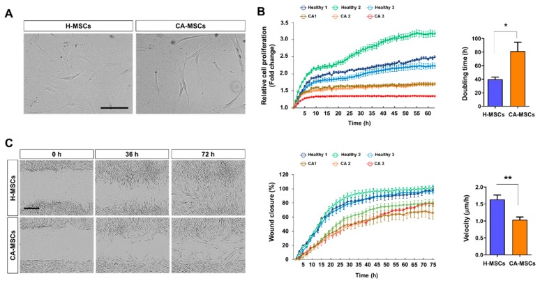
Mesenchymal stem cell (MSC) therapy is a promising alternative approach for the treatment of neurodegenerative diseases, according to its neuroprotective and immunomodulatory potential. Despite numerous clinical trials involving autologous MSCs, their outcomes have often been unsuccessful. Several reports have indicated that MSCs from patients have low capacities in terms of the secretion of neurotrophic or anti-inflammatory factors, which might be associated with cell senescence or disease severity. Therefore, a new strategy to improve their capacities is required for optimal efficacy of autologous MSC therapy. In this study, we compared the secretory potential of MSCs among cerebellar ataxia patients (CA-MSCs) and healthy individuals (H-MSCs). Our results, including secretome analysis findings, revealed that CA-MSCs have lower capacities in terms of proliferation, oxidative stress response, motility, and immunomodulatory functions when compared with H-MSCs. The functional differences were validated in a scratch wound healing assay and neuron-glia co-cultures. In addition, the neuroprotective and immunoregulatory protein follistatin-like 1 (FSTL1) was identified as one of the downregulated proteins in the CA-MSC secretome, with suppressive effects on proinflammatory microglial activation. Our study findings suggest that targeting aspects of the downregulated anti-inflammatory secretome, such as FSTL1, might improve the efficacy of autologous MSC therapy for CA.

Keywords: antiinflammation; cerebellar ataxia; mesenchymal stem cells.


Conflict of interest statement

The authors declare no conflict of interest.

Figures

Figure 1
Figure 1
Comparison of the biological characteristics of mesenchymal stem cells (MSCs) derived from cerebellar ataxia patients (CA-MSCs) and healthy individuals (H-MSCs). (A) Comparison of the morphology of H-MSCs and CA-MSCs. Scale bar = 200 μm. (B) Comparison of the proliferation of H-MSCs and CA-MSCs. The cell proliferation rate was assessed using the IncuCyte Time-lapse Microscopy System and was presented as fold change of cell density. Right panel shows doubling time of MSCs. (C) Scratch-wound healing assay. Representative bright-field images showing the migration potential of MSCs at 0, 36, and 72 h after wound scratch. Scale bar = 400 μm. In vitro cell migration and wound closure were assessed every 2 h. The scratch gap was calculated as a percentage of the relative cell density based on the initial wound area (middle panel). Right panel shows the velocity (μm/h) of wound closure. Error bars indicate the standard deviation of three replicate experiments. Data are presented as mean ± SEM. One-way analysis of variance was followed by the Tukey post-hoc test. * p < 0.05, ** p < 0.01.
Figure 2
Figure 2
Characteristics of the immunomodulatory potential of MSCs derived from CA-MSCs on microglial activation. (A) Schematic drawing of the experimental procedure. The conditioned medium obtained from MSCs culture (MSCs-CM) was added with/without lipopolysaccharide (LPS) (100 ng/mL) to BV-2 microglial cells or primary microglia for 24 h. (B,C) Nitric oxide assay in BV-2 cells and primary microglia cell culture was performed in triplicate. Nitric oxide production was measured using Griess reagents and was used to indicate microglial activation. (D) TNF-α production in BV-2 cells and primary microglia cell culture was measured in triplicate. The TNF-α level was measured using an enzyme-linked immunosorbent assay. Results are presented as mean ± SEM obtained from MSC culture (MSC-CM) from healthy subjects (n = 3) or CA patients (n = 3). One-way analysis of variance was followed by the Tukey post-hoc test. *** p < 0.001 vs. control; ## p < 0.01, ### p < 0.001 vs. control + LPS; †† p < 0.01, ††† p < 0.001 vs. healthy MSC-CM + LPS. ns, not significant.
Figure 3
Figure 3
Characteristics of the neuroprotective potential of MSCs derived from CA-MSCs on oxidative stress or microglial toxicity. (A) Schematic drawing of the experimental procedure. The conditioned medium obtained from MSC culture was added with/without H2O2 (200 µM) or microglial conditioned medium (M-CM). M-CM was generated by BV-2 microglial cell culture stimulated with LPS (100 ng/mL) for 24 h. (B) Cell viability assay in cerebellar granule neurons (CGNs) was performed in triplicate. Cell viability was measured using the MTT assay. Results are presented as mean ± SEM obtained from MSC-CM from healthy subjects (n = 3) or CA patients (n = 3). One-way analysis of variance was followed by the Tukey post-hoc test. *** p < 0.001 vs. control; # p < 0.05, ### p < 0.001 vs. control + H2O2 or M-CM; p < 0.05 vs. healthy MSC-CM + H2O2 or M-CM. ns, not significant. DW, distilled water.
Figure 4
Figure 4
Characteristics of the secretome of MSCs derived from CA-MSCs. (A) Schematic drawing of the secretome analysis procedure. (B) Venn diagrams showing the number of identified proteins in the secretome of MSCs derived from healthy individuals (H-MSCs) and CA-MSCs. Overall, 50 proteins are exclusively identified in H-MSCs, 17 are identified in CA-MSCs, and 76 are identified in both MSCs. (C) Pie chart illustrates the functional analysis using the biological processes of the downregulated secretome in CA-MSCs.
Figure 5
Figure 5
Validation of the secretome analysis results. (A) Validation of the identified proteins in secretome analysis according to mRNA expression by reverse transcription polymerase chain reaction. (B) Protein validation of FSTL1 in the conditioned media of mesenchymal stem cells (MSCs) derived from H-MSCs (n = 9) and MSCs derived from cerebellar ataxia patients (CA-MSCs) (n = 12). Data are presented as mean ± SEM. Comparisons of the two groups were carried out using an unpaired two-tailed t-test. ** p < 0.01, *** p < 0.001 vs. H-MSCs.
Figure 6
Figure 6
FSTL1 has a suppressive potential for microglial activation. (A) FSTL1 inhibited LPS-induced nitric oxide production in BV-2 microglial cells. BV-2 cells were stimulated with LPS (100 ng/mL) for 24 h after 2-h pretreatment with different concentrations of FSTL1 (n = 3 each). (B) Suppressive effect of FSTL1 on LPS-induced nitric oxide production in primary microglia. Data are presented as mean ± SEM. One-way analysis of variance was followed by the Tukey post-hoc test. * p < 0.05, ** p < 0.01, *** p < 0.001 vs. control; # p < 0.05, ## p < 0.01 vs. vehicle control (phosphate-buffered saline, PBS).

References

    1. Gamez J., Carmona F., Raguer N., Ferrer-Sancho J., Martin-Henao G.A., Marti-Beltran S., Badia M., Gratacos M., Rodriguez-Gonzalez E., Seoane J.L., et al. Cellular transplants in amyotrophic lateral sclerosis patients: An observational study. Cytotherapy. 2010;12:669–677. doi: 10.3109/14653241003774037. - DOI - PubMed
    1. Lee J.S., Hong J.M., Moon G.J., Lee P.H., Ahn Y.H., Bang O.Y. A long-term follow-up study of intravenous autologous mesenchymal stem cell transplantation in patients with ischemic stroke. Stem Cells. 2010;28:1099–1106. doi: 10.1002/stem.430. - DOI - PubMed
    1. Venkataramana N.K., Kumar S.K., Balaraju S., Radhakrishnan R.C., Bansal A., Dixit A., Rao D.K., Das M., Jan M., Gupta P.K., et al. Open-labeled study of unilateral autologous bone-marrow-derived mesenchymal stem cell transplantation in Parkinson’s disease. Transl. Res. 2010;155:62–70. doi: 10.1016/j.trsl.2009.07.006. - DOI - PubMed
    1. Staff N.P., Jones D.T., Singer W. Mesenchymal Stromal Cell Therapies for Neurodegenerative Diseases. Mayo Clin. Proc. 2019;94:892–905. doi: 10.1016/j.mayocp.2019.01.001. - DOI - PMC - PubMed
    1. Ullah I., Subbarao R.B., Rho G.J. Human mesenchymal stem cells—Current trends and future prospective. Biosci. Rep. 2015;35:e00191. doi: 10.1042/BSR20150025. - DOI - PMC - PubMed

Publication types

Substances

LinkOut - more resources