

Biomechanics of the Human Brain during Dynamic Rotation of the Head
- PMID: 31952465
- PMCID: PMC7307677
- DOI: 10.1089/neu.2019.6847
Biomechanics of the Human Brain during Dynamic Rotation of the Head
Abstract
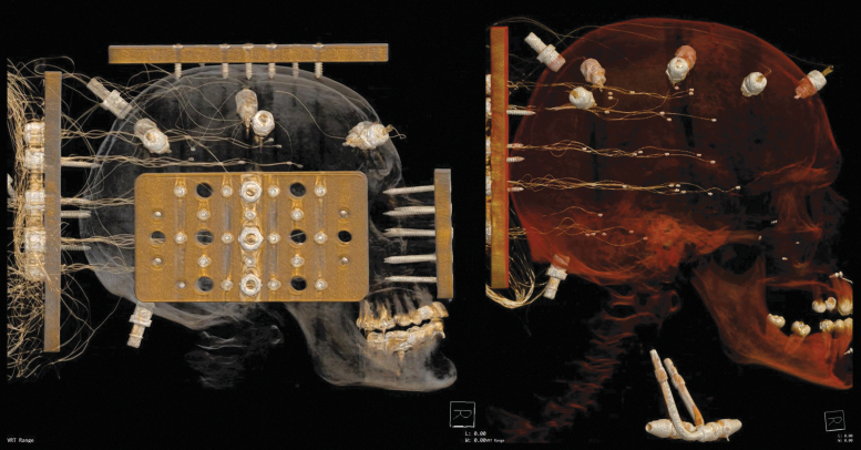
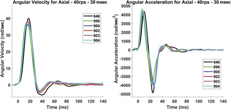
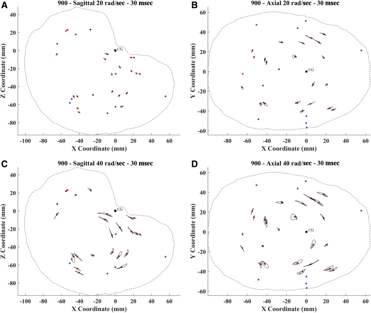
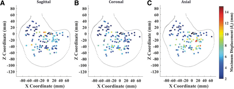
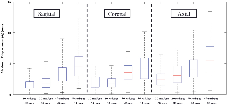
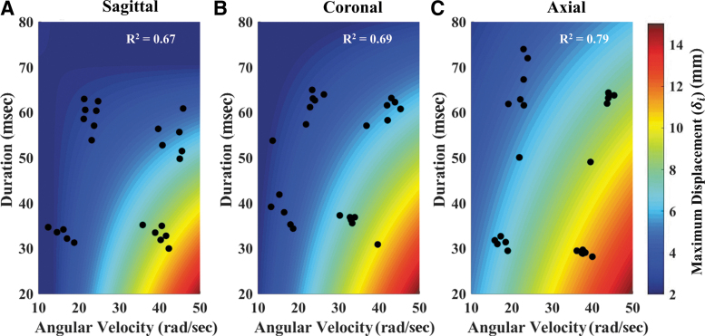
Traumatic brain injuries (TBI) are a substantial societal burden. The development of better technologies and systems to prevent and/or mitigate the severity of brain injury requires an improved understanding of the mechanisms of brain injury, and more specifically, how head impact exposure relates to brain deformation. Biomechanical investigations have used computational models to identify these relations, but more experimental brain deformation data are needed to validate these models and support their conclusions. The objective of this study was to generate a dataset describing in situ human brain motion under rotational loading at impact conditions considered injurious. Six head-neck human post-mortem specimens, unembalmed and never frozen, were instrumented with 24 sonomicrometry crystals embedded throughout the parenchyma that can directly measure dynamic brain motion. Dynamic brain displacement, relative to the skull, was measured for each specimen with four loading severities in the three directions of controlled rotation, for a total of 12 tests per specimen. All testing was completed 42-72 h post-mortem for each specimen. The final dataset contains approximately 5,000 individual point displacement time-histories that can be used to validate computational brain models. Brain motion was direction-dependent, with axial rotation resulting in the largest magnitude of displacement. Displacements were largest in the mid-cerebrum, and the inferior regions of the brain-the cerebellum and brainstem-experienced relatively lower peak displacements. Brain motion was also found to be positively correlated to peak angular velocity, and negatively correlated with angular velocity duration, a finding that has implications related to brain injury risk-assessment methods. This dataset of dynamic human brain motion will form the foundation for the continued development and refinement of computational models of the human brain for predicting TBI.
Keywords: FE model validation; brain biomechanics; sonomicrometry; traumatic brain injury.
Conflict of interest statement
No competing financial interests exist.
Figures
References
-
- Coronado V.G., McGuire L.C., Sarmiento K., Bell J., Lionbarger M.R., Jones C.D., Geller A.I., Khoury N., and Xu L. (2012). Trends in traumatic brain injury in the US and the public health response: 1995-2009. J. Safety Res. 43, 299–307 - PubMed
-
- Santiago L.A., Oh B.C., Dash P.K., Holcomb J.B., and Wade C.E. (2012). A clinical comparison of penetrating and blunt traumatic brain injuries. Brain Inj. 26, 107–125 - PubMed
-
- Faul M., Xu L., Wald M.M., and Coronado V.G.; National Center for Injury and Control (US), Division of Injury Response. (2010). Traumatic brain injury in the United States. Atlanta, GA: CDC Stacks Public Health Publications. CDC-INFO Pub ID 211298
-
- Gennarelli T.A. (1993). Mechanisms of brain injury. J. Emerg. Med. 11, 5–11 - PubMed
Publication types
MeSH terms
LinkOut - more resources
Full Text Sources
Medical
