Review
. 2020 Apr;18(2):326-334.
doi: 10.1016/j.jtos.2019.12.006. Epub 2020 Jan 14.

Immune - Goblet cell interaction in the conjunctiva

Affiliations
Review

Immune - Goblet cell interaction in the conjunctiva

Jehan Alam et al. Ocul Surf. 2020 Apr.

Abstract

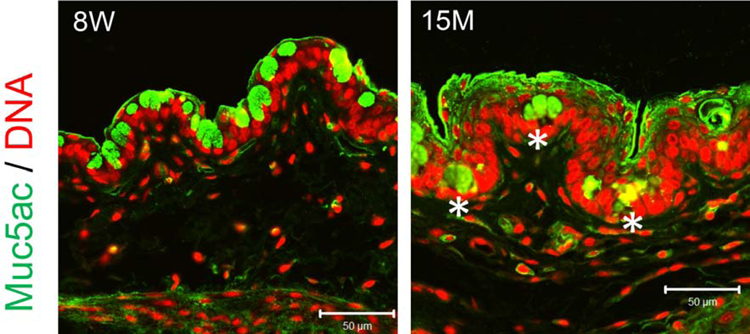
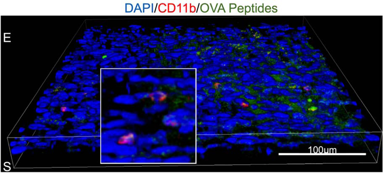
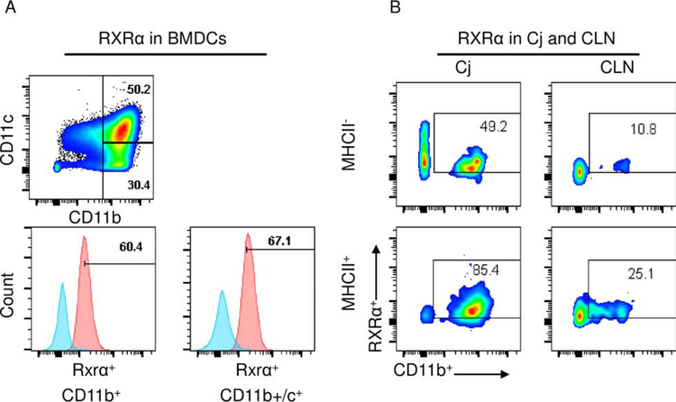
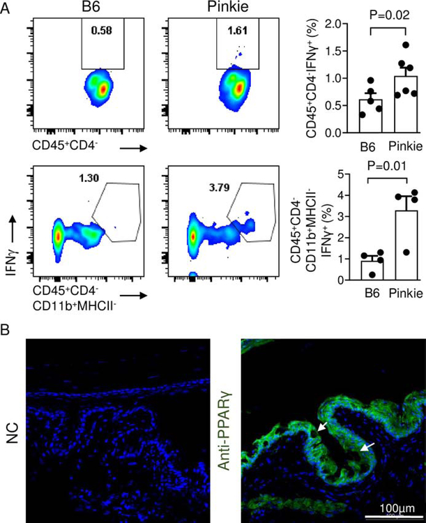
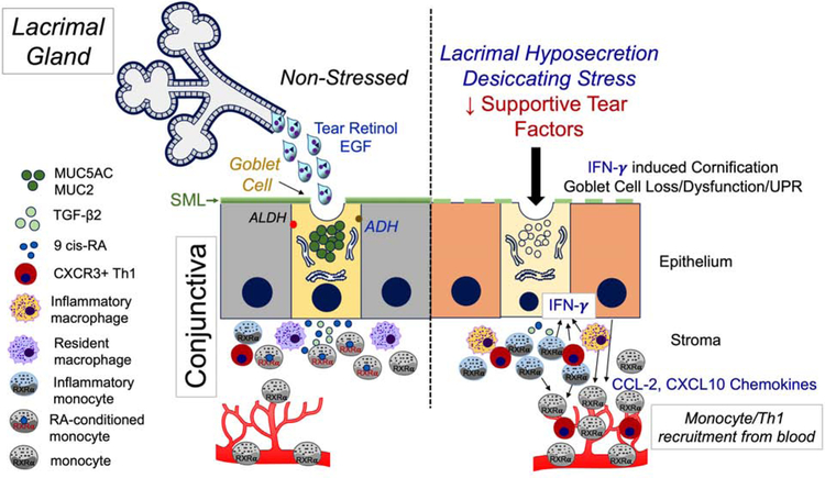
The conjunctiva is a goblet cell rich mucosal tissue. Goblet cells are supported by tear growth factors and IL-13 produced by resident immune cells. Goblet cell secretions are essential for maintaining tear stability and ocular surface homeostasis. In addition to producing tear stabilizing mucins, they also produce cytokines and retinoic acid that condition monocyte-derived phagocytic cells in the conjunctiva. Aqueous tear deficiency from lacrimal gland disease and systemic inflammatory conditions results in goblet cell loss that amplifies dry eye severity. Reduced goblet cell density is correlated with more severe conjunctival disease, increased IFN-γ expression and antigen presenting cell maturation. Sterile Alpha Motif (SAM) pointed domain epithelial specific transcription factor (Spdef) gene deficient mice that lack goblet cells have increased infiltration of monocytes and dendritic cells with greater IL-12 expression in the conjunctiva. Similar findings were observed in the conjunctiva of aged mice. Reduced retinoic acid receptor (RXRα) signaling also increases conjunctival monocyte infiltration, IFN-γ expression and goblet cell loss. Evidence suggests that dry eye therapies that suppress IFN-γ expression preserve conjunctival goblet cell number and function and should be considered in aqueous deficiency.

Keywords: Conjunctiva; Goblet cell; Immune response; Immunoregulation; Interferon gamma; Retinoic acid; Retinoid receptor.


Conflict of interest statement

Declaration of competing interest None of the authors have any financial or personal relationships to disclose that would cause a conflict of interest regarding this article.

Figures

Figure 1.
Figure 1.
Representative images of palpebral conjunctival cryosections stained for Muc5ac (green) and propidium counterstaining (DNA, in red) of 8-week-old (8W) and 15-months-old (15M) female C57BL/6 mice. Note that some Muc5ac+ cells are buried in the aged conjunctival epithelium (asterisks) and therefore unable to discharge to the ocular surface.
Figure 2.
Figure 2.
Confocal microscopy of whole mount conjunctiva 2 hours after topical application of fluorescent OVA peptide showing CD11b+ cells (red) beneath conjunctival epithelium that have phagocytosed the OVA peptide (green). Conjunctival epithelium is labeled E and the stroma S. Nuclei are stained blue with DAPI.
Figure 3.
Figure 3.
A. Flow cytometry was performed on cultured bone marrow derived cells (BMDCs) gated on CD11c and CD11b and the percentage of cells positive for the retinoid X receptor alpha (RXRα) was evaluated. Over 60% of CD11b+ and CD11b+CD11c+ cells were RXRα+; B. The percentage of CD11b+RXRα+ MHCII positive and negative cells in the conjunctiva and draining cervical lymph nodes was evaluated by flow cytometry. The percentage of RXRα+ cells was higher in the conjunctiva than the cervical nodes.
Figure 4.
Figure 4.
A. The percentages of CD45+CD4IFN-γ+ (top) and CD45+CD4-CD11b+ IFN-γ+ (bottom) cell populations in conjunctival tissue obtained from C57BL/6 and Pinkie mouse strains were evaluated by flow cytometry. Both cell populations were significantly higher in the Pinkie strain (bar graphs, right side). B. Mouse conjunctival sections stained for RXRα partner nuclear receptor peroxisome proliferator-activated receptor gamma (PPARγ). Secondary antibody negative control (NC) on the left and PPARγ antibody staining on the right. Arrows indicate goblet cells.
Figure 5.
Figure 5.
Retinoid X receptors (RXRs) dimerize with other partner nuclear receptors. Active RXRs regulate gene transcription by forming permissive heterodimers with fernesoid X receptor (FXR), pregnan X receptor (PXR), peroxisome proliferator-activated receptor (PPARs), Nurr1 and Nurr7, and liver X receptors (LXRs) and non-permissive heterodimers with thyroid receptors (TRs), retinoic acid receptor (RAR) and vitamin D receptor (VDR).
Figure 6.
Figure 6.
Immune - goblet cell interaction in the conjunctiva. During normal non-stressed conditions (left side), the lacrimal gland secretes tears containing epidermal growth factor (EGF) and vitamin A in the form of retinol. EGF supports goblet cell protein synthesis and proliferation and retinol is taken up by the goblet cells and metabolized by alcohol (ADH) and aldehyde (ALDH) dehyrogenases into the biologically active form retinoic acid (RA) that exists in equilibrium between the all trans- and 9-cis isomers. RA and TGF-β2 condition mononuclear phagocytic cells, including monocytes and macrophages in the conjunctiva. RA signaling through nuclear receptors, including the nuclear receptor RXRα in monocyte-derived cells, suppresses differentiation to inflammatory phenotypes that have higher expression levels of IFN-γ, IL-12 and antigen presenting cell maturation markers such as CD86. When lacrimal gland secretory function is reduced and the ocular surface is exposed to desiccation or other danger signals (right side), goblet cell number and function decreases and there is reduced conditioning of resident and recruited monocyte-derived cells resulting in increased expression of IFN-γ and IL-12 and monocyte and Th1 chemokines, such as CCL-2 and CXCL10, respectively, by the surface epithelium and monocytes. IFN-γ stimulates expression of cornifying genes, inhibits cholinergic signaling and induces an unfolded protein response (UPR) and apoptosis in the conjunctival goblet cells, reducing the secretory mucin layer (SML) and further amplifying ocular surface inflammation and epithelial disease.

References

    1. Farrand KF, Fridman M, Stillman IO, Schaumberg DA. Prevalence of Diagnosed Dry Eye Disease in the United States Among Adults Aged 18 Years and Older. American journal of ophthalmology. 2017;182:90–98. - PubMed
    1. Pflugfelder SC, Gumus K, Feuerman J, Alex A. Tear Volume-based Diagnostic Classification for Tear Dysfunction. International ophthalmology clinics. 2017;57(2):1–12. - PMC - PubMed
    1. Dartt DA. Regulation of mucin and fluid secretion by conjunctival epithelial cells. Prog Retin Eye Res 2002;21(6):555–576. - PubMed
    1. de Paiva CS, Raince JK, McClellan AJ, et al. Homeostatic control of conjunctival mucosal goblet cells by NKT-derived IL-13. Mucosal Immunol 2011;4(4):397–408. - PMC - PubMed
    1. Tukler Henriksson J, Coursey TG, Corry DB, De Paiva CS, Pflugfelder SC. IL-13 Stimulates Proliferation and Expression of Mucin and Immunomodulatory Genes in Cultured Conjunctival Goblet Cells. Investigative ophthalmology & visual science. 2015;56(8):4186–4197. - PMC - PubMed

Publication types