. 2020 Feb 21;295(8):2438-2448.
doi: 10.1074/jbc.RA119.011983. Epub 2020 Jan 17.

Protein secondary structure determines the temporal relationship between folding and disulfide formation

Affiliations

Protein secondary structure determines the temporal relationship between folding and disulfide formation

Philip J Robinson et al. J Biol Chem. .

Abstract

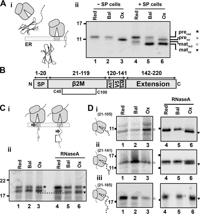
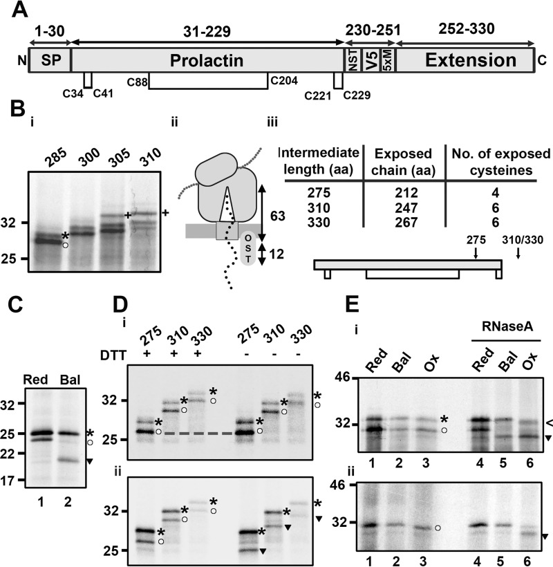
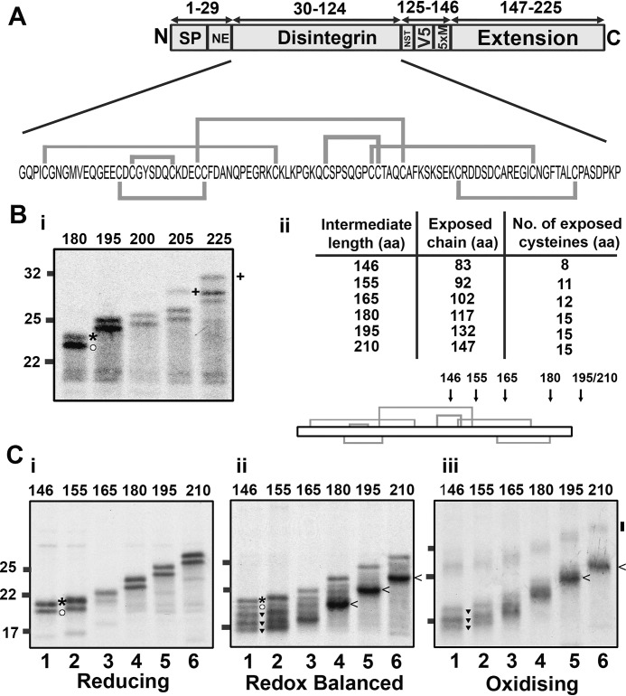
How and when disulfide bonds form in proteins relative to the stage of their folding is a fundamental question in cell biology. Two models describe this relationship: the folded precursor model, in which a nascent structure forms before disulfides do, and the quasi-stochastic model, where disulfides form prior to folding. Here we investigated oxidative folding of three structurally diverse substrates, β2-microglobulin, prolactin, and the disintegrin domain of ADAM metallopeptidase domain 10 (ADAM10), to understand how these mechanisms apply in a cellular context. We used a eukaryotic cell-free translation system in which we could identify disulfide isomers in stalled translation intermediates to characterize the timing of disulfide formation relative to translocation into the endoplasmic reticulum and the presence of non-native disulfides. Our results indicate that in a domain lacking secondary structure, disulfides form before conformational folding through a process prone to nonnative disulfide formation, whereas in proteins with defined secondary structure, native disulfide formation occurs after partial folding. These findings reveal that the nascent protein structure promotes correct disulfide formation during cotranslational folding.

Keywords: disulfide; endoplasmic reticulum (ER); folded precursor model; nascent chain; protein disulfide isomerase; protein folding; protein homeostasis; protein secretion; protein structure; protein translocation; quasi-stochastic model.


Conflict of interest statement

The authors declare that they have no conflicts of interest with the contents of this article

Figures

Figure 1.
Figure 1.
An experimental system to monitor disulfide formation and rearrangement in translation intermediates. A, ribbon diagrams representing the substrates used in this study: i, human β2M (PDB code 1A1M); ii, human prolactin (PDB code 1RW5); iii, the disintegrin domain of human ADAM10 (PDB code 6BE6). Disulfide bonds are highlighted in yellow. B, schematic illustrating the C-terminal extension added to each folding domain to produce the extended constructs. The position of the signal peptide, glycosylation site (NST), V5 epitope, and five methionine residues (5×M) are indicated. C, schematic of the ribosome–Sec complex expressing an ER-exposed nascent polypeptide (i). Phase 1 shows how the C-terminal extension retains the polypeptide attached to the ribosome to allow phase 2. In phase 2, the N-terminal folding domain of the polypeptide is fully exposed to the ER lumen. G6Pi drives the cytosolic reducing pathway (ii), which is the source of reducing equivalents for disulfide rearrangements in the ER (iii). PGL, phosphoglucolactone; G6PDH, glucose 6-phosphate dehydrogenase; TrxR1, thioredoxin reductase; TrX, thioredoxin.
Figure 2.
Figure 2.
β2M-disulfide formation is folding-driven. A, i, diagram illustrating signal peptide cleavage, which occurs following translocation of β2M. ii, nonreducing SDS-PAGE of immunoisolated WT β2M translated with or without SP cells in reducing (Red), redox-balanced (Bal), or oxidizing (Ox) lysate. The migration of preprotein (pre) and mature protein (mat) when oxidized (ox) or reduced (red) is indicated by the associated symbols (asterisks, >, circles, and inverted triangles). B, organization of the extended β2M construct with the single disulfide (Cys45-Cys100) displayed. C, i, diagram illustrating the dependence of disulfide formation on domain exposure; arrows highlight the end of the β2M folding domain. ii, nonreducing SDS-PAGE shows isolated 175 intermediates translated with SP cells in redox-balanced, oxidizing, and reducing lysate. RNaseA treatment following translation is indicated. D, nonreducing SDS-PAGE analysis of isolated ribosome/nascent chain complexes representing β2M 21–105 (i), 21–141 (ii), and 21–165 (iii) translated in redox-balanced, oxidizing, or reducing lysate without SP cells. Samples treated with RNaseA following translation are indicated. Schematics next to each panel indicate the predicted cysteine exposure at each intermediate length. All gels in this figure are representative of at least three repeats.
Figure 3.
Figure 3.
Disulfide formation in prolactin only occurs after ribosome release. A, organization of the extended prolactin construct with disulfide bond positions indicated. B, i, reducing SDS-PAGE of extended prolactin intermediates (285–310 aa in length), translated with SP cells. Preprotein (asterisk) and mature protein (circle) are highlighted for the 285 intermediate, and glycosylated protein is highlighted for the 305 and 310 intermediates (+). ii, schematic of the extension length (63 aa) required for ER exposure. iii, table of prolactin intermediate lengths containing the predicted exposure of amino acids and cysteine residues to the ER lumen and a topology diagram with ER exposure of specific intermediates, indicated by arrows. C, nonreducing SDS-PAGE of WT prolactin translated with SP cells in reducing (Red) or redox-balanced (Bal) lysate. D, extended prolactin intermediates (275–330) lacking a glycosylation site were translated with SP cells in redox-balanced lysate and either stalled (i) or released through RNaseA treatment (ii). Samples were immunoisolated (V5) and run under reducing (+DTT) and non-reducing (−DTT) conditions. E, non-reducing SDS-PAGE of the 310 intermediate translated in reducing, redox-balanced, or oxidizing (Ox) lysate in the presence (i) or absence (ii) of SP cells. Stalled samples were compared with released (RNaseA-treated) samples. In C–E, bands corresponding to reduced preprotein (asterisks), reduced mature protein (circles), oxidized preprotein (<), and oxidized mature protein (inverted triangles) are highlighted. Representative data from at least three experimental repeats in each case are shown.
Figure 4.
Figure 4.
Disulfide formation occurs in a partially ER-exposed disintegrin domain. A, organization of the disintegrin construct with the sequence and native disulfide bonding pattern highlighted. B, i, reducing SDS-PAGE to identify glycosylation (+) in extended disintegrin intermediates of lengths of 180–225 (preprotein (asterisk) and mature protein (circle) are highlighted for the 180 intermediate) (i), with the table and topology diagram (ii) detailing expected exposure of amino acids and cysteine residues to the ER lumen. C, non-reducing SDS-PAGE showing radiolabeled intermediates of increasing length (146–210 preprotein length) translated in reducing (i), redox-balanced (ii), and oxidizing lysate (iii). For the 146 samples, gel bands representing reduced preprotein (asterisks), reduced mature protein (circles) and oxidized mature protein (inverted triangles) are indicated. All gels represent translation reactions with SP cells immunoisolated to the V5 epitope and are representative of three independent repeats.
Figure 5.
Figure 5.
ER-specific disulfide rearrangements occur in a partially ER-exposed disintegrin intermediate. A–C, non-reducing SDS-PAGE shows either total translation product (V5) or ER-specific species (NE) for the 146 intermediate of the disintegrin domain when translated in reducing (Red), redox-balanced (Bal), and oxidizing (Ox) lysate (A); when translated in oxidizing lysate with G6Pi added after translation and samples taken at specific time points after G6Pi treatment (B, lanes 4–6); and when translated in oxidizing lysate to which G6Pi was added after translation in the presence or absence of auranofin (Aur) (C, lanes 4–7). Control samples for B and C were translated in reducing, redox-balanced, and oxidizing lysates (lanes 1–3 in both cases) for gel mobility comparison. The experiments in A and B were repeated three times and those in C twice, with representative data shown. Symbols indicate reduced preprotein (asterisks), reduced mature protein (circles), and oxidized mature protein (inverted triangles).
Figure 6.
Figure 6.
Stochastic disulfide formation occurs even when the cysteine density decreases. A, topology diagram showing the location of disulfides in the WT sequence of the disintegrin domain. B–D, non-reducing SDS-PAGE showing disulfide formation in stalled intermediates translated under reducing (Red), redox-balanced (Bal), or oxidizing (Ox) conditions for either the N-term cluster (B) or triple (C) or single (D) constructs immunoisolated using either a V5 or NE antibody as indicated. For each case, partially ER-exposed (146 or 165) intermediates (i) are compared with fully exposed (210) intermediates (ii). Topology diagrams above each panel show the position of disulfides in the relevant construct, with an arrow indicating the degree of ER exposure expected. Each condition was repeated three times, and representative data are shown. Symbols indicate the gel position of reduced preprotein (asterisks), reduced mature protein (circles), and oxidized mature protein bands (inverted triangles).

References

    1. Fass D., and Thorpe C. (2018) Chemistry and enzymology of disulfide cross-linking in proteins. Chem. Rev. 118, 1169–1198 10.1021/acs.chemrev.7b00123 - DOI - PMC - PubMed
    1. Hudson D. A., Gannon S. A., and Thorpe C. (2015) Oxidative protein folding: from thiol-disulfide exchange reactions to the redox poise of the endoplasmic reticulum. Free Radic. Biol. Med. 80, 171–182 10.1016/j.freeradbiomed.2014.07.037 - DOI - PMC - PubMed
    1. Oka O. B., and Bulleid N. J. (2013) Forming disulfides in the endoplasmic reticulum. Biochim. Biophys. Acta 1833, 2425–2429 10.1016/j.bbamcr.2013.02.007 - DOI - PubMed
    1. Welker E., Wedemeyer W. J., Narayan M., and Scheraga H. A. (2001) Coupling of conformational folding and disulfide-bond reactions in oxidative folding of proteins. Biochemistry 40, 9059–9064 10.1021/bi010409g - DOI - PubMed
    1. Qin M., Wang W., and Thirumalai D. (2015) Protein folding guides disulfide bond formation. Proc. Natl. Acad. Sci. U.S.A. 112, 11241–11246 10.1073/pnas.1503909112 - DOI - PMC - PubMed

Publication types

MeSH terms

LinkOut - more resources