. 2020 Mar 13;295(11):3614-3634.
doi: 10.1074/jbc.RA119.011527. Epub 2020 Jan 17.

The small molecule GAT1508 activates brain-specific GIRK1/2 channel heteromers and facilitates conditioned fear extinction in rodents

Affiliations

The small molecule GAT1508 activates brain-specific GIRK1/2 channel heteromers and facilitates conditioned fear extinction in rodents

Yu Xu et al. J Biol Chem. .

Abstract

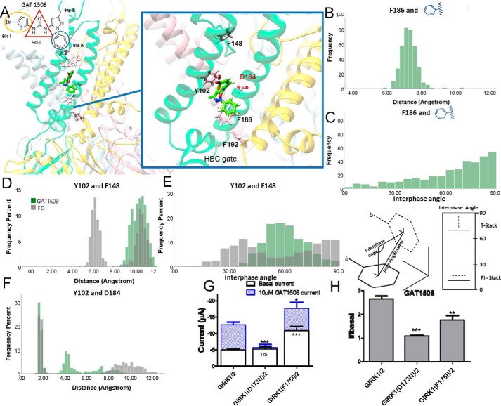
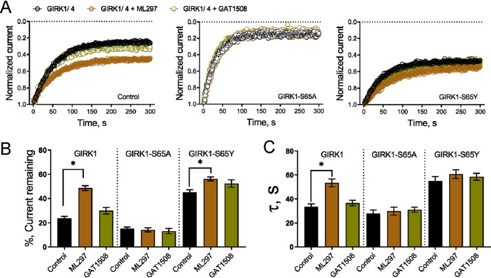
G-protein-gated inwardly-rectifying K+ (GIRK) channels are targets of Gi/o-protein-signaling systems that inhibit cell excitability. GIRK channels exist as homotetramers (GIRK2 and GIRK4) or heterotetramers with nonfunctional homomeric subunits (GIRK1 and GIRK3). Although they have been implicated in multiple conditions, the lack of selective GIRK drugs that discriminate among the different GIRK channel subtypes has hampered investigations into their precise physiological relevance and therapeutic potential. Here, we report on a highly-specific, potent, and efficacious activator of brain GIRK1/2 channels. Using a chemical screen and electrophysiological assays, we found that this activator, the bromothiophene-substituted small molecule GAT1508, is specific for brain-expressed GIRK1/2 channels rather than for cardiac GIRK1/4 channels. Computational models predicted a GAT1508-binding site validated by experimental mutagenesis experiments, providing insights into how urea-based compounds engage distant GIRK1 residues required for channel activation. Furthermore, we provide computational and experimental evidence that GAT1508 is an allosteric modulator of channel-phosphatidylinositol 4,5-bisphosphate interactions. Through brain-slice electrophysiology, we show that subthreshold GAT1508 concentrations directly stimulate GIRK currents in the basolateral amygdala (BLA) and potentiate baclofen-induced currents. Of note, GAT1508 effectively extinguished conditioned fear in rodents and lacked cardiac and behavioral side effects, suggesting its potential for use in pharmacotherapy for post-traumatic stress disorder. In summary, our findings indicate that the small molecule GAT1508 has high specificity for brain GIRK1/2 channel subunits, directly or allosterically activates GIRK1/2 channels in the BLA, and facilitates fear extinction in a rodent model.

Keywords: GIRK channels; PIP2; basolateral amygdala; medicinal chemistry; neurophysiology; phosphoinositide; potassium channel; small molecule; specific activator.


Conflict of interest statement

The authors declare that they have no conflicts of interest with the contents of this article

Figures

Figure 1.
Figure 1.
ML297 derivatives that specifically activate the brain GIRK1/2 over the cardiac GIRK1/4. A, shown are the rational design and the systematic focused approach to a preliminary SAR study on ML297. Assessing Site I, which is an important pharmacophore for selectivity and efficacy on GIRK1-containing channels, several analogs of ML297 were synthesized using rapid, tandem, and one-pot methodology for urea synthesis to probe their selectivity for GIRK1/2 over GIRK1/4 channels. The bioisosteric replacement strategy of the phenyl ring of Site I with a thiophene ring was adopted and found to be most tolerable and GIRK1/2 channel-selective. A thiophene ring was used as a prototype for further optimization revealing 5-bromo and 5-methyl substituents as optimal pharmacophores for GIRK1/2 selectivity, efficacy, and potency that proved better than ML297. B, concentration-response curves for ML297 and the selective compounds GAT1508 and GAT1521 were assessed in HEK293 cells expressing GIRK1/2 WT (blue open circle) and GIRK1/4 WT (solid red diamond) using whole-cell patch-clamp recordings. Data are mean ± S.E. for 7–12 cells per condition. The EC50 of ML297 at GIRK1/2 was 110 ± 13 nm, and the EMAX was 5.1 ± 0.1 (considered here to be 100%); at GIRK1/4 the EC50 was 1.2 ± 0.5 μm, and EMAX was 2.3 ± 0.2 (100%). At GIRK1/2 channels, GAT1508 had an EC50 of 75 ± 10 nm, and an EMAX of 8.4 ± 0.2 (165% relative to the EMAX of ML297), whereas GAT1521 had an EC50 of 350 ± 58 nm, and an EMAX of 4.1 ± 0.13 (81% relative to the EMAX of ML297).
Figure 2.
Figure 2.
Basis for selective binding of GAT1508 between brain and cardiac heteromeric channels. A, percentage of ML297 or GAT1508 contacts with the transmembrane M1 (light blue bars) and M2 (pink bars) helices of the GIRK2/2FD and GIRK4/4FD channels during the MD trajectories. Contact was defined as drug–Cα distance ≤7 Å. Snapshots of GAT1508 binding in GIRK2/2FD (B) and GIRK4/4FD (C) are shown. The flipping of the GAT1508 position in the GIRK4/4FD system and the proximity of the thiofuran group to the Thr-94 of the M1 helix of the WT GIRK4 subunit are shown. D, concentration-response curves of GAT1508 in GIRK1/2 (solid blue circle) and GIRK1/4 (solid red square) WT or the double mutant channels GIRK1/2(IV-VT) (purple triangle) or GIRK1/4(VT-IV) (pink inverted triangle). Data are mean ± S.E. for 16 cells (four oocytes × four frogs) per concentration. GAT1508 stimulated brain (GIRK1/2) heteromeric channel currents with an EC50 of 0.37 ± 0.11 μm and an EMAX of 2.42 ± 0.07 (considered here to be 100%). Double mutants in the GIRK2 WT subunits to corresponding GIRK4 residues showed reduced potency and efficacy for the GAT1508-induced currents with an EC50 of 0.94 ± 0.15 μm and an EMAX of 1.68 ± 0.04 (72% of the control GIRK1/2). Double mutants in GIRK4 WT subunits to corresponding GIRK2 residues conferred stimulation of the heteromeric channel by GAT1508 with an EC50 of 0.55 ± 0.11 μm and an EMAX of 1.76 ± 0.03 (75% of the control GIRK1/2). E, single mutants between brain and cardiac channels switch GAT1508 sensitivity. Concentration-response curves of GAT1508 in GIRK1/2 (solid blue circle) and GIRK1/4 (solid red square) WT or single mutant channels between unique M1 residues in GIRK2 and GIRK4: GIRK1/2(I-V) (open blue circle), GIRK1/2(V-T) (cyan star), GIRK1/4(V-I) (pink open diamond), and GIRK1/4(T-V) (blue plus). Data are mean ± S.E. for eight cells (four oocytes × two frogs) per concentration. GAT1508 stimulated brain (GIRK1/2) heteromeric channel currents with an EC50 of 0.37 ± 0.11 μm and an EMAX of 2.36 ± 0.23 (considered here to be 100%). Single mutants in the GIRK2 WT subunits to corresponding GIRK4 residues significantly reduced the potency and efficacy of the GAT1508-induced selective currents: GIRK1/2(V-T) with an EC50 of 0.53 ± 0.07 μm and an EMAX of 1.92 ± 0.03 (65% of the control GIRK1/2) and GIRK1/2(I-V) with an EC50 of 0.50 ± 0.11 μm and an EMAX of 1.61 ± 0.03 (42% of the control GIRK1/2). Single mutants in GIRK4 WT subunits to corresponding GIRK2 residues conferred stimulation of the heteromeric channel by GAT1508: GIRK1/4(T-V) with an EC50 of 0.50 ± 0.47 μm and an EMAX of 1.12 ± 0.03 (7% of the control GIRK1/2); GIRK1/4(V-I) with an EC50 of 0.53 ± 0.15 μm and an EMAX of 1.29 ± 0.03 (11% of the control GIRK1/2). F, impact of mutating GIRK2 or GIRK4 side chains predicted to lead to selective binding of the GAT1508 molecule on basal current and 20 μm drug-induced current-enhancing effects are shown. ns signifies no significant changes in basal or drug-induced currents. Symbols denote statistical significance by one-way ANOVA. *, p < 0.05 compares WT GIRK1/2 and GIRK1/4 with GIRK1/2(V-T) and GIRK1/4(V-I), respectively. ***, p < 0.0005 compares WT GIRK1/2 or GIRK1/4 with their respective double mutants, n = 8.
Figure 3.
Figure 3.
Site I of GAT1508 binding joins into the Pi network system around pore helix of GIRK1-like subunit and single mutant functional effects. A, right, binding site of GAT1508 in the GIRK2FD subunit (FD subunit is shown in green; adjacent WT subunit is shown in yellow, and nondrug-binding subunits are shown in gray). Left, zooming in on Site I of the GAT1508-binding site (yellow outlines) reveals Pi interactions of the compound with residues Phe-108 and Phe-109 in the M1 helix of the GIRK2FD subunit. The Pi network was monitored during the MD simulation and displayed by plotting the distance and interphase angle frequency distributions between GAT1508 and Phe-108 (B and C) between GAT1508 and Phe-109 (E and F) between Phe-108 and Phe-109 (D and G), and between Phe-109 and Phe-148 (H and I) in GIRK2/2FD in the absence of the compound (black) or with GAT1508 (olive green). The plane interphase angle was calculated as the cross-product of the normal vectors of each aromatic ring. The impact of mutations of GIRK1 side chains predicted to bind the GAT1508 molecule on basal currents and on the 10 μm drug-induced current-enhancing effect (J) and normalized drug-induced current (K). Symbols denote statistical significance by one-way ANOVA. **, p < 0.005 compares WT GIRK1/2 with GIRK1(F97I)/2. n = 8.
Figure 4.
Figure 4.
Predictions of GAT1508 Site II interactions with residues in the middle of the M1 helix in the GIRK1-like subunit probed by point mutations. A, right, binding site of GAT1508 in the GIRK2FD subunit (FD subunit is shown in green; adjacent WT subunit is shown in yellow, and nondrug-binding subunits are shown in gray). Left, zooming in on the GAT1508-binding Site II (red outlines) shows hydrophobic interactions of the compound with residues Val-104 and Val-101 (on the M1 helix) and Leu-179 (on the M2 helix). B, impacts of these GIRK2FD side chains on basal current and 10 μm drug-induced current were probed with corresponding mutations on GIRK1 (numbers of residues 11 less that GIRK2: Val-93, Thr-90, and Leu-168) that either preserved the hydrophobic or hydrophilic property of the residue or removed the side chain with a Gly residue (C), normalized drug-induced current over basal current. Results from GIRK1(Val-93L)/2, GIRK1(Val-93S)/2, GIRK1(Val-93G), GIRK1(T90G)/2, GIRK1(T90S)/2, GIRK1(L168G)/2, and GIRK1(L168V) are shown. Symbols denote statistical significance by one-way ANOVA. *, p < 0.05 compares WT GIRK1/2 and GIRK1(Val-93G)/2 for basal current and drug-induced current, and GIRK1(Val-93S)/2 for drug-induced current, respectively. **, p < 0.005 compares WT GIRK1/2 with GIRK1(V93S)/2 and GIRK1(T90G)/2 for normalized drug-induced current, and GIRK1(L168G)/2 for drug-induced current. ***, p < 0.0005 compares WT GIRK1/2 with GIRK1(L168V)/2 for basal current and drug-induced current, and with GIRK1(L168G)/2 for normalized drug-induced current. n = 8.
Figure 5.
Figure 5.
Probing predicted GAT1508 Site IV interactions with the intracellular side of M2 helix in the GIRK2FD subunit with point mutants. A, right, binding site of GAT1508 in the GIRK2FD subunit (FD subunit is shown in green; adjacent WT subunit is shown in yellow, and nondrug-binding subunits are shown in gray). Left, zooming in on the GAT150-binding Site IV (blue outlines) reveals Pi interactions of the compound with GIRK2FD residue Phe-186. The interaction network was monitored during the MD simulation (distance and interface angle) as frequency distributions between GAT1508 Site IV and Phe-186 (B and C), between Tyr-102 and Phe-148 (D and E), and between Tyr-102 and Asp-184 in GIRK2/2FD in the absence of the compound (black) or with GAT1508 (olive green). The plane interphase angle is calculated as the cross-product of the normal vectors of each aromatic ring. When this interphase angle is between 600 and 900, the two aromatic rings form a T stack. When the interphase angle is less than 30, they form traditional π-stacking interactions. B and C, angles between the GAT1508 benzene ring of Site IV and Phe-186 show a broad distribution with a peak at 900, suggesting T-stacking interactions. D–F, GAT1508 binding destabilizes the Phe-148 interaction with Tyr-102, releasing it to form H-bond interactions with Asp-184. G and H, impact of probing these predictions with corresponding mutations on GIRK1 (e.g. F175I and D173N) is significant. Symbols denote statistical significance by one-way ANOVA. *, p < 0.05 compares WT GIRK1/2 and GIRK1(F175I)/2 in drug-induced current. **, p < 0.005 compares WT GIRK1/2 and GIRK1(F175I)/2 in normalized drug-induced current. ***, p < 0.0005 compares WT GIRK1/2 with GIRK1(D173N)/2 drug-induced current and normalized drug-induced current and with GIRK1(F175I)/2 basal current. n = 8.
Figure 6.
Figure 6.
Specific activation by GAT compounds increases brain GIRK2 but not cardiac GIRK4 channel–PIP2 interactions. A and B, normalized salt-bridge formation between the head group PIP2 and positively-charged channel residues are calculated during the last 25 ns of the 100-ns MD simulation runs in the absence of ligand or with ML297 and GAT1508 bound in the GIRK2/2FD (blue) and GIRK4/4FD (red) systems. C, GIRK1/2 channel NPo was assessed by diC8–PIP2 concentration–response curves using inside-out macropatches from Xenopus oocytes in the presence and absence of 10 μm ML297 or GAT1508; data are means ± S.E. for 5–6 patches per condition. When studied under control conditions (black solid circle), GIRK1/2 channels showed an apparent affinity to diC8–PIP2 of 36 ± 7 μm and an EMAX of 5.7 ± 0.3 (100%). ML297 (brown solid triangle) increases the apparent affinity to 23 ± 5 μm and the EMAX to 7.5 ± 0.4 (100%). GAT1508 (olive green inverted solid triangle) increased the apparent affinity and EMAX further to 5.5 ± 7 and 10.5 ± 0.3 μm (184%), respectively. D, under control conditions, GIRK1/4 channels had an apparent affinity for diC8-PIP2 of 48.4 ± 8 μm and an EMAX of 4.4 ± 0.2 (100%). ML297 increased the apparent affinity to 17.4 ± 2 μm and raised the EMAX to 5.4 ± 0.2 (123% of the control). In contrast, treatment with GAT1508 did not change the apparent affinity for diC8-PIP2 from control (46 ± 7.4 μm) or the efficacy (98.6 and 103%, respectively.
Figure 7.
Figure 7.
ML297 and GAT1508 protect the increasing channel activity from inhibition by PIP2-dephosphorylating light-sensitive phosphatase. Data are currents recorded from HEK293 cells using patch-clamp in the whole-cell mode and are shown as means ± S.E. for 6–8 cells per group. Statistical significance was calculated using unpaired t tests. *, p < 0.01. A, representative plots of normalized current magnitude against time show that GIRK1/2 channel current decreases in response to light-activated dephosphorylation of PIP2 by the phosphatase 5-ptaseOCRL (control, black open circle). The decrease in current is reduced when GIRK1/2 channels are studied in the presence of 10 μm GAT1508 (olive green open circle). B, 5-ptaseOCRL-mediated decrease in GIRK1/2 current is characterized by mono-exponential fits in the presence and absence of 10 μm ML297 (brown open circle) or GAT1508 (olive green open circle). ML297 and GAT1508 increase to a different degree the percentage of GIRK1/2 channel current remaining following activation of 5-ptaseOCRL (C) and increase the τ of current inhibition to a different extent (D). ML297, but not GAT1508, increases the percentage of residual GIRK1/4 channel current (E), and the τ of the current decrease, following activation of 5-ptaseOCRL (F).
Figure 8.
Figure 8.
GAT1508 binding to the FD-containing subunits induces a smaller increase in the adjacent (non-FD or WT) subunit in GIRK4 than in GIRK2 via a critical Slide helix-specific polar interaction. A, based on the PIP2-binding area, we classified PIP2 into two types. P-WT (yellow) represents the two PIP2 molecules interacting with WT (nondrug binding on non-FD containing) subunits around the area that drugs bind when present. P-FD (green) represents the two PIP2-interacting molecules with the FD-mutated drug-binding subunits (analogous to GIRK1) in the absence of drugs bound. B, percentage of salt-bridge formation between each type of PIP2 (P-WT and P-FD) and positively-charged channel residues in GIRK2/2FD and GIRK4/4FD systems. The binding of ML297 or GAT1508 shifts the PIP2-binding site and increases P-WT interactions with the GIRK2 more so than GIRK4 WT subunits. The interaction between GAT1508 and the FD-mutated subunits decreases the channel P-FD interaction in both GIRK2 and GIRK4 heteromeric channels. C, average snapshots of PIP2 interacting with GIRK2/GIRK4 Tyr-76/71 residues in the channel slide helix region with ML297 (top) and GAT1508 (bottom) binding in GIRK2/2FD (left panels) and GIRK4/4FD (right panels). PCA suggests large conformational changes induced by GAT1508 binding in GIRK2FD or ML297 binding in GIRK2FD and GIRK4FD. A strong H-bond interaction is formed between Tyr residue in the slide helix of FD-mutated subunit and P4/5 of PIP2, which can position the PIP2 head group toward the junction of the M2 and TM–CTD (where CTD is cytoplasmic domain) linker and induce the increase in channel P-WT interactions. However, GAT1508 binding around the M2 helix of GIRK4/4FD results in loss of the H-bond interaction between GIRK4FD(Tyr-71) and PIP2 and failure to position the head group for optimal interactions. D, minimum distance between the O atom of Tyr-76/71 in the FD-mutated subunits and P4,5-PIP2. E, sequence alignment of the GIRK channel Slide helix region. The key residue interactions proposed by the simulations for the GAT1508-induced changes in GIRK activation are highlighted. As polar residues, both Ser and Tyr could form H-bond interactions with PIP2.
Figure 9.
Figure 9.
GIRK1 subunit Slide helix residue Ser-65 is required for the drug-induced enhancement of GIRK activity. Data are currents recorded from HEK293 cells using patch-clamp in the whole-cell mode and are shown as means ± S.E. for 5–6 cells per group. Statistical significance was calculated using unpaired t tests. *, p < 0.01. ML297 and GAT1508 were used at 10 μm. A, left, representative plots of normalized current magnitude against time show that GIRK1/4 channel current decreases in response to light-activated metabolism of PIP2 by the phosphatase 5-ptaseOCRL (control, black open circles). The decrease in current is reduced when GIRK1/4 channels are studied in the presence of ML297 (brown open circle) but not with GAT1508 (olive green open circle). Middle, channels containing GIRK1-S65A subunit currents are more sensitive to 5-ptaseOCRL and are not protected by either compound. Right, in contrast, channels with GIRK1-S65Y subunits are more protected than WT. B, bar graph representing the mean increase in the percentage of GIRK1/4 channel current remaining following activation of 5-ptaseOCRL in the absence (control) or presence of ML297 or GAT1508. C, 5-ptaseOCRL–mediated decrease in GIRK1/4 current is characterized by mono-exponential fits in the presence and absence of the compound indicated. The bar graph shows the mean τ of current inhibition is increased when WT channels are studied with ML297 but not GAT1508. This effect is decreased when GIRK1-S65A subunits are expressed. Channels with GIRK1-S65Y subunits have an increased τ of current inhibition that is not statistically increased with ML297 or GAT1508.
Figure 10.
Figure 10.
Optical mapping of atrial electrical propagation in the isolated Langendorff-perfused mouse heart. A, and D, fluorescence images of the mapped field. SVC, superior vena cava; RAA, right atrial appendage; RV, right ventricle; Stim, bipolar stimulation electrode. B, and E, APD70 maps at 10 Hz stimulation in control. C and F, APD70 maps at 10 Hz stimulation 10 min after perfusion with 2.4 μm ML297 and GAT1508, respectively. G, quantification of APD70, where ML297 at 2× IC50 significantly shortened the duration from 23.93 ± 2.28 ms to 16.53 ± 2.36 ms, n = 3, p < 0.01 (paired t test). GAT1508, at concentrations severalfold higher than its IC50 for activation of GIRK1/2 did not affect the atrial APD, where the duration was 23.1 ± 1.21 ms in control and 23.57 ± 1.96 ms in the presence of the drug, p is not significant (N.S.) (paired t test).
Figure 11.
Figure 11.
GAT1508 demonstrates GIRK agonist and GIRK allosteric modulation in the BLA. A, representative current responses from BLA neurons in response to 30 s of GAT1508 perfusion from a −50-mV holding potential. B, summary data depicting significant current response following 30 μm GAT1508. C, representative current responses from BLA neurons in response to 100 ms, 100 μm, baclofen administered via pressure micropipette. Responses to each condition were recorded from the same cells. D, summary data depicting significant current response to baclofen and significant potentiation of baclofen response by GAT1508 that was blocked by Ba2+. Symbols denote statistical significance by one-way ANOVA and Tukey's post hoc. *, p < 0.05 versus all other conditions; #, p < 0.05 compares baclofen responses before and during GAT1508 treatment, n = 9 cells.
Figure 12.
Figure 12.
Systemic pretreatment with GIRK1/2 allosteric modulators enhanced extinction of fear memories. A, schematic diagram of the cue-induced fear-conditioning procedure (see under “Experimental procedures” for details). B, all groups of animals that had tone/shock pairings showed normal cue-induced fear acquisition. Animals were injected with vehicle (black solid circle) or GAT1508 at 10 mg/kg (green square), or 30 mg/kg (olive green triangle). C, pretreatment with GAT1508 had no effects on consolidation of fear memories. D, animals injected with either 10 or 30 mg/kg of GAT1508 showed significantly faster fear extinction compared with vehicle control rats. E–G, pretreatment with ML297 (n; 30 mg/kg, light brown squares) did not affect acquisition (E) and consolidation (F) or extinction (G) of fear memories, while higher concentrations did (60 mgKg, dark brown triangles). *, p < 0.05, ANOVA with Tukey's post hoc within group analysis. #, p < 0.05, ANOVA with Sidak's post hoc between groups analysis. n = 9 in all groups.

References

    1. Dascal N., and Kahanovitch U. (2015) The roles of Gβγ and Gα in gating and regulation of GIRK channels. Int. Rev. Neurobiol. 123, 27–85 10.1016/bs.irn.2015.06.001 - DOI - PubMed
    1. Doupnik C. A. (2015) RGS redundancy and implications in GPCR-GIRK signaling. Int. Rev. Neurobiol. 123, 87–116 10.1016/bs.irn.2015.05.010 - DOI - PubMed
    1. Glaaser I. W., and Slesinger P. A. (2015) Structural insights into GIRK channel function. Int. Rev. Neurobiol. 123, 117–160 10.1016/bs.irn.2015.05.014 - DOI - PubMed
    1. Hibino H., Inanobe A., Furutani K., Murakami S., Findlay I., and Kurachi Y. (2010) Inwardly rectifying potassium channels: their structure, function, and physiological roles. Physiol. Rev. 90, 291–366 10.1152/physrev.00021.2009 - DOI - PubMed
    1. Logothetis D. E., Mahajan R., Adney S. K., Ha J., Kawano T., Meng X. Y., and Cui M. (2015) Unifying mechanism of controlling Kir3 channel activity by G proteins and phosphoinositides. Int. Rev. Neurobiol. 123, 1–26 10.1016/bs.irn.2015.05.013 - DOI - PubMed

Publication types

MeSH terms

Associated data

LinkOut - more resources