. 2020 Jan 17;11(1):351.
doi: 10.1038/s41467-019-14257-0.

Polypeptide templating for designer hierarchical materials

Affiliations

Polypeptide templating for designer hierarchical materials

Hui Sun et al. Nat Commun. .

Abstract

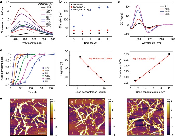
Despite advances in directing the assembly of biomacromolecules into well-defined nanostructures, leveraging pathway complexity of molecular disorder to order transition while bridging materials fabrication from nano- to macroscale remains a challenge. Here, we present templated crystallization of structural proteins to nanofabricate hierarchically structured materials up to centimeter scale, using silk fibroin as an example. The process involves the use of ordered peptide supramolecular assemblies as templates to direct the folding and assembly of silk fibroin into nanofibrillar structures. Silk polymorphs can be engineered by varying the peptide seeds used. Modulation of the relative concentration between silk fibroin and peptide seeds, silk fibroin molecular weight and pH allows control over nanofibrils morphologies and mechanical properties. Finally, facile integration of the bottom-up templated crystallization with emerging top-down techniques enables the generation of macroscopic nanostructured materials with potential applications in information storage/encryption, surface functionalization, and printable three-dimensional constructs of customized architecture and controlled anisotropy.


Conflict of interest statement

The authors declare no competing interests.

Figures

Fig. 1
Fig. 1. Supramolecular assembly of dodecapeptide (GAGSGA)2.
a Hydrophobicity index of Bombyx mori silk fibroin heavy chain, which is composed of a non-repetitive (NR) N-terminal domain (red), 12 highly repetitive (HR) hydrophobic domains (orange) separated by eleven NR short hydrophilic domains (blue) in the middle, and a NR C-terminal domain (green). The dodecapeptide of sequence (GAGSGA)2 composes ~ 40% of the silk fibroin heavy chain sequence and the repeats were highlighted in pink, indicating its quintessential role in silk fibroin primary structure. b Representative atomic force micrograph of (GAGSGA)2 self-assembled in water, showing formation of regular nanowhisker-like supramolecular oligomers. Scale bar, 400 nm. c Representative negative-stain transmission electron micrograph of the (GAGSGA)2 nanowhiskers. Scale bar, 200 nm. d ATR-FTIR spectra of (GAGSGA)2 powders as synthesized and upon assembly from an aqueous suspension. A sharp peak centered at 1619 cm−1 and a strong shoulder at 1698 cm−1 in the Amide I band indicate a highly ordered β-sheet conformation. e 2-D WAXS pattern of the (GAGSGA)2 nanowhiskers, depicting multiple characteristic d-spacings associated with inter-strand and inter-sheet distances. f CD spectra of (GAGSGA)2 over time, revealing a typical β-sheet structure of the assembled nanowhiskers.
Fig. 2
Fig. 2. Templated crystallization of silk fibroin on (GAGSGA)2 nanowhiskers.
a Fluorescence emission spectra of 8-anilinonaphthalene-1-sulfonic acid (ANS) bound to silk fibroin titrated with (GAGSGA)2 nanowhiskers. The increase in the fluorescence emission of ANS indicates a positive correlation between (GAGSGA)2 concentration and silk fibroin assembly driven by hydrophobic interactions. b Effective hydrodynamic diameters of silk fibroin, (GAGSGA)2 nanowhiskers, and silk fibroin seeded by (GAGSGA)2 nanowhiskers at 10%, measured by DLS over time. Error bars represent standard deviation. c CD spectra of silk fibroin seeded by 7.5% (GAGSGA)2 nanowhiskers, showing an increase in the β-sheet content of silk fibroin over time. d Evaluation of the kinetics of templated crystallization: fraction completion of silk fibroin assembly as a function of time at different (GAGSGA)2 concentrations (left), the data points were calculated from time-series CD spectra and fitted with a logistic function (solid lines); Lag time and growth rate extracted from the model fitting and plotted against (GAGSGA)2 concentration (middle and right, respectively). e Atomic force micrographs depicting a directed assembly of silk fibroin on the (GAGSGA)2 nanowhiskers. Thicker and longer (GAGSGA)2-silk nanocomplexes formed shortly after seeding (left), followed by growth of thinner silk nanofibrils branching out from the thicker (GAGSGA)2-silk nanocomplexes (middle and left). Scale bars, 400 nm.
Fig. 3
Fig. 3. Mechanism of templated crystallization.
a Schematic of an intact Bombyx mori silk fibroin heavy chain and randomly chopped chain fragments resulting from protein degradation during the regeneration process. Both native and regenerated silk fibroin were used for templated crystallization study. b Self-assembly of silk fibroin into micelles in water as a result of the hydrophilic–hydrophobic multi-block copolymer primary structure. c Micelle globule formation driven by increased silk fibroin concentration. d Fluorescence emission spectra of pyrene exposed to silk fibroin of increasing concentrations, from which the intensity ratio of the first (I1) and third (I3) pyrene emission peaks was calculated and plotted against silk fibroin concentration (inset). e Exposure to (GAGSGA)2 nanowhiskers decreases the activation energy for the disassembly of silk fibroin micelles (native state). Once the micelles disassemble, the extended silk fibroin chains fold into a more compact transition state (which is a β-sheet dominated state according to the CD data) to minimize the hydrophobic surfaces exposed to water. The folded silk fibroin chains are then able to build up on the (GAGSGA)2 nanowhiskers to form nanocomplexes. As silk fibroin concentration increases, micelles become more aggregated in the native state, therefore increasing the activation energy between the native and transition state, making random coils to β-sheet transition less favorable even in the presence of (GAGSGA)2 nanowhiskers. f Schematic free energy diagram of the templated crystallization process. g Negative-stain TEM images of silk nanofibrils seeded by (GAGSGA)2 nanowhiskers at 1 h and 48 h (left and right, respectively), for widths and lengths measurement and comparison with silk nanofibrils formed in the absence of templates (Supplementary Fig. 6). Scale bars, 200 nm. h Histograms of widths of (GAGSGA)2 nanowhiskers and (GAGSGA)2-silk nanocomplexes at different assembly time points.
Fig. 4
Fig. 4. Structural characterization of HBSP and silk fibroin seeded by HBSP.
a Representative atomic force micrograph of HBSP self-assembled in water, showing that the peptide self-assembles into oligomers of irregular shapes and dimensions. Scale bar, 400 nm. b CD spectra of HBSP depicting its structural evolution. HBSP is mostly unordered right after dissolution in water and then assembles into a β-sheet dominant structure through intermediate α-helical dominant states (e.g., at 2 h). c ATR-FTIR spectra of HBSP powders as synthesized and upon assembly from an aqueous suspension, showing a combination of β-sheet (main peaks at 1621 and 1615 cm−1) and α-helix (shoulders at 1652 and 1659 cm−1) conformations. d CD spectra of silk fibroin seeded by 10% HBSP, depicting an increase in the molecular order over time, but with a lower β-sheet content compared to silk fibroin seeded by (GAGSGA)2. e Effective hydrodynamic diameters of silk fibroin, HBSP nanoassemblies, and silk fibroin seeded by 10% HBSP, measured by DLS over time. Error bars represent standard deviation. f Representative atomic force micrograph of silk fibroin seeded by HBSP nanoassemblies, showing mature nanofibrils at 48 h. Scale bar, 400 nm. g CD spectra of naturally aged silk fibroin in the absence of templates, (GAGSGA)2- and HBSP-templated silk fibroin, revealing different β-sheet contents (i.e., molecular order). h 1-D WAXS spectra of (GAGSGA)2 and HBSP nanoassemblies, showing distinct intra-/intermolecular distances. i 1-D WAXS spectra of naturally aged silk fibroin, (GAGSGA)2- and HBSP-templated silk fibroin, depicting different molecular packing (i.e., d-spacings) of silk fibroin, owing to the templates’ effects.
Fig. 5
Fig. 5. Nanomechanical characterization of the peptide seeds and associated silk nanofibrils.
Young’s modulus maps overlaid on 3D topography (left) and histograms of the Young’s moduli (right) for: a (GAGSGA)2 nanowhiskers. b (GAGSGA)2-templated silk nanofibrils. c HBSP nanoassemblies. d HBSP-templated silk nanofibrils. e Silk nanofibrils seeded by (GAGSGA)2 nanowhiskers at intermediate time points. The Young’s moduli in each histogram (typically of ~ 10,000 data points) were fitted with a log-normal distribution and reported as mean ± standard deviation.
Fig. 6
Fig. 6. Engineering silk nanofibrils into macroscopic materials.
a Schematic of the process for epitaxial-like growth of silk fibroin on substrates modified with peptide seeds. Yellow and red denote (GAGSGA)2 and HBSP seeds, respectively. b ATR-FTIR map of a patterned silk film (inset in the right panel, scale bar, 3 mm) fabricated by the process shown in a revealing the hidden Arabic numeral “1”, which is indiscernible by visual or microscopic inspection. The color scale represents the ratio of absorbance intensity at 1523 cm−1 to that at 1536 cm−1 (Amide II peaks). Representative spectra at the three asterisks in the FTIR map were given in the right panel, showing phase transformation of silk fibroin from random coils to β-sheet in the patterned areas, and (GAGSGA)2 seeds template silk fibroin into a higher β-sheet content compared to HBSP seeds. c SEM characterization of a surface functionalized with mesoporous and nanofibrillar silk fibroin that grows on the pre-deposited seeds. Scale bars, 200 μm (left) and 300 nm (right). d Inkjet printing of a silk nanofibrils suspension obtained through templated crystallization. Scale bars, 1 mm (left) and 1 μm (right). e Fluorescence excitation–emission matrix (EEM) of suspensions of silk nanofibrils seeded by (GAGSGA)2 (upper panel) and HBSP (lower panel), where the excitation and emission maxima at 355 and 438 nm, respectively (i.e., fluorescence in the visible range) indicate the presence of highly β-sheeted (i.e., hydrogen bond rich) protein fibrils. The color scale represents fluorescence intensity (arbitrary unit). f Evaluation of the rheological properties of silk nanofibrils gels: shear viscosity and stress as a function of shear rate, depicting a shear-thinning behavior (upper panel); Storage and loss moduli (G’ and G”, respectively) obtained by oscillation strain sweep at fixed frequency, as a measurement of viscoelasticity (middle panel); continuous printing of the silk nanofibrils gel into a three-dimensional construct (lower panel, inset) and a representative SEM image of the internal structure, showing an anisotropic nanofibrillar network. Scale bars, 5 mm (inset) and 200 nm (SEM).

References

    1. Dunlop JWC, Fratzl P. Biological composites. Annu. Rev. Mater. Res. 2010;40:1–24. doi: 10.1146/annurev-matsci-070909-104421. - DOI
    1. Wegst UGK, Bai H, Saiz E, Tomsia AP, Ritchie RO. Bioinspired structural materials. Nat. Mater. 2015;14:23–36. doi: 10.1038/nmat4089. - DOI - PubMed
    1. Tagliazucchi M, Szleifer I. Transport mechanisms in nanopores and nanochannels: can we mimic nature? Mater. Today. 2015;18:131–142. doi: 10.1016/j.mattod.2014.10.020. - DOI
    1. Adler-Abramovich L, et al. Self-assembled arrays of peptide nanotubes by vapour deposition. Nat. Nanotechnol. 2009;4:849–854. doi: 10.1038/nnano.2009.298. - DOI - PubMed
    1. Stuart MAC, et al. Emerging applications of stimuli-responsive polymer materials. Nat. Mater. 2010;9:101–113. doi: 10.1038/nmat2614. - DOI - PubMed

Publication types