. 2020 Jan 17;10(1):569.
doi: 10.1038/s41598-019-56930-w.

Heavy metals contaminating the environment of a progressive supranuclear palsy cluster induce tau accumulation and cell death in cultured neurons

Affiliations

Heavy metals contaminating the environment of a progressive supranuclear palsy cluster induce tau accumulation and cell death in cultured neurons

Carolina Alquezar et al. Sci Rep. .

Abstract

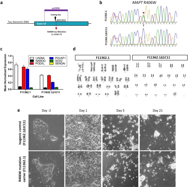
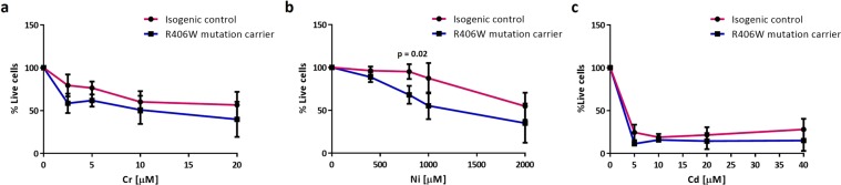
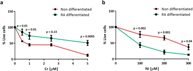
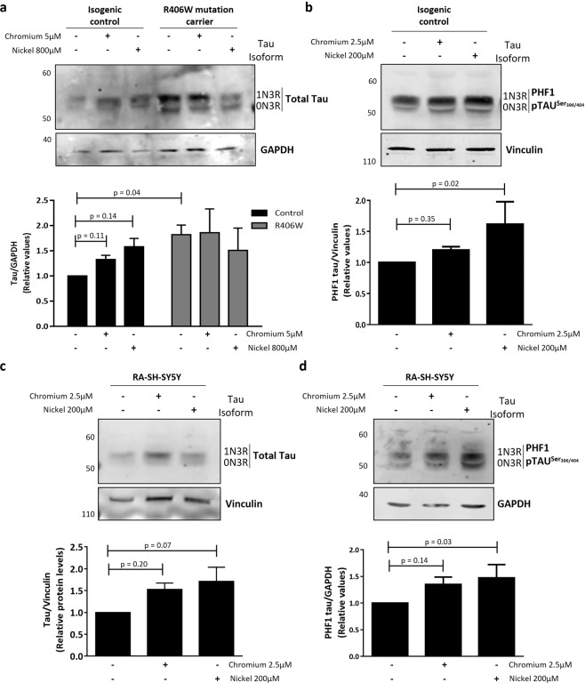
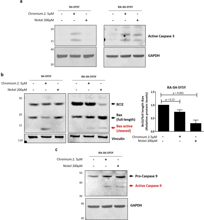
Progressive supranuclear palsy (PSP) is a neurodegenerative disorder characterized by the presence of intracellular aggregates of tau protein and neuronal loss leading to cognitive and motor impairment. Occurrence is mostly sporadic, but rare family clusters have been described. Although the etiopathology of PSP is unknown, mutations in the MAPT/tau gene and exposure to environmental toxins can increase the risk of PSP. Here, we used cell models to investigate the potential neurotoxic effects of heavy metals enriched in a highly industrialized region in France with a cluster of sporadic PSP cases. We found that iPSC-derived iNeurons from a MAPT mutation carrier tend to be more sensitive to cell death induced by chromium (Cr) and nickel (Ni) exposure than an isogenic control line. We hypothesize that genetic variations may predispose to neurodegeneration induced by those heavy metals. Furthermore, using an SH-SY5Y neuroblastoma cell line, we showed that both heavy metals induce cell death by an apoptotic mechanism. Interestingly, Cr and Ni treatments increased total and phosphorylated tau levels in both cell types, implicating Cr and Ni exposure in tau pathology. Overall, this study suggests that chromium and nickel could contribute to the pathophysiology of tauopathies such as PSP by promoting tau accumulation and neuronal cell death.


Conflict of interest statement

The authors declare no competing interests.

Figures

Figure 1
Figure 1
Genetic correction and characterization of the patient-derived iPSC lines. (a) Schematic representation of the CRISPR/Cas9 gene engineering protocol used to correct the mutant allele (c.1216 C > T) in a human iPSC line carrying the R406W tau mutation (F11362.1) to generate an isogenic control iPSC line (F11362.1Δ1C11). (b) Sanger sequencing confirming the presence of a c.1216 C > T substitution in a single allele of exon 13 in the MAPT gene corresponding to the R406W tau mutation (F11362.1) that was corrected in the isogenic control iPSC line (F11362.1Δ1C11). (c) qPCR showing the relative expression of pluripotency markers from the embryonic stem cell lines. GAPDH was used as gene of reference (d) G-band karyotyping showing that both R406W tau mutation carrier (F11362.1) and CRISPR/Cas9-corrected control lines (F11362.1Δ1C11) display no chromosomal abnormalities. (e) Representative microscopy images (10x) showing the neuronal differentiation of both R406W mutant and isogenic control iPSC lines. Cells presented neuronal phenotype 21 days after starting the differentiation process. No differences were found in cell viability and neuronal differentiation between isogenic control (F11362.1Δ1C11) and R406W tau mutant (F11362.1) iPSC lines.
Figure 2
Figure 2
Cr and Ni treatments in iPSC-derived iNeurons 30,000 iPSC from a R406W mutation carrier individual (F11362.1) and isogenic control line (F11362.1Δ1C11) were seeded in triplicate in 96 well plates and then differentiated into iNeurons for 3 weeks. After differentiation, iNeurons were treated with increasing doses of chromium (0–20 µM) (a), nickel (0–2000 µM) (b) and cadmium (0–40 µM) (c) for 72 hours and the (3‐(4,5‐dimethylthiazol‐2‐yl)‐2,5‐diphenyltetrazolium bromide) (MTT) assay was performed to assess the cell death induced by these three heavy metals. Results represent the percentage of live cells in treated iNeurons compared to untreated ones. Data shown are the mean ± SEM of 3 independent experiments for each heavy metal. Treatment with 800 µM Ni induced statistical significant cell death in R406W mutation carrier iNeurons compare with isogenic control (p-value < 0.05).
Figure 3
Figure 3
Non-differentiated SH-SY5Y cells are highly sensitive to Cr treatment but RA-differentiated ones are more vulnerable to Ni exposure. Non differentiated and RA-differentiated SH-SY5Y cells seeded in triplicate in 96 well plates and were treated with increasing concentrations of Cr (0–5 µM) (a) and Ni (0–300 µM) (b) for 24 hours. Plots represent the percentage of live cells after heavy metal treatments relative to untreated ones determined using the MTT assay. Data show the mean ± SEM of 5 independent experiments. Statistical significance was determined by two‐way analysis of variance (anova) followed by Bonferroni’s test for multiple comparisons using GraphPad Prism 6. p-values comparing non-differentiated vs RA-differentiated cells after Cr and Ni treatments are included in the graphs. Statistical significance was considered when p-values were minor or equal to 0.05.
Figure 4
Figure 4
Cr and Ni exposure increases tau levels and phosphorylation in iNeurons and RA-differentiated SH-SY5Y cells. (a,b) iPSCs carrying the R406W tau mutation and isogenic controls were differentiated into iNeurons and treated with Cr (5 µM) and Ni (800 µM) for 72 hours. The levels of total (a) and phospho-tauSer396/404 (PHF-1) (b) tau were measured by western blot. GAPDH and vinculin were used as loading controls. Images show representative immunoblots comparing tau and phospho-tau levels in control and mutant iNeurons before and after Cr and Ni treatment. Plots represents the average ± SEM of 3 independent experiments. (c,d) RA-differentiated SH-SY5Y cells were treated with Cr (2.5 µM) and Nickel (200 µM) for 24 hours before protein extraction. Images show representative immunoblots comparing the levels total tau (c) and phospho-tauSer396/404 (PHF-1) tau (d) before and after heavy metals treatment. GAPDH and vinculin were used as loading control. Plots represent the average ± SEM of 4 independent experiments. Statistical significance was determined by two‐ANOVA followed by Bonferroni’s test for multiple comparisons or one-way ANOVA using GraphPad Prism 6. p-values comparing untreated and heavy metal treated cells are included in the graphs. Statistical significance was considered when p-value ≤ 0.05.
Figure 5
Figure 5
Cr and Ni exposure induces apoptotic cell death in SH-SY5Y cells. Non-differentiated and RA-differentiated SH-SY5Y cells were treated with Cr (2.5 µM) and Ni (200 µM) for 24 hours. Whole cell lysates were collected in order to analyze by western blot the levels of cleaved caspase-3 protein (a), the ratio of anti-apoptotic protein Bcl2 to pro-apoptotic protein Bax (b) and the cleavage and activation of caspase-9 (c). Vinculin and GAPDH were used as loading controls. (a) Representative immunoblot showing the presence of the 17/19KD fragment of caspase-3 after Cr and Ni exposure. (b) Representative immunoblots showing the levels of Bcl2 and Bax proteins after heavy metal exposure. The activation of the intrinsic/mitochondrial apoptosis pathway was assessed by the decrease of the ratio of Bcl2 to full length Bax and the presence of a cleavage form of Bax protein. The plot represent the average ± SEM of 3 independent experiments. Statistical significance was determined by one-way analysis of variance (anova) followed by Bonferroni’s test for multiple comparisons using GraphPad Prism 6. p-values comparing untreated and heavy metal treated cells are included. Statistical significance was considered when p-value ≤ 0.05. (c) Immunoblot showing the presence of the fragment (43KD) of active caspase-9 after Cr and Ni exposure to confirm the activation of the intrinsic/mitochondrial pathway. All experiments were performed in triplicates.

References

    1. Boxer AL, et al. Advances in progressive supranuclear palsy: new diagnostic criteria, biomarkers, and therapeutic approaches. Lancet Neurol. 2017;16:552–563. doi: 10.1016/S1474-4422(17)30157-6. - DOI - PMC - PubMed
    1. Murphy KE, Karaconji T, Hardman CD, Halliday GM. Excessive dopamine neuron loss in progressive supranuclear palsy. Mov. Disord. 2008;23:607–610. doi: 10.1002/mds.21907. - DOI - PubMed
    1. Lee G, Leugers CJ. Tau and tauopathies. Prog. Mol. Biol. Transl. Sci. 2012;107:263–93. doi: 10.1016/B978-0-12-385883-2.00004-7. - DOI - PMC - PubMed
    1. Wang Y, Mandelkow E. Tau in physiology and pathology. Nat. Rev. Neurosci. 2016;17:22–35. doi: 10.1038/nrn.2015.1. - DOI - PubMed
    1. Golbe LI. In Handbook of clinical neurology. 2008;89:457–459. doi: 10.1016/S0072-9752(07)01242-0. - DOI - PubMed

Publication types

MeSH terms