. 2020 Jan 17;10(1):594.
doi: 10.1038/s41598-019-57396-6.

Ultrasensitive nano-aptasensor for monitoring retinol binding protein 4 as a biomarker for diabetes prognosis at early stages

Affiliations

Ultrasensitive nano-aptasensor for monitoring retinol binding protein 4 as a biomarker for diabetes prognosis at early stages

Raheleh Torabi et al. Sci Rep. .

Abstract

Prognosis of diabetes risk at early stages has become an important challenge due to the prevalence of this disease. Retinol binding protein 4 (RBP4), a recently identified adipokine, has been introduced as a predictor for the onset of diabetes type 2 in coming future. In the present report a sensitive aptasensor for detection of RBP4 is introduced. The immune sandwich was prepared by immobilizing biotinylated RBP4 aptamers on streptavidin coated polystyrene micro-wells and then incubation of RBP4 as target and finally addition of luminol-antibody bearing intercross-linked gold nanoparticles as reporter. The chemiluminescence intensity was recorded in the presence of hydrogen peroxide as oxidant agent and Au3+ as an efficient catalyst for luminol oxidation. The aptasensor responded to RBP4 in the linear concentration range from 0.001 to 2 ng/mL and detection limit was slightly less than 1 pg/mL. The proposed method has successfully applied to determine the RBP4 in patient real serums. By using the intercross-linked gold nanoparticles, it is possible to provide more accessible surface for immobilizing luminol and enhance the chemiluminescence signal. Therefore, the analytical parameters such as sensitivity, specificity, detection limit and linear range were improved in compare to the biosensors reported in the literature.


Conflict of interest statement

The authors declare no competing interests.

Figures

Figure 1
Figure 1
Transmission electron microscopy (TEM) images of three types of GNPs: (a) single GNPs and intercross-linked GNPs at (b) lower and (c) higher concentration of cysteine bearing GNPs. As shown in the lower part of images, the mean diameter obtained by DLS for (a, b and c) are 22.5, 31.7 and 67 nm, respectively. (d) UV-Visible spectroscopy of 20 nm-GNPs (Dashed line, Peak: 530 nm), cross-linked GNPs (Dotted line, Peak: 560 nm) and luminol-antibody bearing cross-linked GNPs (Dash-dotted line, Peak: 570 nm). (e) FT-IR spectrum of intercross-linked GNPs.
Figure 2
Figure 2
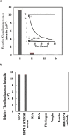
(a) Comparison between the chemiluminescence intensity obtained by aptamer/RBP4/luminol-antibody bearing intercross-linked gold nanoparticles (i) and aptamer/RBP4/luminol-antibody bearing gold nanoparticles (ii). In both tests the concentration of RBP4 was1 ng/mL. iii and iv are negative controls (Same as i and ii but, in the absence of RBP4) (b) Specificity of RBP4 biosensor towards different targets. Artificial serum was PBS containing 1 ng/mL of either main target (RBP4) or the other potentially interfering proteins: BSA, HSA, fibrinogen, insulin and anti-RBP4 antibody and vaspin.
Figure 3
Figure 3
Steps for preparation of nano-aptasensor to monitor RBP4: (A) Carboxylation of GNPs via self-assembly of MUA on GNPs. (B) Activation of carboxylate functional group of GNPs, (C) Immobilization of antibody on GNPs (Ab-GNPs). (D) Immobilization of luminol on Ab-GNPs (L-GNP-Ab). (E) Self-assembly of cysteine on GNPs (Cys-GNPs). (F) Intercross-linking of Cys-GNPs and Ab-GNPs (GNP-Cys-GNP-Ab). (G) Activation of carboxylate functional group of cysteine on GNPs. (H) Immobilization of luminol on GNP-Cys-GNP-Ab. (I) Immobilization of biotinylated aptamer on streptavidin coated polystyrene micro well plate and then RBP4 protein binding to aptamer.
Figure 4
Figure 4
(a) Chemiluminescence intensity measurements by aptasensor in the presence of different concentrations of RBP4 (From down to up: 0, 0.001, 0.025, 0.05, 0.1, 0.5, 1 and 2 ng/mL). (b) Calibration curve for determination of RBP4. Each point stands for the mean value of three independent experiments.

References

    1. Kiernan UA, Phillips DA, Trenchevska O, Nedelkov D. Quantitative mass spectrometry evaluation of human retinol binding protein 4 and related variants. PloS one. 2011;6:e17282. doi: 10.1371/journal.pone.0017282. - DOI - PMC - PubMed
    1. Deshpande AD, Harris-Hayes M, Schootman M. Epidemiology of Diabetes and Diabetes-Related Complications. Physical Therapy. 2008;88:1254–1264. doi: 10.2522/ptj.20080020. - DOI - PMC - PubMed
    1. Kwanbunjan K, et al. Association of retinol binding protein 4 and transthyretin with triglyceride levels and insulin resistance in rural thais with high type 2 diabetes risk. JBMC Endocrine Disorders. 2018;18:26. doi: 10.1186/s12902-018-0254-2. - DOI - PMC - PubMed
    1. Wei, Y. et al. Serum retinol binding protein 4 is associated with insulin resistance in patients with early and untreated rheumatoid arthritis. Joint Bone Spine, 10.1016/j.jbspin.2018.07.002 (2018). - PubMed
    1. Javanbakht M, et al. Cost-of-Illness Analysis of Type 2 Diabetes Mellitus in Iran. PloS one. 2011;6:e26864. doi: 10.1371/journal.pone.0026864. - DOI - PMC - PubMed

Publication types