Meta-Analysis
. 2020 May:112:300-323.
doi: 10.1016/j.neubiorev.2020.01.004. Epub 2020 Jan 15.

Convergent neural representations of experimentally-induced acute pain in healthy volunteers: A large-scale fMRI meta-analysis

Affiliations
Meta-Analysis

Convergent neural representations of experimentally-induced acute pain in healthy volunteers: A large-scale fMRI meta-analysis

Anna Xu et al. Neurosci Biobehav Rev. 2020 May.

Abstract

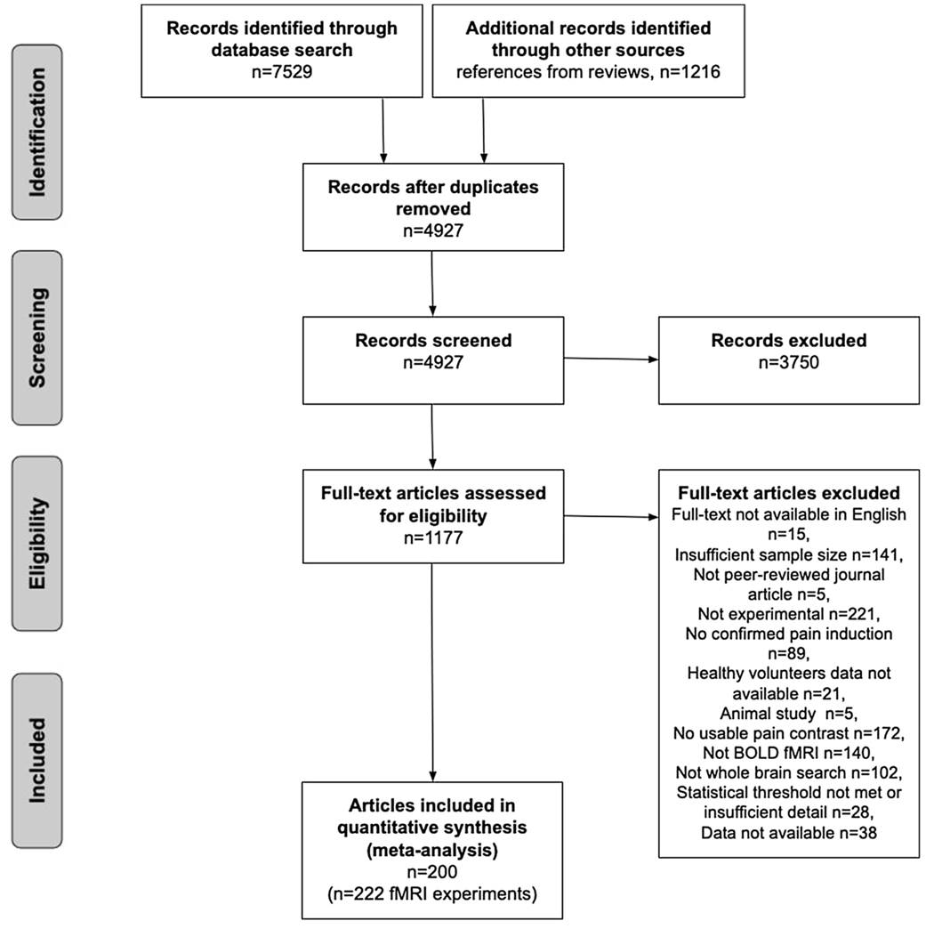
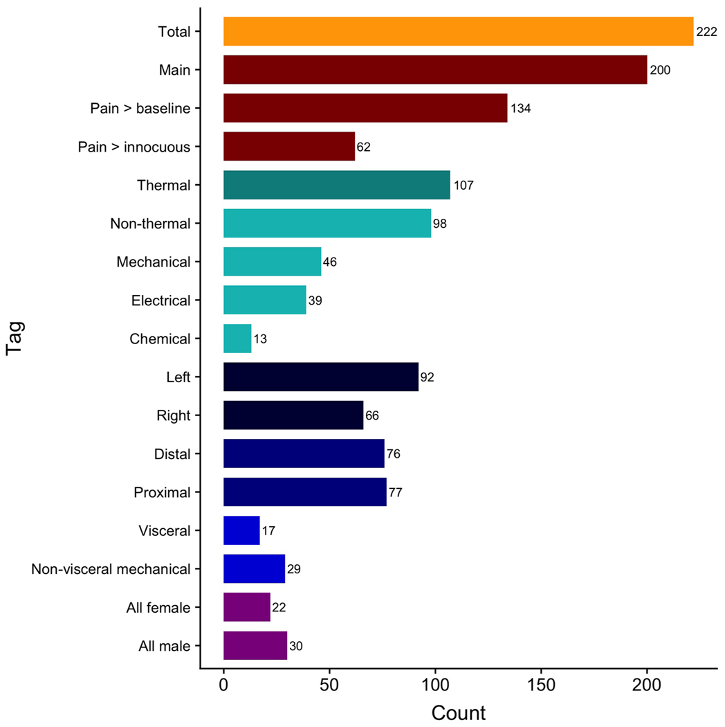
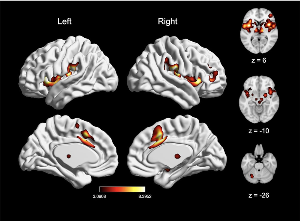
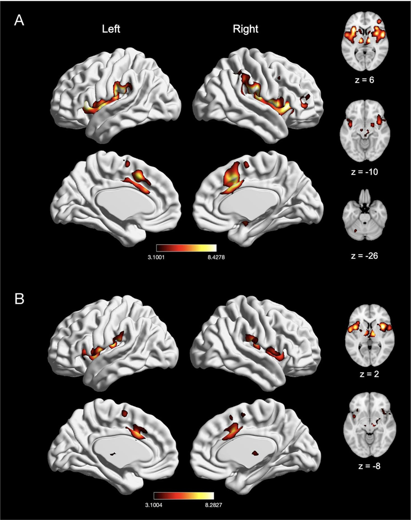
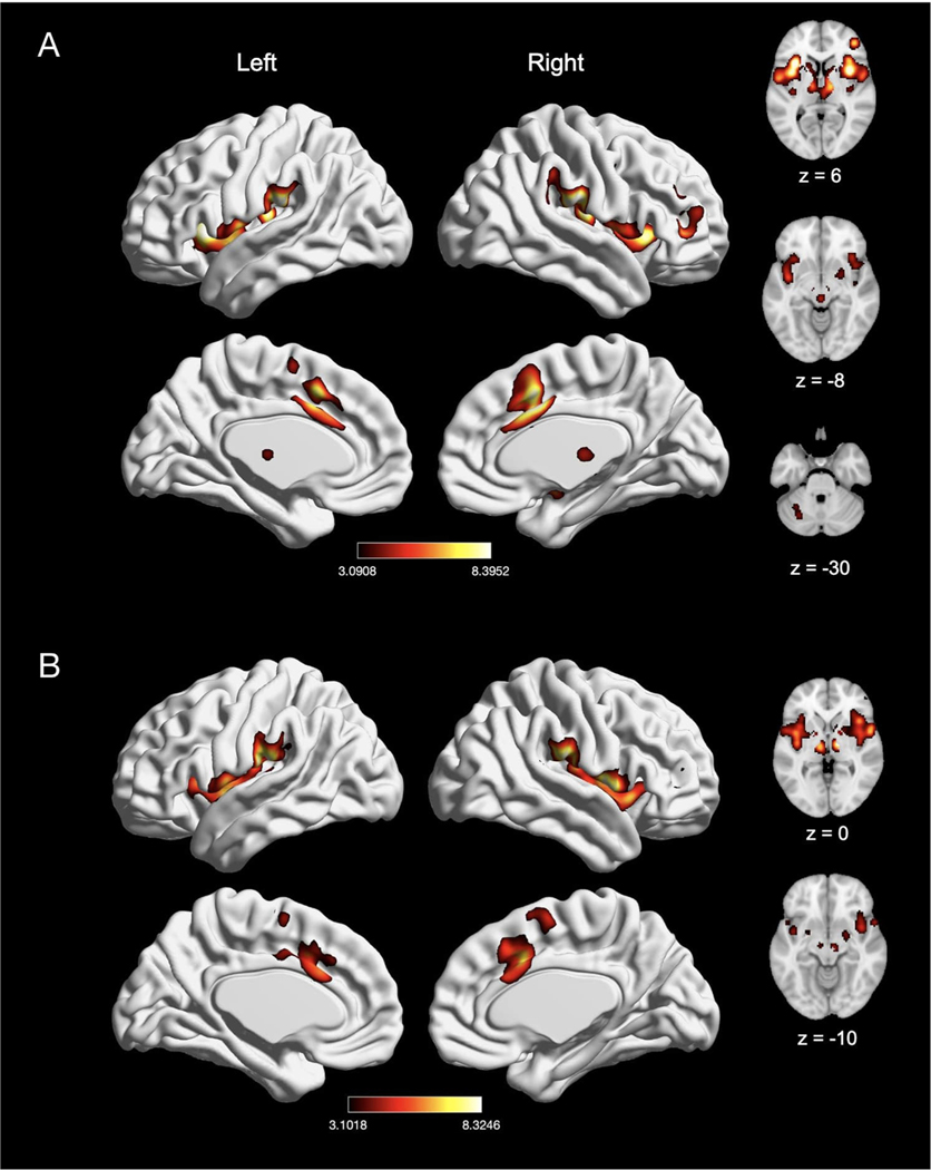
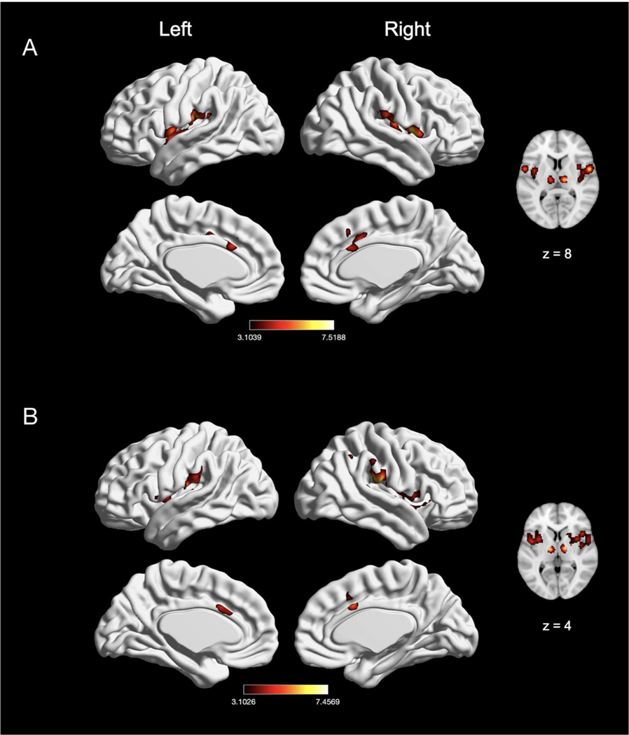
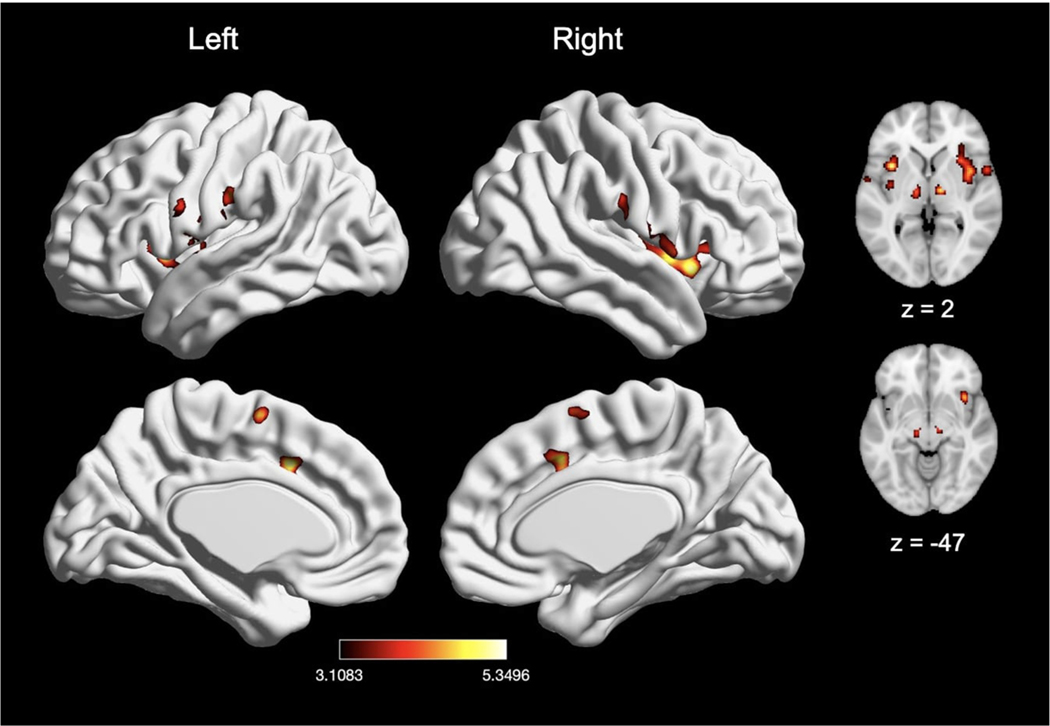
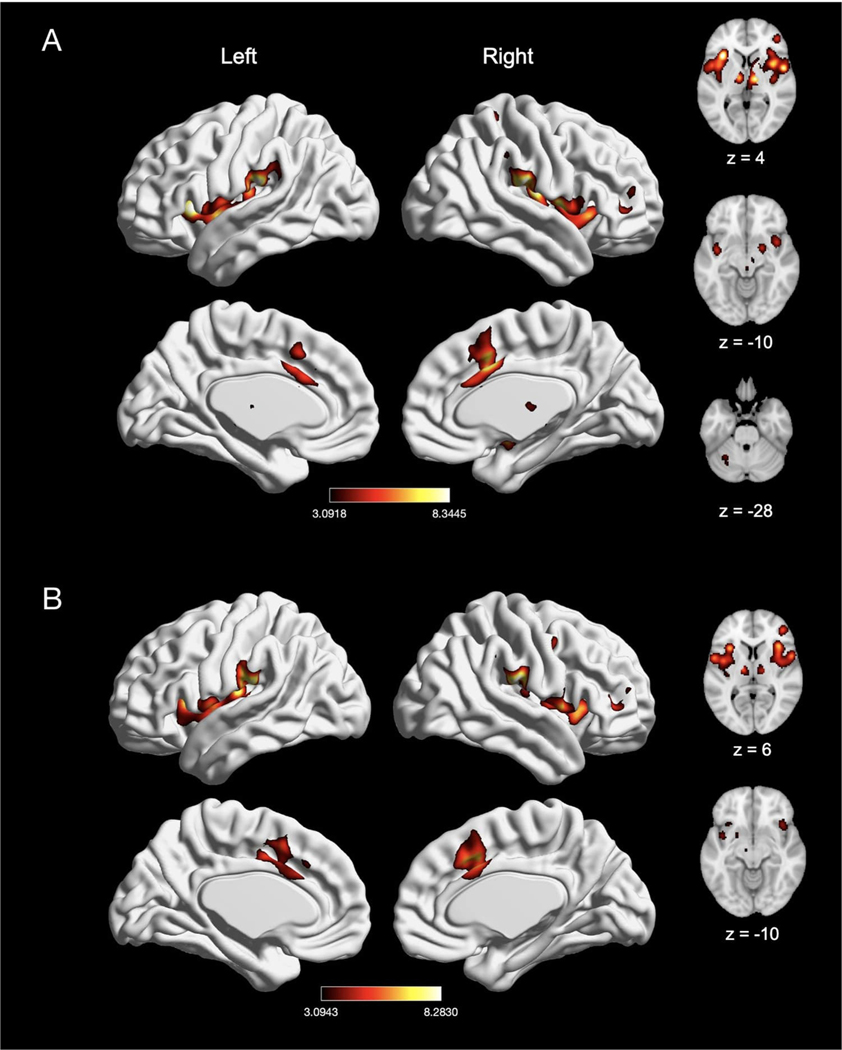
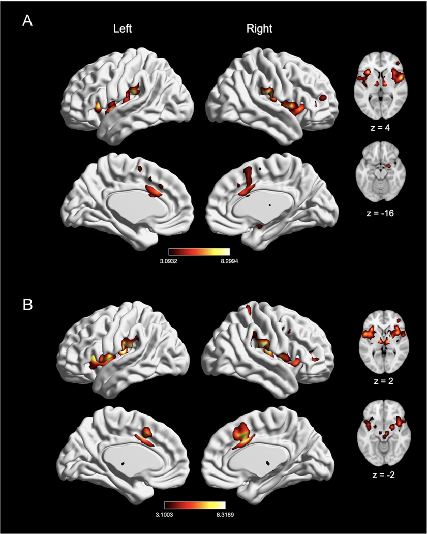
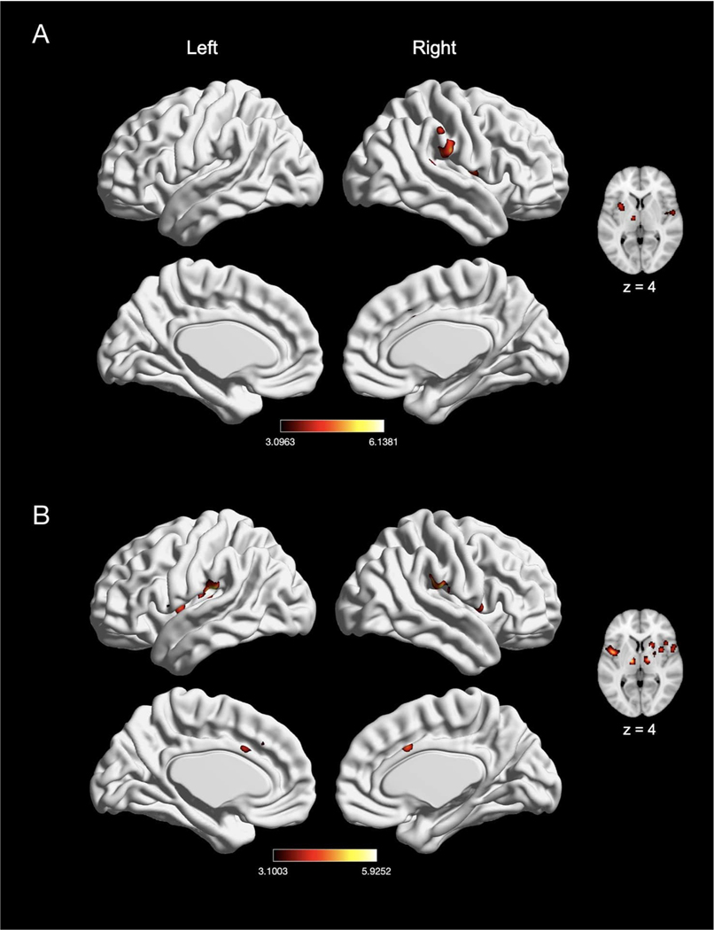
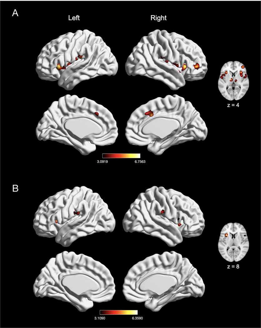
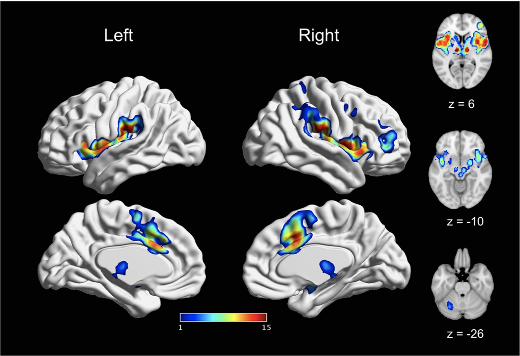
Characterizing a reliable, pain-related neural signature is critical for translational applications. Many prior fMRI studies have examined acute nociceptive pain-related brain activation in healthy participants. However, synthesizing these data to identify convergent patterns of activation can be challenging due to the heterogeneity of experimental designs and samples. To address this challenge, we conducted a comprehensive meta-analysis of fMRI studies of stimulus-induced pain in healthy participants. Following pre-registration, two independent reviewers evaluated 4,927 abstracts returned from a search of 8 databases, with 222 fMRI experiments meeting inclusion criteria. We analyzed these experiments using Activation Likelihood Estimation with rigorous type I error control (voxel height p < 0.001, cluster p < 0.05 FWE-corrected) and found a convergent, largely bilateral pattern of pain-related activation in the secondary somatosensory cortex, insula, midcingulate cortex, and thalamus. Notably, these regions were consistently recruited regardless of stimulation technique, location of induction, and participant sex. These findings suggest a highly-conserved core set of pain-related brain areas, encouraging applications as a biomarker for novel therapeutics targeting acute nociceptive pain.

Keywords: Meta-analysis; Neuroimaging; Pain; fMRI.


Conflict of interest statement

Declaration of Competing Interest Robert H. Dworkin, PhD, has received in the past 36 months research grants and contracts from the US Food and Drug Administration and the US National Institutes of Health, and compensation for consulting on clinical trial methods from Abide, Acadia, Adynxx, Analgesic Solutions, Aptinyx, Aquinox, Asahi Kasei, Astellas, AstraZeneca, Biogen, Biohaven, Boston Scientific, Braeburn, Celgene, Centrexion, Chromocell, Clexio, Concert, Decibel, Dong-A, Eli Lilly, Eupraxia, Glenmark, Grace, Hope, Immune, Lotus Clinical Research, Mainstay, Neumentum, NeuroBo, Novaremed, Novartis, Olatec, Pfizer, Phosphagenics, Quark, Reckitt Benckiser, Regenacy (also equity), Relmada, Sanifit, Scilex, Semnur, Sollis, Teva, Theranexus, Trevena, and Vertex.

Figures

Fig. 1.
Fig. 1.
PRISMA flowchart for study inclusion.
Fig. 2.
Fig. 2.
Number of experiments included in each analysis of interest.
Fig. 3.
Fig. 3.
Main effect of experimental induction of acute pain (n = 200). Coordinates and statistics for significant clusters are shown in Table 2.
Fig. 4.
Fig. 4.
Meta-analytic effect of experiments using “pain > rest” and “pain > innocuous” contrast. (A) Main effect of experiments using “pain > rest” contrast (n = 134). (B) Main effect of experiments with “pain > innocuous” contrast (n = 62). Coordinates and statistics for significant clusters are shown in Table 3.
Fig. 5.
Fig. 5.
The meta-analytic effects of thermal and non-thermal nociceptive pain induction. (A) Main effect of thermal pain experiments (n = 107). (B) Main effect of non-thermal pain experiments (n = 98). Coordinates and statistics for significant clusters associated with the main effect of thermal and non-thermal pain (as well as the between-experiment contrast and conjunction of thermal and non-thermal pain experiments) are shown in Table 4.
Fig. 6.
Fig. 6.
Effects of induction of electrically-evoked and mechanical nociceptive pain. (A) Main effect of electrically-evoked pain experiments (n = 39). (B) Main effect of mechanical pain experiments (n = 46). Coordinates and statistics for significant clusters associated with the main effect of electrical and mechanical pain (as well as the between-experiment contrast and conjunction of electrical and mechanical pain experiments) are shown in Table 5.
Fig. 7.
Fig. 7.
Main effect of meta-analysis of nociceptive chemical pain experiments (n = 13). Coordinates and statistics for significant clusters associated with the main effect of chemical pain are shown in Table 6.
Fig. 8.
Fig. 8.
The meta-analytic effects of left-sided and right-sided pain induction. (A) Main effect of left-sided pain experiments (n = 92). (B) Main effect of right-sided pain experiments (n = 66). Coordinates and statistics for significant clusters associated with the main effect of left-sided and right-sided pain (as well as the between-experiment contrast and conjunction of left-sided and right-sided pain experiments) are shown in Table 7.
Fig. 9.
Fig. 9.
The meta-analytic effects of distal and proximal nociceptive pain. (A) Main effect of experiments inducing acute nociceptive pain in the distal extremities (n = 68). (B) Main effect of experiments inducing pain in the proximal extremities (n = 85). Coordinates and statistics for significant clusters associated with the main effect of distal and proximal pain (as well as the between-experiment contrast and conjunction of distal and proximal pain experiments) are shown in Table 8.
Fig. 10.
Fig. 10.
The meta-analytic effects of visceral and non-visceral mechanical pain. (A) Main effect of visceral pain experiments (n = 17). (B) Main effect of non-visceral mechanical pain experiments (n = 29). Coordinates and statistics for significant clusters associated with the main effect of visceral and non-visceral pain (as well as the between-experiment contrast and conjunction of visceral and non-visceral pain experiments) are shown in Table 9.
Fig. 11.
Fig. 11.
Effect of pain in males and females. (A) Main effect of experiments with an all-female sample (n = 22). (B) Main effect of experiments with an all-male sample (n = 30). Coordinates and statistics for significant clusters are shown in Table 10.
Fig. 12.
Fig. 12.
A core set of bran regions recruited by acute pain. This figure depicts the spatial consistency of above-threshold activation convergence cross all reported main effects meta-analyses. The value assigned to each voxel reflects the number of main effects analyses in which it was reported as significant. Values range from 1 (reported significant in only one main effects meta-analyses) to 15 (reported as significant in all main effects meta-analyses). The most consistently activated areas include bilateral thalamus, bilateral insula, bilateral SII, and bilateral MCC.

References

    1. Ab Aziz CB, Ahmad AH, 2006. The role of the thalamus in modulating pain. Malays. J. Med. Sci 13 (2), 11–18. - PMC - PubMed
    1. Apkarian AV, Bushnell MC, Treede R-D, Zubieta J-K, 2005. Human brain mechanisms of pain perception and regulation in health and disease. Eur. J. Pain 9 (4). 10.1016/j.ejpain.2004.11.001. 463–463. - DOI - PubMed
    1. Basbaum AI, Fields HL, 1978. Endogenous pain control mechanisms: review and hypothesis. Ann. Neurol 4 (5), 451–462. 10.1002/ana.410040511. - DOI - PubMed
    1. Behrens TEJ, Johansen-Berg H, Woolrich MW, Smith SM, Wheeler-Kingshott CA, Behrens TEJ, Johansen-Berg H, Woolrich MW, Smith SM, Wheeler-Kingshott CAM, Boulby PA, et al., 2003. Non-invasive mapping of connections between human thalamus and cortex using diffusion imaging. Nat. Neurosci 6 (7), 8. - PubMed
    1. Bingel U, Lorenz J, Glauche V, Knab R, Gläscher J, Weiller C, Büchel C, 2004. Somatotopic Organization of human somatosensory cortices for pain: a single trial fMRI study. NeuroImage 23 (1), 224–232. 10.1016/j.neuroimage.2004.05.021. - DOI - PubMed

Publication types

MeSH terms