Review
. 2020 Feb;72(2):288-306.
doi: 10.1016/j.jhep.2019.09.026.

mRECIST for HCC: Performance and novel refinements

Affiliations
Review

mRECIST for HCC: Performance and novel refinements

Josep M Llovet et al. J Hepatol. 2020 Feb.

Abstract

In 2010, modified RECIST (mRECIST) criteria were proposed as a way of adapting the RECIST criteria to the particularities of hepatocellular carcinoma (HCC). We intended to overcome some limitations of RECIST in measuring tumour shrinkage with local and systemic therapies, and also to refine the assessment of progression that could be misinterpreted with conventional RECIST 1.1, due to clinical events related to the natural progression of chronic liver disease (development of ascites, enlargement of lymph nodes, etc.). mRECIST has served its purpose since being adopted or included in clinical practice guidelines (European, American and Asian) for the management of HCC; it has also been instrumental for assessing response and time-to-event endpoints in several phase II and III investigations. Nowadays, mRECIST has become the standard tool for measurement of radiological endpoints at early/intermediate stages of HCC. At advanced stages, guidelines recommend both methods. mRECIST has been proven to capture higher objective response rates in tumours treated with molecular therapies and those responses have shown to be independently associated with better survival. With the advent of novel treatment approaches (i.e. immunotherapy) and combination therapies there is a need to further refine and clarify some concepts around the performance of mRECIST. Similarly, changes in the landscape of standard of care at advanced stages of the disease are pointing towards progression-free survival as a potential primary endpoint in some phase III investigations, as effective therapies applied beyond progression might mask overall survival results. Strict recommendations for adopting this endpoint have been reported. Overall, we review the performance of mRECIST during the last decade, incorporating novel clarifications and refinements in light of emerging challenges in the study and management of HCC.

Keywords: Endpoints; Liver cancer; Systemic therapies; mRECIST, RECIST, trial design.


Conflict of interest statement

Conflict of interest

JML is receiving research support from Bayer HealthCare Pharmaceuticals, Eisai Inc, Bristol-Myers Squibb and Ipsen, and consulting fees from Bayer HealthCare Pharmaceuticals, Bristol-Myers Squibb, Eisai Inc, Celsion Corporation, Eli Lilly, Exelixis, Merck, Ipsen, Glycotest, Navigant, Leerink Swann LLC, Midatech Ltd, Fortress Biotech, Sprink Pharmaceuticals, Nucleix and Can-Fite and Roche. RL has nothing to declare with respect to this manuscript.

Please refer to the accompanying ICMJE disclosure forms for further details.

Figures

Fig. 1.
Fig. 1.. Representation of EASL Guidelines: recommendations for treatment according to levels of evidence and strength of recommendation (adaptation of the GRADE system).
Adapted from EASL Guidelines. J Hep 2018. In green, recommended treatments based on level 1–2 evidence. In orange, treatments with promising results based on phase II data. In red, treatments not recommended because of negative clinical trial investigations. AFP, alpha-fetoprotein; BCLC, Barcelona Clinic Liver Cancer; LDLT, living donor liver transplantation; LT, orthotopic liver transplantation; MW, microwave.
Fig. 2.
Fig. 2.. Correlation between progression-free survival and overall survival (adapted from Llovet JM et al., J Hep 2019).
Trial level correlation between endpoints. R and R2 refers to the weighted Pearson coefficient between the HR of PFS and the HR of TTP. Each dot represents one of the phase III clinical trials conducted on advanced HCC. Size of the dot is proportional to the total number of patients enrolled in the trial. Trials are coloured based on whether the final result was positive, negative or non-inferior for the primary endpoint (OS). X and Y axis depict the value of the HR for the surrogate TTP and PFS, respectively. Gray shaded areas represent the upper and lower limits of the 95% CIs for the regression. HCC, hepatocellular carcinoma; HR, hazard ratio; mRECIST, modified RECIST; OS, overall survival; PFS, progression-free survival; TTP, time-to-progression.
Fig. 3.
Fig. 3.. Measurement of the longest tumour diameter in a target hepatic lesion: mRECIST vs. standard RECIST.
Arterial-phase CT scan obtained after systemic targeted therapy. According to standard RECIST, the overall longest diameter of the tumour is captured (white arrow), regardless of the presence of a large area of intratumoural treatment-induced necrosis (‘‘N”). In contrast, mRECIST measurement (red arrow) only includes the longest diameter of the viable portion of the tumour, as recognized by contrast enhancement. mRECIST, modified RECIST.
Fig. 4.
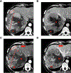
Fig. 4.. Differentiation between spontaneous tumour necrosis and viable tumour with reduced arterial perfusion.
Baseline (A) arterial phase and (B) portal venous phase CT scans: 2 lesions with typical vascular pattern are selected as mRECIST target lesions and the longest viable tumour diameters are measured in the (A) arterial-phase scan by avoiding the inclusion of major intervening areas of spontaneous necrosis (‘‘N”). In the (C) arterial phase and (D) portal venous phase CT scans obtained after treatment with systemic targeted therapy, the viable portion of the larger tumour shows reduced arterial perfusion (* in C) with respect to baseline; however, it still shows unequivocal contrast uptake (the level of arterial enhancement is similar to that of liver parenchyma) and can be distinguished from the non-enhancing areas of necrosis (‘‘N”). (C) The longest viable tumour diameter of both lesions is measured. Note also the appearance of an unequivocal 1 cm new lesion (arrows in C and D). mRECIST, modified RECIST.
Fig. 5.
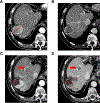
Fig. 5.. Differentiation between treatment-induced tumour necrosis and viable tumour with reduced arterial perfusion.
Baseline (A) arterial phase and (B) portal venous phase CT scans. The tumour shows typical vascular profile, with arterial enhancement and portal venous washout. The longest viable tumour diameter is measured in the arterial phase, in which the tumour is better delineated (A). In the (C) arterial phase and (D) portal venous phase CT scans obtained after treatment with systemic targeted therapy, the tumour shows an area of treatment-induced necrosis (‘‘N”) that fails to show any contrast enhancement. The area of necrosis can be distinguished from the area indicated by the asterisk, that, despite the reduced arterial perfusion compared to baseline, still shows unequivocal contrast uptake, consistent with viable tumour (C). The longest viable tumour diameter is obtained in the portal venous phase, in which viable tumour is better delineated, and includes the area of reduced perfusion (C). Note also the appearance of an unequivocal new lesion with typical vascular pattern (arrows in C and D).
Fig. 6.
Fig. 6.. Equivocal new lesion determined to be unequivocal at subsequent time point.
(A) Arterial phase and (B) portal venous phase CT scans obtained at post-baseline time point 2. A new lesion is detected (arrow in A). However, the lesion is smaller than 1 cm and shows (A) arterial hypervascularization without (B) venous washout. The new lesion must be considered equivocal. (C) Arterial phase and (D) portal venous phase CT scans obtained at post-baseline timepoint 3: the lesion is now larger than 1 cm and shows (C) arterial hypervascularisation with (D) portal venous washout. Thus, it meets the criteria for unequivocal diagnosis of HCC, and can be considered as evidence of progressive disease. However, the time point of progression will be time point 2, the time point that the lesion was first noted as equivocal. HCC, hepatocellular carcinoma.

References

    1. European Association for the Study of the Liver. EASL clinical practice guidelines: management of hepatocellular carcinoma. J Hepatol 2018;69:182–236. - PubMed
    1. Llovet JM, Montal R, Sia D, Finn RS. Molecular therapies and precision medicine for hepatocellular carcinoma. Nat Rev Clin Oncol 2018;15:599–616. - PMC - PubMed
    1. Torre L, Bray F, Siegel R, Ferlay J, Lortet-Tieulent J, Jemal A. Global cancer statistics, 2012. CA Cancer J Clin 2015;65:87–108. - PubMed
    1. Llovet JM, Montal R, Villanueva A. Randomized trials and endpoints in advanced HCC: role of PFS as a surrogate of survival. J Hepatol 2019;70:1262–1277. - PMC - PubMed
    1. Beaver JA, Howie LJ, Pelosof L, Kim T, Liu J, Goldberg KB, et al. A 25-year experience of US food and drug administration accelerated approval of malignant hematology and oncology drugs and biologics. JAMA Oncol 2018;4:849–856. - PubMed

Publication types

MeSH terms

Substances