. 2020:25:102165.
doi: 10.1016/j.nicl.2020.102165. Epub 2020 Jan 11.

Tractographic description of major subcortical projection pathways passing the anterior limb of the internal capsule. Corticopetal organization of networks relevant for psychiatric disorders

Affiliations

Tractographic description of major subcortical projection pathways passing the anterior limb of the internal capsule. Corticopetal organization of networks relevant for psychiatric disorders

Volker A Coenen et al. Neuroimage Clin. 2020.

Abstract

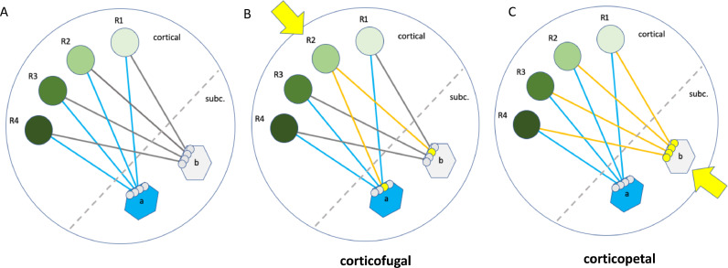
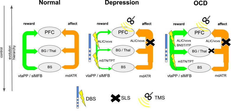
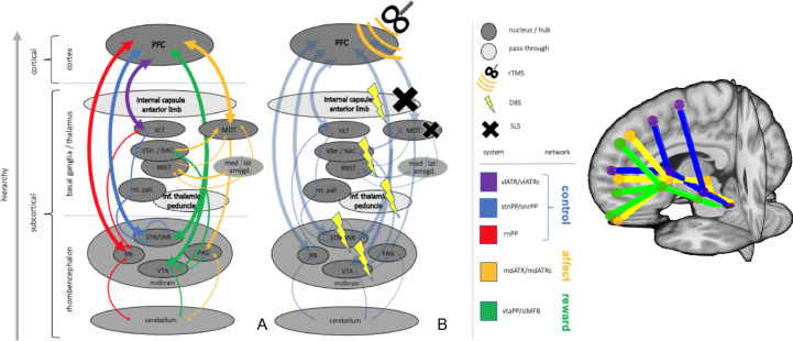
Background: Major depression (MD) and obsessive-compulsive disorder (OCD) are psychiatric diseases with a huge impact on individual well-being. Despite optimal treatment regiments a subgroup of patients remains treatment resistant and stereotactic surgery (stereotactic lesion surgery, SLS or Deep Brain Stimulation, DBS) might be an option. Recent research has described four networks related to MD and OCD (affect, reward, cognitive control, default network) but only on a cortical and the adjacent sub-cortical level. Despite the enormous impact of comparative neuroanatomy, animal science and stereotactic approaches a holistic theory of subcortical and cortical network interactions is elusive. Because of the dominant hierarchical rank of the neocortex, corticofugal approaches have been used to identify connections in subcortical anatomy without anatomical priors and in part confusing results. We here propose a different corticopetal approach by identifying subcortical networks and search for neocortical convergences thereby following the principle of phylogenetic and ontogenetic network development.

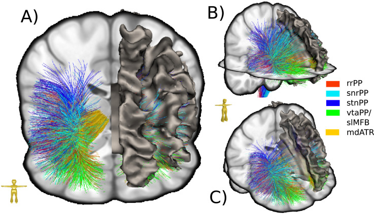
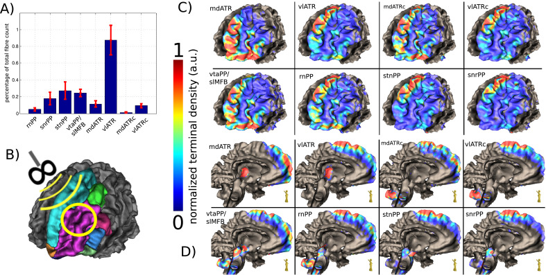
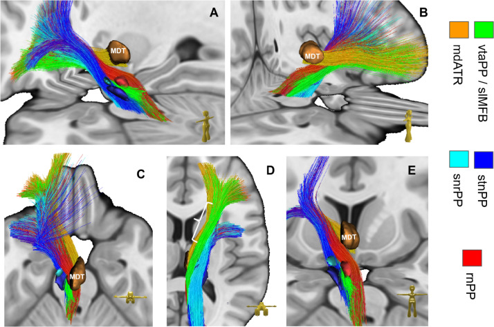
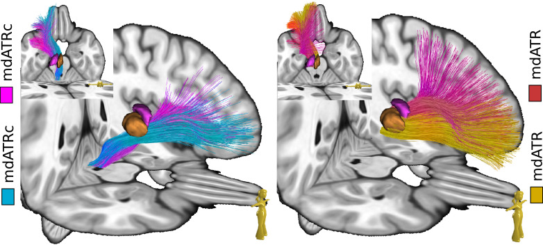
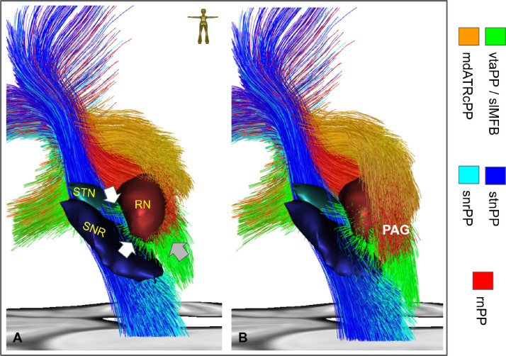
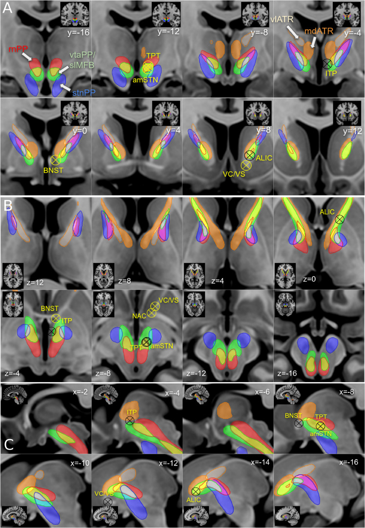
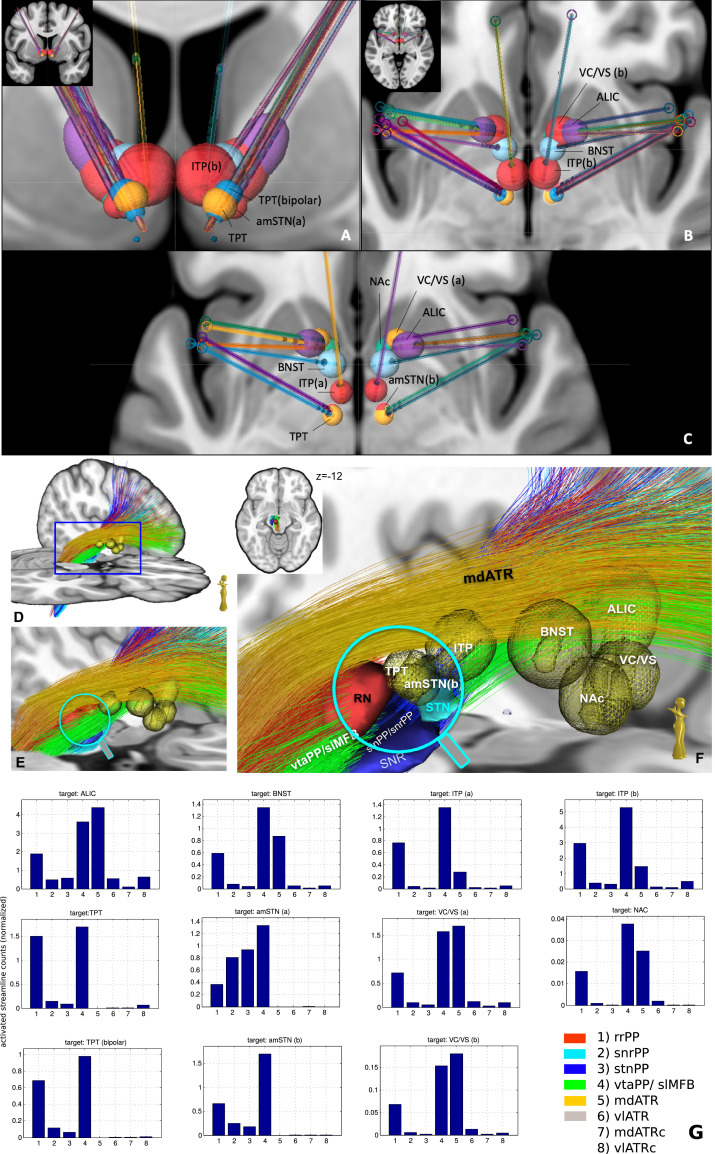
Material and methods: This work used a diffusion tensor imaging data from a normative cohort (Human Connectome Project, HCP; n = 200) to describe eight subcortical fiber projection pathways (PPs) from subthalamic nucleus (STN), substantia nigra (SNR), red nucleus (RN), ventral tegmental area (VTA), ventrolateral thalamus (VLT) and mediodorsal thalamus (MDT) in a normative space (MNI). Subcortical and cortical convergences were described including an assignment of the specific pathways to MD/OCD-related networks. Volumes of activated tissue for different stereotactic stimulation sites and procedures were simulated to understand the role of the distinct networks, with respect to symptoms and treatment of OCD and MD.

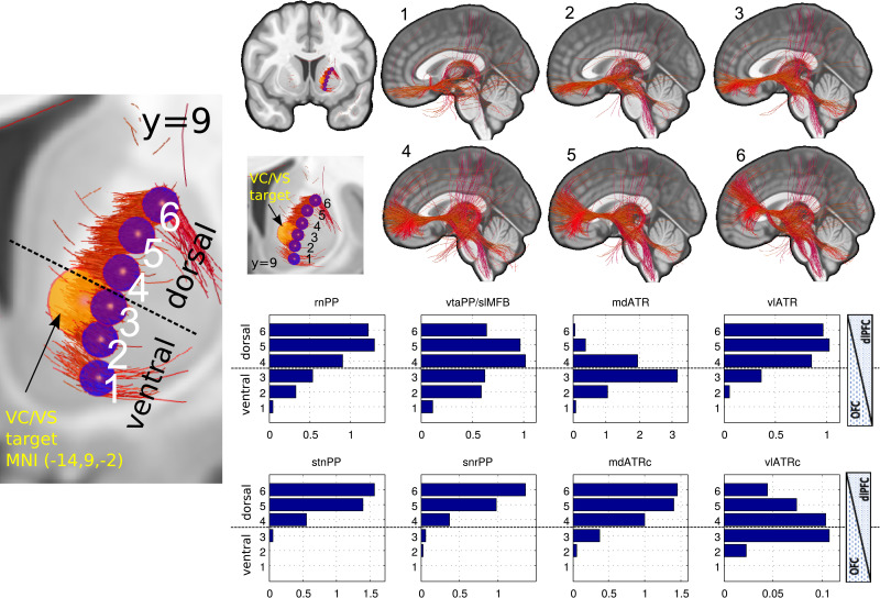
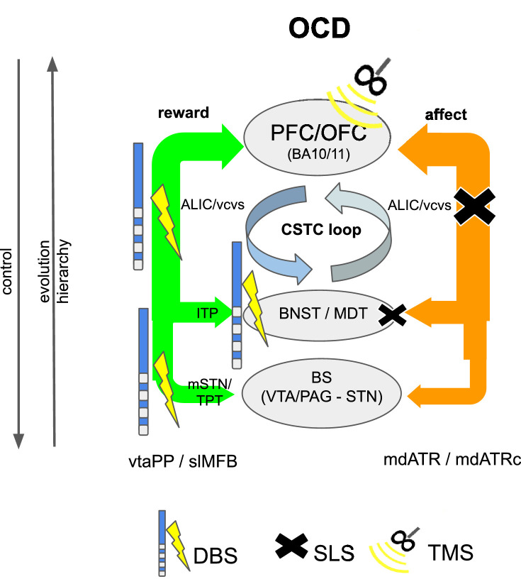
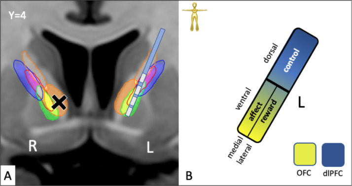
Results: The detailed course of eight subcortical PPs (stnPP, snrPP, rnPP, vlATR, vlATRc, mdATR, mdATRc, vtaPP/slMFB) were described together with their subcortical and cortical convergences. The anterior limb of the internal capsule can be subdivided with respect to network occurrences in ventral-dorsal and medio-lateral gradients. Simulation of stereotactic procedures for OCD and MD showed dominant involvement of mdATR/mdATRc (affect network) and vtaPP/slMFB (reward network).

Discussion: Corticofugal search strategies for the evaluation of stereotactic approaches without anatomical priors often lead to confusing results which do not allow for a clear assignment of a procedure to an involved network. According to our simulation of stereotactic procedures in the treatment of OCD and MD, most of the target regions directly involve the reward (and affect) networks, while side-effects can in part be explained with a co-modulation of the control network.

Conclusion: The here proposed corticopetal approach of a hierarchical description of 8 subcortical PPs with subcortical and cortical convergences represents a new systematics of networks found in all different evolutionary and distinct parts of the human brain.

Keywords: Anterior limb of internal capsule; DBS; Depression; Functional networks; Hyperdirect pathway, Midbrain; MD; Neocortex; OCD; Prefrontal cortex; Projection pathways; Stereotactic lesion surgery.


Conflict of interest statement

Declaration of Competing Interest The authors declare that they have no known competing financial interests or personal relationships that could have appeared to influence the work reported in this paper.

Figures

Fig 1
Fig. 1
Identification of cortical / subcortical networks with different tractographic approaches. Schematic representation. A, anatomical situation: Cortical functional regions (R1-R4, green spheres) are connected with distinct subcortical hub regions (a, b). These hub regions are subcortical nuclei. Hub regions with distinct functions converge onto the same cortical functional regions. A hub together with its fiber connections and the cortical functional regions constitutes a network. B, corticofugal tractographic approach: Seeding from a single cortical functional region (R2, yellow arrow) leads to an only partial identification of the involved hubs (a,b) and their attached network but shows the overall connectedness of the cortical region with subcortical structures. The network as a whole cannot be appreciated, nor can the convergence of subcortical networks (as a whole) onto cortical regions be understood. C, corticopetal approach (as used in this paper): Seeding from a subcortical hub region (b, yellow arrow) identifies the entire network which consists of the hub region (b), the fiber connections (yellow lines) and the cortical projection fields (R1-R4). The other hub (a) is not part of this network (For interpretation of the references to color in this figure legend, the reader is referred to the web version of this article.).
Fig. 2
Fig. 2
Interaction of two networks (exemplarily: reward, affect) in an evolutionary and hierarchical context (normal, depression, OCD). Legend: DBS, deep brain stimulation; SLS, stereotactic lesion surgery; rTMS, repetitive transcranial magnetic stimulation; PFC, prefrontal cortex; Thal, thalamus; BG, basal ganglia; BS, brain stem; slMFB, superolateral medial forebrain bundle; mdATR, anterior thalamic radiation from dorsomedial thalamus; ALIC, anterior limb of internal capsule (DBS target); VC, ventral capsule (DBS target); mSTN, medial subthalamic nucleus; TPT, target point for slMFB DBS.
Fig. 3
Fig. 3
Terminals of subcortical projection pathways (PP) in streamline rendition. Right side shown only. The convergence of PP fibers can nicely be seen. Note how fibesr from mediodorsal thalamus (mdATR, copper) converge together with fibers from the ventral tegmental area (vtaPP/slMFB, green) on the same frontopolar and orbitofrontal regions. For quantification of terminals see Fig. 4 (For interpretation of the references to color in this figure legend, the reader is referred to the web version of this article.).
Fig. 4
Fig. 4
Cortical convergences of distinct projection pathways which belong to distinguishable networks. A, histogram showing the relative distribution of fibers over the considered cohort. error bars indicate the inter-individual deviations. B, Definition of the prefrontal cortex according to (Desikan et al., 2006). Left side shown only. C, density of cortical terminals in a view from anterolateral left. .D, view from midsagittal. Legend: PP, projection pathway; rnPP, red nucleus PP; snrPP, substantia nigra PP; stnPP, subthalamic nucleus PP (hyperdirect pathway); vtaPP, ventral tegmental area PP; slMFB, superolateral branch of the medial forebrain bundle; ATR, anterior thalamic radiation; mdATR, mediodorsal nucleus ATR; vlATR, ventrolateral nucleus ATR; mdATRc, mdATR with extension to cerebellum; vlATRc, vlATR with extension to cerebellum (For interpretation of the references to color in this figure legend, the reader is referred to the web version of this article.).
Fig. 5
Fig. 5
Subcortical anatomical course of distinct subcortical projection pathways. Left side shown only. Note how fibers of reward network (green) and affect network (copper) run in the ventral half of the anterior limb of the internal capsule on their way to the OFC. Fibers of the control network (rnPP, stnPP, snrPP) are located more dorsal and head toward dlPFC. vlATR and vlATRc not shown. Left white parenthesis in D (view from ventral) shows anterior-posterior dimension of anterior limb of internal capsule. Legend: see Fig. 3; MDT, mediodorsal nucleus of thalamus (For interpretation of the references to color in this figure legend, the reader is referred to the web version of this article.).
Fig. 6
Fig. 6
Topographic positions of mdATR/mdATRC and vlATR/vl ATRc. Left side shown only.
Fig. 7
Fig. 7
Midbrain region viewed from posterior. Left side shown only. A, fibers end in a coronal cut just posterior of the red nucleus (RN). White arrows indicate fiber which originate from antero-medial STN (upper) or from medial SNR (lower). Together they constitute the mesocortical projections of the vtaPP/slMFB. Grey arrow indicates fiber of the mesolimbic projections which extend into the dorsal raphe nucleus (not shown). B, coronal cut further posterior in the periaqueductal grey (PAG) shows intermingling/connection of mdATRC and vtaPP/slMFB fibers. For a better view streamlines of the vtaPP, rnPP and mdATRc are cut coronally at the levels of y = -22 (a) and y = -25 (b), respectively. The STN, SNR and RN are additionally shown for better orientation. Legend: STN, subthalamic nucleus; SNR, substantia nigra; PAG, periaqueductal grey (For interpretation of the references to color in this figure legend, the reader is referred to the web version of this article.).
Fig. 8
Fig. 8
Approaching the anterior limb of the internal capsule with sub-segmentation based on distinct PPs. A, coronal; B, axial; C, sagittal. The anterior limb of the internal capsule is a fiber pass-through for different fiber pathways which run parallel and in part overlap. Targets for DBS and SLS are roughly indicated (sometimes the slice coordinate of the target does not perfectly match the imaging slice). Note that pathways assigned to reward and affect networks are located in the ventral/inferior anterior limb of the internal capsule, pathways assigned to control network are located dorsally. Stereotactic targets: ALIC, anterior limb of internal capsule; VC/VS, ventral capsule ventral striatum; NAc, nucleus accumbens; amSTN(b), medial subthalamic nucleus; ITP, inferior thalamic peduncle; BNST, bed-nucleus of stria terminalis; TPT, target point of vtaPP/slMFB DBS.
Fig. 9
Fig. 9
Simulation of DBS approaches for MD and OCD in different target regions. A-C; Definition of VATs (volumes of activated tissue) in MNI space, specific for distinct electrode geometries and significantly different stimulation amplitudes and settings (see methods). A, coronal overview; B, C axial views. Note: amSTN(b) and TPT are almost identical in coordinates and VAT size. D-F, simulation of same VATs in MNi152 space D, tractographic view of vtaPP, stnPP, snrPP, rnPP and mdATR; for the close-up view fibers of vtaPP, stnPP and rnPP are cut at levels y > -9 and y < -19. G, plots of all pairwise fiber activations as the sum of fiber visits within the simulated VATs. All target regions significantly recruit fibers from the reward network (vtaPP/slMFB system, column 4). ALIC (anterior limb of internal capsule DBS target) recruits almost 3-fold the fiber count from reward system than amSTN (b) and TPT (a) but needs 5-fold higher amplitude setting (10mA) and a larger electrode geometry (3 mm contact, 4 mm spacing).
Fig. 10
Fig. 10
Connectomic assessment of the anterior limb of the internal capsule. Left overview, coronal (left side shown only); upper right, resultant fiber tracts from spherical seed regions (A) without anatomical priors projected on mid sagittal view; lower right, resulting histograms now including anatomical priors. Distinct fiber systems can be addressed in distinct parts of the anterior limb of the internal capsule. Note how maximal likelihood of mdATR perturbation is at level 3 while likelihood for vtaPP/slMFB is maximal in 4 and 5, for vlATRc maximal in 3 and 4. Fibers reaching the precentral region have recently been described (Hosp et al., 2019) (Legend: 1-6, ventral-dorsal gradient. PP, projection pathways compare Figs. 4–6)
Fig. 11
Fig. 11
Detailed subcortical projection pathways in a hierarchical network perspective. A, Hub-regions represent network connections or stations in networks, pass-through -regions allow anatomical proximity of networks but no direct physiological functional connection and interaction. Default network not shown. Legend: STN=subthalamic nucleus; SNR=substantia nigra, VTA=ventral tegmental area, RN = red nucleus, PAG = peri-aquaeductal grey, MDT=mediodorso nucleus of thalamus, VS/NAC = ventral striatum/nucleus accumbens, BNST=bed nucleus of stria terminalis, ITP=inferior thalamic peduncle, VLT = ventrolateral thalamus; PFC = prefrontal cortex) (For interpretation of the references to color in this figure legend, the reader is referred to the web version of this article.).
Fig. 12
Fig. 12
Detailed Integration of subcortical projection pathways and the cortico-striato-thalamo-cortical (CSTC) loop theory in OCD. DBS potentially works largely over a modulation of the reward system (vtaPP/slMFB) and changes a top down pathological synchrony while SLS will largely inhibit affect system fibers (mdATR). DBS and SLS will have effect on both systems and act on both arms of the same CSTC loop. Both systems in combination have also been named the “salience network” (Peters et al., 2016). Legend: BS, brainstem; STN = subthalamic nucleus; VTA = ventral tegmental area; MDT=mediodorsal nucleus of thalamus; BNST = bed nucleus of stria terminalis, ITP=inferior thalamic peduncle, ALIC=anterior limb of internal capsule (DBS target), vc, ventral capsule (DBS target); PFC=prefrontal cortex; mdATR=medial anterior thalamic radiation; mdATRc= mdATR with cerebellar extension; vtaPP= VTA projection pathway; slMFB=superolateral medial forebrain bundle).
Fig. 13
Fig. 13
Proposed mode of action for SLS (cross) and DBS of the ICa in OCD and MD. A, overview applying the network model. B, schematic. DBS and SLS are most effective in the ventral part. Further dorsal application will likely result in cognitive effects (confusion and deficiency of cognitive control of emotions in SLS, changes in decision making and cognitive emotion control in DBS). DBS (electrode) in tendency more lateral, SLS (cross) more medial.

References

    1. Abler B., Greenhouse I., Ongur D., Walter H., Heckers S. Abnormal reward system activation in mania. Neuropsychopharmacology. 2008;33:2217–2227. - PMC - PubMed
    1. Alexander G.E., DeLong M.R., Strick P.L. Parallel organization of functionally segregated circuits linking basal ganglia and cortex. Annu. Rev. Neurosci. 1986;9:357–381. - PubMed
    1. Alves-Pinto A., Rus O.G., Reess T.J., Wohlschläger A., Wagner G., Berberich G., Koch K. Altered reward-related effective connectivity in obsessive-compulsive disorder: an fMRI study. J. Psychiatry Neurosci. 2019;44:1–12. - PMC - PubMed
    1. Anisman H., Matheson K. Stress, depression, and anhedonia: caveats concerning animal models. Neurosci. Biobehav. Rev. 2005;29:525–546. - PubMed
    1. Apergis-Schoute A.M., Bijleveld B., Gillan C.M., Fineberg N.A., Sahakian B.J., Robbins T.W. Hyperconnectivity of the ventromedial prefrontal cortex in obsessive-compulsive disorder. Brain Neurosci. Adv. 2018;2 239821281880871–10. - PMC - PubMed

MeSH terms