. 2020 May 12;26(6):885-897.
doi: 10.1093/ibd/izz330.

Cathelicidin Mediates a Protective Role of Vitamin D in Ulcerative Colitis and Human Colonic Epithelial Cells

Affiliations

Cathelicidin Mediates a Protective Role of Vitamin D in Ulcerative Colitis and Human Colonic Epithelial Cells

John Gubatan et al. Inflamm Bowel Dis. .

Abstract

Background: Vitamin D plays a protective role in ulcerative colitis (UC) patients through unclear mechanisms. Cathelicidin is an antimicrobial peptide induced by 1,25(OH)D2. Our goal was to evaluate the link between cathelicidin and vitamin D-associated clinical outcomes in UC patients, explore vitamin D induction of cathelicidin in human colon cells, and evaluate the effects of intrarectal human cathelicidin on a murine model of colitis.

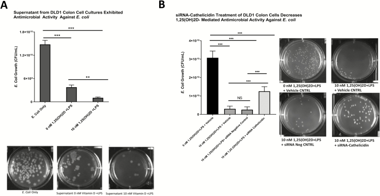
Methods: Serum and colonic cathelicidin levels were measured in UC patients and correlated with clinical and histologic outcomes. Human colon cells were treated with 1,25(OH)2D and production of cathelicidin and cytokines were quantified. Antimicrobial activity against Escherichia coli from cell culture supernatants was measured. Mice were treated with intrarectal cathelicidin, and its effects on DSS colitis and intestinal microbiota were evaluated.

Results: In UC patients, serum 25(OH)D positively correlated with serum and colonic cathelicidin. Higher serum cathelicidin is associated with decreased risk of histologic inflammation and clinical relapse but not independent of 25(OH)D or baseline inflammation. The 1,25(OH)2D treatment of colon cells induced cathelicidin and IL-10, repressed TNF-α, and suppressed Escherichia coli growth. This antimicrobial effect was attenuated with siRNA-cathelicidin transfection. Intrarectal cathelicidin reduced the severity of DSS colitis but did not mitigate the impact of colitis on microbial composition.

Conclusions: Cathelicidin plays a protective role in 25(OH)D-associated UC histologic outcomes and murine colitis. Cathelicidin is induced by vitamin D in human colonic epithelial cells and promotes antimicrobial activity against E. coli. Our study provides insights into the vitamin D-cathelicidin pathway as a potential therapeutic target.

Keywords: cathelicidin; colonic epithelium; cytokines; ulcerative colitis; vitamin D.


Figures

FIGURE 1.
FIGURE 1.
Summarizes the association of cathelicidin with vitamin D and clinical outcomes in ulcerative colitis patients. A, Higher serum 25(OH)D levels is associated with higher serum cathelicidin protein levels (Pearson r = 0.4138; P < 0.001) .B, Serum 25(OH)D level >20 ng/mL is associated with a 10.6-fold increase in cathelicidin mRNA expression compared with UC patients with a 25(OH)D level ≤20 ng/mL (P < 0.01). C, Serum cathelicidin levels are higher in UC patients with histologic mucosal healing compared with UC patients with histologic inflammation (245.2 ng/mL vs 196.5 ng/mL; P < 0.01). Serum cathelicidin levels are higher in UC patients who remained in clinical remission compared with UC patients who had a clinical relapse at 1-year follow-up (238.0 ng/mL vs 195.1 ng/mL; 0.01). D, The area under the curve (AUC) for serum cathelicidin in detecting the presence of histologic inflammation was 0.70 (P < 0.01). *P < 0.05, **P < 0.01, ***P < 0.001.
FIGURE 2.
FIGURE 2.
Summarizes the effect of 1,25(OH)D2 on cathelicidin mRNA and protein levels by DLD1 colonic epithelial cells. A, In the absence of LPS, 1,25(OH)2D induced cathelicidin mRNA 3.46-fold (P < 0.01) and 3.04-fold (P < 0.01) at 0.01 nM and 0.1 nM, respectively, compared with controls. In the presence of LPS, 1,25(OH)2D induced cathelicidin mRNA 6.25-fold (P < 0.01), 3.46-fold (P < 0.01), 2.53-fold (P < 0.05), and 2.36-fold (P < 0.05) at 0.01 nM, 0.1 nM, 1 nM, and 10 nM, respectively, compared with controls. B, Treatment of 1,25(OH)D2 at lower concentrations (0 nM, 0.01 nM, 0.1 nM, 1 nM) with or without LPS did not increase cathelicidin protein concentrations measured from cell culture supernatants compared with controls. At 10 nM of 1,25(OH)D2 without LPS, cathelicidin protein concentration increased to 701.7 pg/mL (P < 0.01) vs 540.3 pg/mL from controls. At 10 nM of 1,25(OH)D2 with LPS, cathelicidin protein concentration increased to 735 pg/mL (P < 0.001) vs 530 pg/mL from controls. At 10 nM of 1,25(OH)D2, there was no difference in cathelicidin protein concentration between the LPS and non-LPS treatment groups. *P < 0.05, **P < 0.01, ***P < 0.001.
FIGURE 3.
FIGURE 3.
A, Summarizes the antimicrobial effect of supernatants from 1,25(OH)2D-treated DLD1 colonic epithelial cell cultures on E. coli. Treatment of E. coli with supernatants from DLD1 cell cultures pretreated with 0 nM of 1,25(OH)D and LPS (supernatant cathelicidin concentration 530 pg/mL) resulted in 4-fold decrease in E. coli growth (1.48 × 1010 CFU/mL vs 3.77 × 109 CFU/mL; P < 0.001). Treatment of E. coli with supernatants from DLD1 cell cultures pretreated with 10 nM of 1,25(OH)D and LPS (supernatant cathelicidin concentration 735 pg/mL) resulted in 14.4-fold decrease in E. coli growth (1.48 × 1010 CFU/mL vs 1.03 × 109 CFU/mL; P < 0.001). Supernatants from DLD1 cell cultures pretreated with 10 nM of 1,25(OH)D suppressed E. coli growth 3.7-fold more (P < 0.01). B, Summarizes the effect of siRNA-cathelicidin treatment on antimicrobial activity of supernatants from DLD1 cell cultures. Supernatants from cell cultures treated with 10 nM of 1,25(OH)2D+ LPS + vehicle (transfection reagent) resulted in a 10.2-fold decrease in E. coli growth (3.07 × 1010 CFU/mL vs 3.0 × 109 CFU/mL; P < 0.001) compared with controls. Supernatants from cell cultures treated with 10 nM of 1,25(OH)2D+ LPS + siRNA negative control resulted in a 12.3-fold decrease in E. coli growth (3.07 × 1010 CFU/mL vs 2.5 × 109 CFU/mL; P < 0.001) compared with controls. Supernatants from cell cultures treated with 10 nM of 1,25(OH)2D+ LPS + siRNA-cathelicidin resulted in a 2.5-fold decrease in E. coli growth (3.07 × 1010 CFU/mL vs 1.25 × 1010 CFU/mL; P < 0.001) compared with controls. Transfection of DLD1 cells with siRNA-cathelicidin attenuated the vitamin D-induced antimicrobial activity against E. coli compared with siRNA negative controls (2.5-fold decrease vs 12.3-fold decrease in E. coli growth; P < 0.001). *P < 0.05, **P < 0.01, ***P < 0.001.
FIGURE 4.
FIGURE 4.
A, Summarizes the treatment allocation (IR cathelicidin or PBS) and timeline of DSS colitis experiments. B, summarizes the effect of intrarectal cathelicidin treatment on DSS colitis disease activity index. After 7 days of 3% DSS, mice treated with cathelicidin had lower disease activity index scores compared with PBS controls (5.8 vs 10.2; P < 0.01 by Mann Whitney U test) Figure 4C summarizes the effect of intrarectal cathelicidin treatment on DSS colitis histologic inflammation scores. Mice treated with cathelicidin had lower histologic inflammation scores (Figure 3C) compared with PBS controls (2.8 vs 5.8; P < 0.05). *P < 0.05, **P < 0.01, ***P < 0.001.

References

    1. Gubatan J, Chou ND, Nielsen OH, et al. . Systematic review with meta-analysis: association of vitamin D status with clinical outcomes in adult patients with inflammatory bowel disease. Aliment Pharmacol Ther. 2019;50:1146–1158. - PubMed
    1. Gubatan J, Mitsuhashi S, Longhi MS, et al. . Higher serum vitamin D levels are associated with protective serum cytokine profiles in patients with ulcerative colitis. Cytokine. 2018;103:38–45. - PMC - PubMed
    1. Gubatan J, Mitsuhashi S, Zenlea T, et al. . Low serum vitamin D during remission increases risk of clinical relapse in patients with ulcerative colitis. Clin Gastroenterol Hepatol. 2017;15:240–246. - PMC - PubMed
    1. Kabbani TA, Koutroubakis IE, Schoen RE, et al. . Association of vitamin D level with clinical status in inflammatory bowel disease: a 5-year longitudinal Study. Am J Gastroenterol. 2016;111:712–719. - PubMed
    1. Nielsen OH, Hansen TI, Gubatan JM, et al. . Managing vitamin D deficiency in inflammatory bowel disease. Frontline Gastroenterol. 2019;10:394–400. - PMC - PubMed

Publication types

MeSH terms