. 2020 Feb 6;180(3):490-501.e16.
doi: 10.1016/j.cell.2019.12.030. Epub 2020 Jan 16.

Cryo-EM Reveals Integrin-Mediated TGF-β Activation without Release from Latent TGF-β

Affiliations

Cryo-EM Reveals Integrin-Mediated TGF-β Activation without Release from Latent TGF-β

Melody G Campbell et al. Cell. .

Abstract

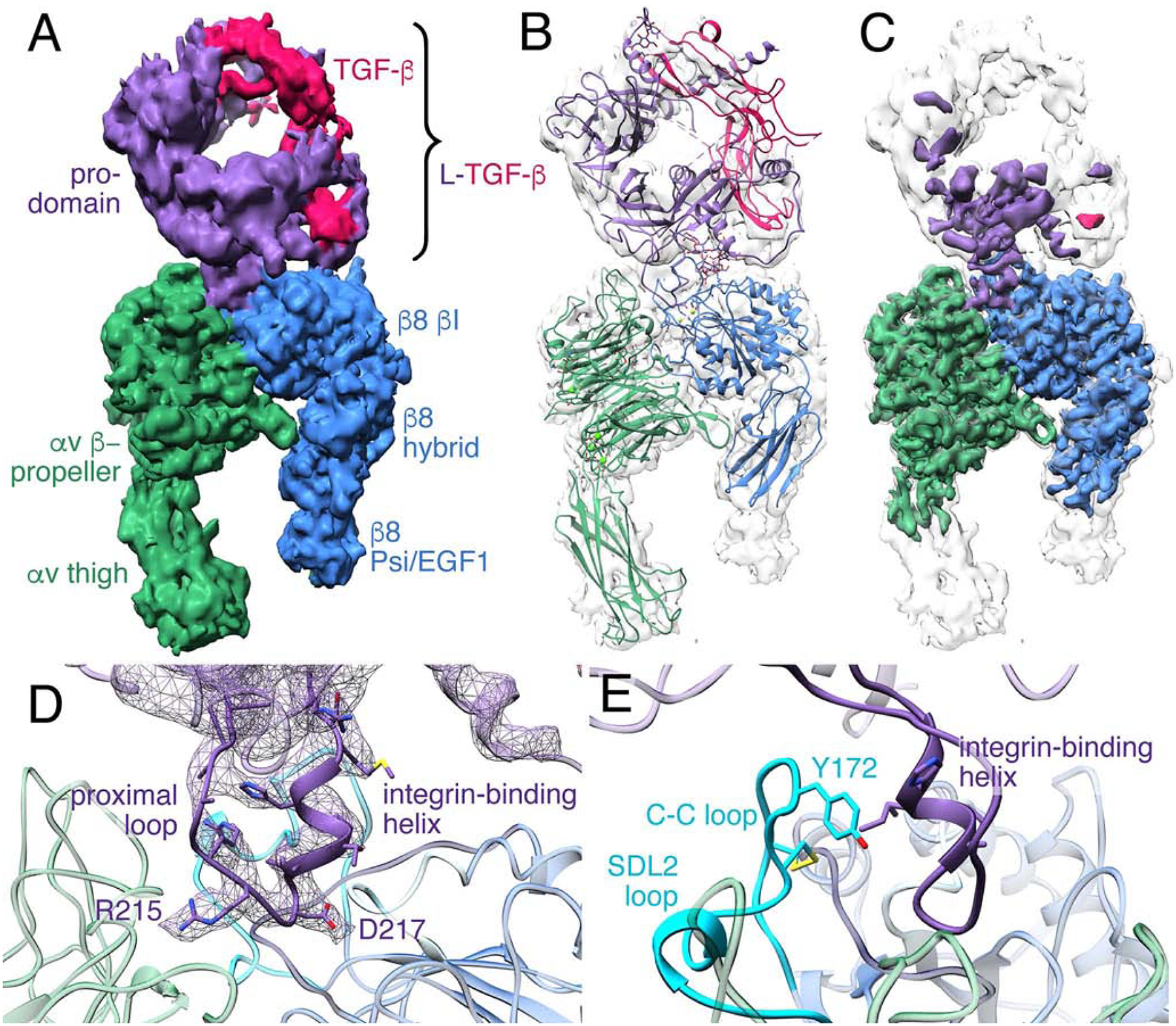
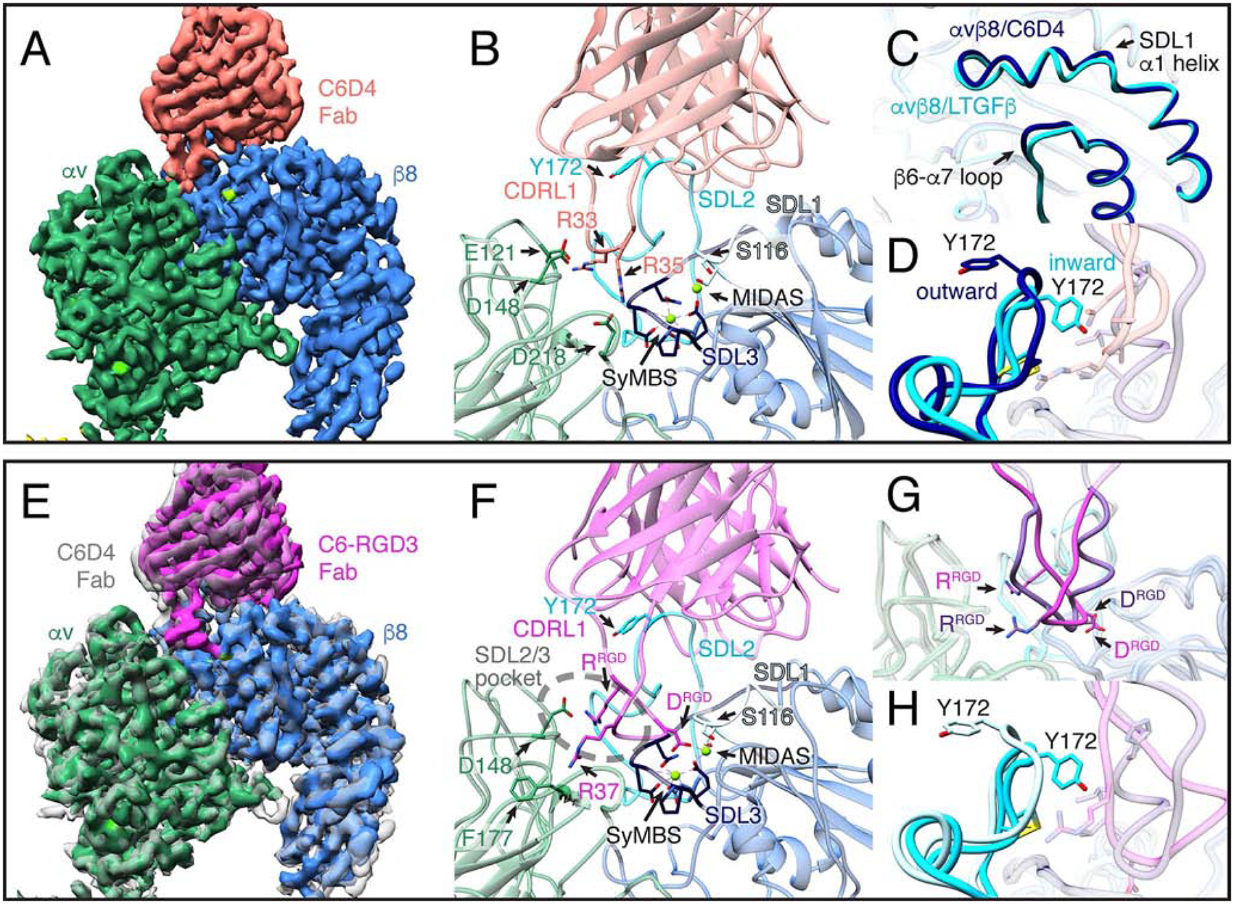
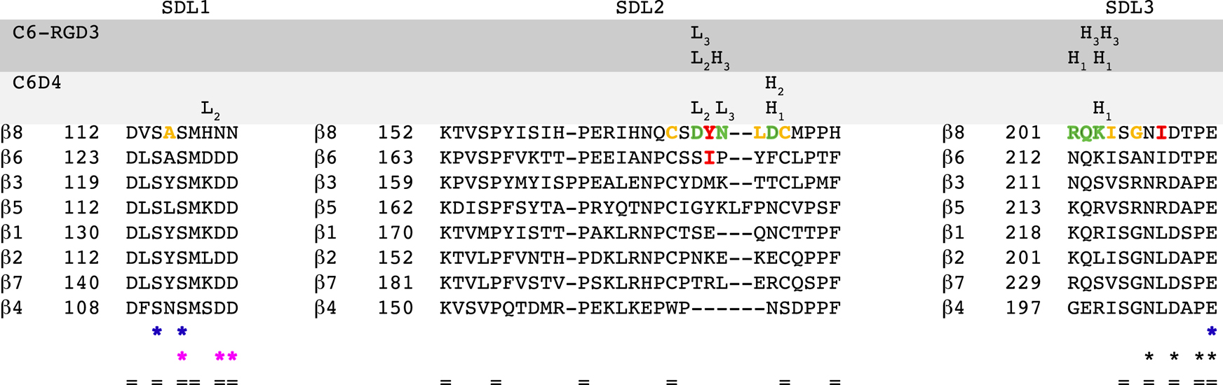
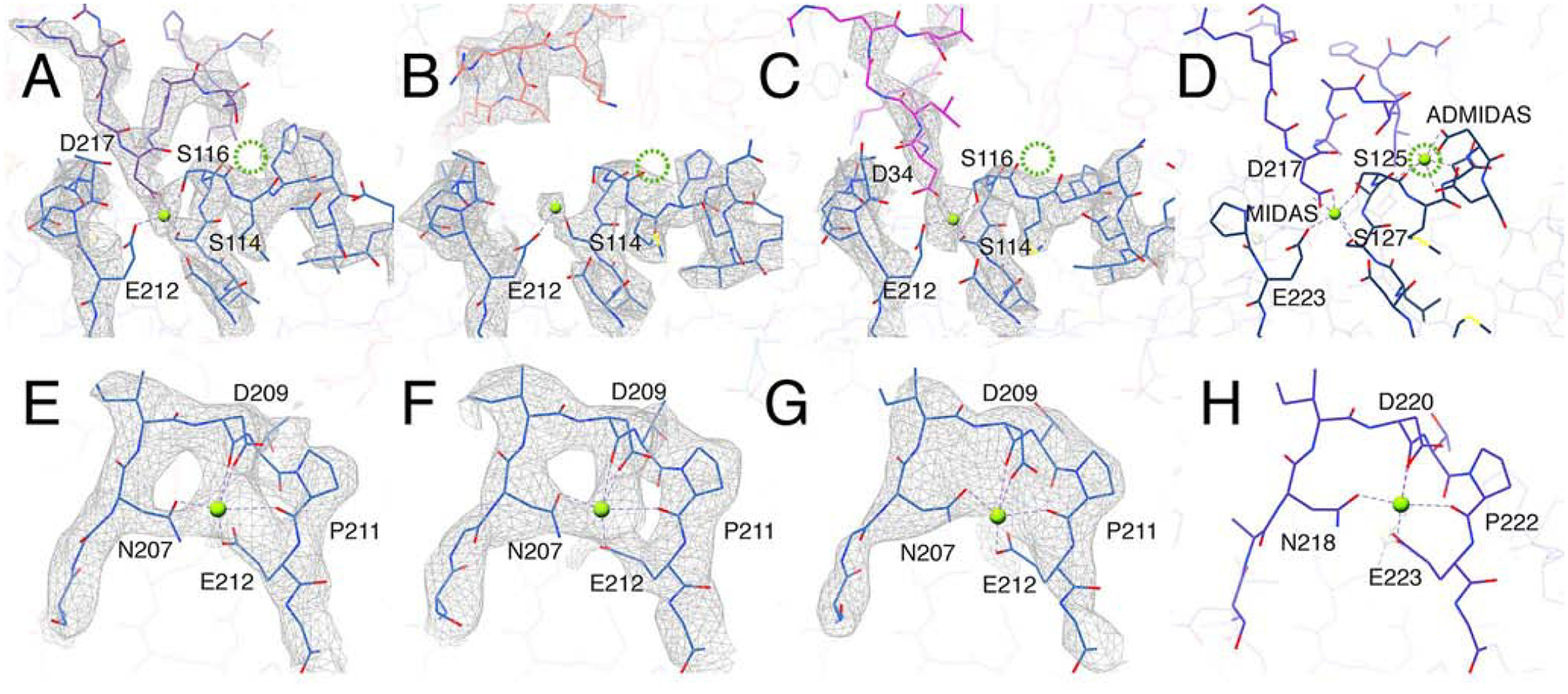
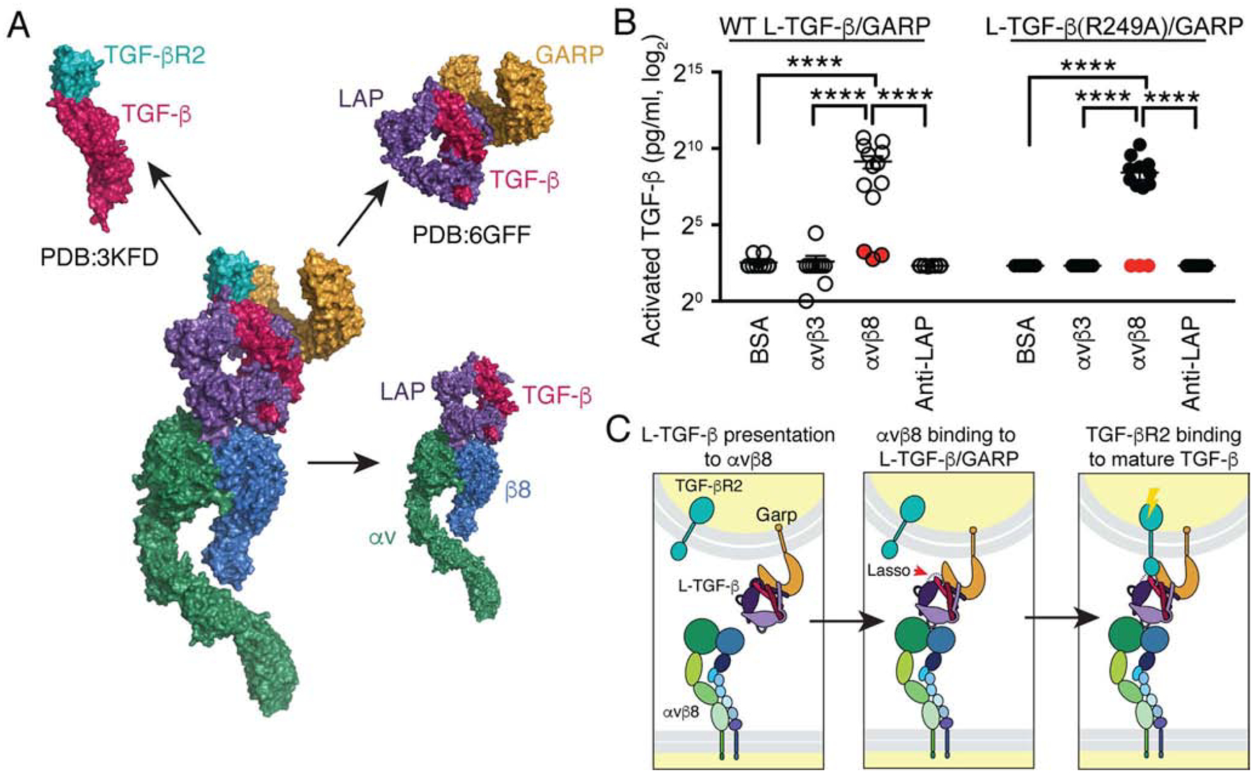
Integrin αvβ8 binds with exquisite specificity to latent transforming growth factor-β (L-TGF-β). This binding is essential for activating L-TGF-β presented by a variety of cell types. Inhibiting αvβ8-mediated TGF-β activation blocks immunosuppressive regulatory T cell differentiation, which is a potential therapeutic strategy in cancer. Using cryo-electron microscopy, structure-guided mutagenesis, and cell-based assays, we reveal the binding interactions between the entire αvβ8 ectodomain and its intact natural ligand, L-TGF-β, as well as two different inhibitory antibody fragments to understand the structural underpinnings of αvβ8 binding specificity and TGF-β activation. Our studies reveal a mechanism of TGF-β activation where mature TGF-β signals within the confines of L-TGF-β and the release and diffusion of TGF-β are not required. The structural details of this mechanism provide a rational basis for therapeutic strategies to inhibit αvβ8-mediated L-TGF-β activation.

Keywords: GARP; TGF-b signaling; TGF-beta; TGF-beta activation; cryo-electron microscopy; integrin; integrin conformation; structural biology.


Conflict of interest statement

Declaration of Interests A.C., J.D.M., J.L., J.L.B., Y.C., and S.L.N. are inventors on a patent on anti-v8 antibodies, which have been out-licensed. S.L.N. is on the Scientific Advisory Board of Venn Therapeutics.

Figures

Fig. 1
Fig. 1. The αvβ8 integrin ectodomain bound to L-TGF-β1
(A) Cryo-EM density map of αvβ8 integrin ectodomain with L-TGF-β1 bound. The map is displayed as unsharpened and at a low-threshold. The color code is as follows: integrin αv-subunit is green, integrin β8-subunit is blue, prodomain of L-TGF-β1 is purple, and mature TGF-β1 is red. (B) Ribbon diagram of αvβ8/L-TGF-β1 displayed within the density map shown in (A). (C) The sharpened cryo-EM map is shown in color, superimposed with the unsharpened map in semi-transparent white. (D) A ribbon diagram shows a close-up view of the binding interface between αvβ8 and L-TGF-β1. The EM density of the integrin-binding motif of L-TGF-β1, including proximal loop, RGD motif, and integrin-binding helix is shown in purple mesh. (E) Ribbon diagram showing the C-C loop within the SDL2 loop of the β8 subunit (cyan) relative to the L-TGF-β1 integrin-binding helix. Related information is in Fig. S1, S3.
Fig. 2.
Fig. 2.. Mechanisms of action of inhibitory C6D4 and C6-RGD3 Fabs
(A) A close-up view of the cryo-EM density map of the αvβ8/C6D4 complex. The full structure is shown in Fig. S2. The αvβ8 ligand-binding cleft is fully occupied by C6D4, demonstrating a steric mode of inhibition. Color code: αv is dark green, β8 is blue, and cations are bright green. (B) Close-up ribbon diagram of the αvβ8/C6D4 interface showing the position of the C6D4 Fab (coral) on the αvβ8 binding-cleft, highlighting interactions between the Fab CDRL1 loop R33 interacting with αv E121 and αv D148, and Fab R35 with αv D218, respectively (for all interactions see Fig. 3, S1, S3 and S4). Additional color code: SyMBS and MIDAS is green, SDL1 is light blue, SDL2 is cyan, SDL3 is dark blue. (C-D) Comparisons are shown between αvβ8/C6D4 and αvβ8/L-TGF-β1 with key changes occurring in β8 upon ligand binding noted. Color code: unliganded C6D4 structure is dark blue, liganded L-TGF-β structure is cyan. There are very subtle ligand-induced changes in the SDL1 α1 helix (~1 Å at the tip) and the β6-α7 loop (C). Y172 of the SDL2 loop is positioned outward relative to the ligand binding site in complex with C6D4 but moves inwards upon ligand binding (D). (E) Cryo-EM density map of αvβ8 (same colors as in A) in complex with the C6-RGD3 ligand-mimetic Fab (magenta) superimposed on the αvβ8/C6D4 map (semi-transparent gray). Aligning the αv-subunit β-propeller domain of the two maps shows a slight shift in the angle of C6-RGD3 Fab binding. (F) Close-up ribbon diagram of the αvβ8/C6-RGD3 binding interface shows the alternative RGD binding mode with C6-RGD3 Fab. Key contacts are: ArgRGD interacting with αv D148 at the edge of the β8 proximal SDL2/3 pocket (dashed ellipse) and C6-RGD3 Fab AspRGD interacting with the MIDAS cation. C6-RGD3 Fab R37 (30-LGRGDLGRL−38) interacts with αv F177 (for all interactions see Fig. 3, S2–4). Integrin color codes are as in B, with C6-RGD3 in magenta. (G) Superimposition of the C6-RGD3 ligand-mimetic RGD loop (magenta) and the native L-TGF-β loop (purple) shows that C6-RGD3 binds in an alternative binding conformation. (H) β8 Y172 in the αvβ8/C6-RGD3 complex (light blue) is in the outward position compared to the αvβ8/LTGFb complex (cyan). Related information is in Fig. S2–4.
Fig. 3.
Fig. 3.. L-TGF-β and inhibitory antibodies contact multiple overlapping residues in β8 SDL loops
Sequence alignments for integrin b subunits are shown in decreasing order of homology to β8. Each SDL region is indicated above the sequence. Each CDR VH and VL loop of C6-RGD3 and C6D4 is shown above the specific amino acids which it contacts. The β8 SDL residues which contact L-TGF-β as well as C6D4 or C6-RGD3 are shown in green letters. The β8 SDL residues which form the hydrophobic patch that interact with the integrin binding helix of L-TGF-β1 are shown in orange letters. The residues that are mutated in Fig. 5 are shown in red letters. The cation binding sites are indicated below the sequence by colored asterisks with MIDAS in blue, ADMIDAS in magenta, and SyMBS in black. Conserved residues are indicated (=). Related information is in Table S1–4.
Fig. 4.
Fig. 4.. The ADMIDAS cation is not present in αvβ8
(A-C) The MIDAS cation binding site formed in αvβ8 integrin (blue) bound with: L-TGF-β1 (purple, A) C6D4 (coral, B) and C6-RGD3 (magenta, C). In all three structures there is clear density for the MIDAS cation (bright green). No density for the ADMIDAS cation is observed (the expected position for ADMIDAS is noted by the dotted green circle). Shown are the cation-coordinating residues of the integrin b subunit or the Asp of RGD of L-TGF-β (D217) or C6-RGD3 (D34). (D) The same binding interface in the αvβ6/LTGFβ crystal structure (5FFO (Dong et al., 2017)). (E-H) Maps and models of the SyMBS cation in αvβ8/L-TGF-β (E), αvβ8/C6D4 (F), αvβ8/C6-RGD3 (G), and αvβ6/LTGFβ crystal structure (5FFO) (H). All panels are color coded as in Fig. 2. Related information is in Fig. S3.
Fig. 5.
Fig. 5.. L-TGF-β is flexible when bound to αvβ8
Seven structures (i-vii) illustrate the conformational variability of the αvβ8/LTGFβ complex. All structures are aligned to each other using the αv-subunit b-propeller domain. Each vertical panel shows two views (A, and B) of the same structure rotated 90˚ around the z-axis and a close-up of the integrin-ligand binding interface with the L-TGF-β integrin-binding helix (C), which is formed in all structures. Subclass (iv) is shown in Figs. 1, 2 and 4. Color coding is the same as for Fig. 2. For (A-B) the primary conformation is shown in colors, as defined in Fig. 1A, and all six other conformations are shown as semi-transparent to provide a reference for L-TGF-β’s range of motion. From left to right, the percentage of particles that went into each class are as follows: 14%, 14%, 14%, 19%, 16%, 11%, 12%. Related information is in Fig. S5.
Fig. 6.
Fig. 6.. Structure based modeling of L-TGF-β binding to αvβ8
(A) A stepwise model of L-TGF-β binding to αvβ8. Our structures were used to define unliganded (αvβ8/C6D4), alternate (αvβ8/C6-RGD3) and canonical RGD binding modes (αvβ8/L-TGF-β). The hypothesized transitions between these binding states suggest possible intermediates where the integrin binding helix either forms before or after engaging the canonical RGD position (color coding as in Fig. 1). (B) TGF-β activation assays test the function of β8- and β6-subunit SDL mutants. Wild-type (WT) αvβ8 or αvβ6 or mutant receptors (β8: I208R, Y172M, Y182N, entire SDL2 deletion mutant (Δ-SDL), or β6: I183N) were stably expressed in CHO cells, sorted for uniform expression, confirmed for proper folding using C6D4 or anti-β6 antibodies (Fig. S6) and tested for ability to support TGF-β activation with TMLC reporter cells. Activation relative to αvβ8 WT expressing CHO cells is shown. (C) Shown is surface rendering of the b-subunit hydrophobic patch (grey) of αvβ8 (upper) and αvβ6 (lower) and the relationship of β8 Y172 and β6 I183 with the L-TGF-β integrin-binding helix (yellow sticks and cartoon). (D) Inhibition of L-TGF-β1 binding to αvβ8 by TGF-β1 and TGF-β3 integrin-binding motif peptides and Ala and Pro mutants was measured. Shown is the inhibitory concentration (IC50) plotted against helical penalty score (Pace and Scholtz, 1998). (E) Sequences of peptides used in (D) are shown. ** p=0.01, *** p=0.001, **** p=0.0001 by unpaired two-sided Student’s t test. Related information is in Fig. S6.
Fig. 7.
Fig. 7.. A structural model of the αvβ8/L-TGF-β/GARP/TGF-βR2 complex predicts that releasing of mature TGF-β is not required for αvβ8-mediated TGF-β activation
(A) Surface representation of a model of the putative complex derived from our αvβ8/L-TGF-β1 structures combined with the crystal structures of the GARP/L-TGF-β1 complex (PDB: 6GFF (Lienart et al., 2018)), and the TGF-β/TGF-βR2 complex (PDB: 3KFD (Radaev et al., 2010)) is shown. Color code: integrin αvβ8 (αv-green, β8-blue), L-TGF-β1 (LAP-purple, TGF-β-red), GARP-orange and TGF-βR2-teal. (B) WT L-TGF-β1/GARP or L-TGFβ1(R249A)/GARP expressed on the surface of TGF-β reporter cells (TMLC) show efficient TGF-β activation when plated on immobilized αvβ8. TMLC are stably transfected with a highly specific TGF-β responsive promoter driving a truncated plasminogen activator inhibitor type I (PAI-1) promoter, which contains SMAD-binding elements driving a luciferase reporter cassette (Abe, et al, 1994). TGF-β activation is then measured as luciferase activity. A R249A mutation in TGF-β prevents proteolytic cleavage by furin of mature TGF-β from LAP during intracellular processing (Shi, et al, 2011). TMLC expressing L-TGF-β1 (R249A)/GARP cannot produce diffusible active TGF-β and thus report only TGF-β activated within the L-TGF-β1 complex (Fig. S7). Minimal or undetectable levels of TGF-β were released into the supernatants from WT L-TGF-β1/GARP or L-TGF-β1 (R249A)/GARP TMLC cells when plated on αvβ8 or control substrates (supernatants were removed and assayed separately using TMLC cells, filled red circles). Luciferase activities are normalized and converted to amount of active TGF-β (pg/ml), as described (Annes et al., 2003). The y-axis is in log2 scale. ****p<0.0001 by one-way ANOVA and Tukey’s multiple comparisons test. (C) Model of TGF-β mediated TGF-β activation is shown. Activation occurs in three steps: Left panel: Initially, αvβ8 in an extended-closed conformation surveys the environment for cells presenting L-TGF-β on their cell surface by co-expression of adaptor molecules such as GARP. Middle panel: Binding of one cell expressing αvβ8 to a L-TGF-β expressing cell increases the flexibility of the latency lasso of TGF-β (red arrow) which exposes the active domain on one fingertip of the mature TGF-β homodimer. Right panel: TGF-βR2 expressed by the same cell presenting L-TGF-β binds to the exposed active domain of mature TGF-β, which initiates the TGF-β signaling cascade. Related information is in Fig. S7.

Comment in

References

    1. Abe M, Harpel JG, Metz CN, Nunes I, Loskutoff DJ, and Rifkin DB (1994). An assay for transforming growth factor-beta using cells transfected with a plasminogen activator inhibitor-1 promoter-luciferase construct. Anal Biochem 216, 276–284. - PubMed
    1. Adams PD, Afonine PV, Bunkoczi G, Chen VB, Davis IW, Echols N, Headd JJ, Hung LW, Kapral GJ, Grosse-Kunstleve RW, et al. (2010). PHENIX: a comprehensive Python-based system for macromolecular structure solution. Acta Crystallogr D Biol Crystallogr 66, 213–221. - PMC - PubMed
    1. Akhurst RJ (2017). Targeting TGF-beta Signaling for Therapeutic Gain. Cold Spring Harb Perspect Biol 9. - PMC - PubMed
    1. Aluwihare P, Mu Z, Zhao Z, Yu D, Weinreb PH, Horan GS, Violette SM, and Munger JS (2009). Mice that lack activity of alphavbeta6- and alphavbeta8-integrins reproduce the abnormalities of Tgfb1- and Tgfb3-null mice. J Cell Sci 122, 227–232. - PMC - PubMed
    1. Annes JP, Munger JS, and Rifkin DB (2003). Making sense of latent TGFbeta activation. J Cell Sci 116, 217–224. - PubMed

Publication types

MeSH terms