Case Reports
. 2019 Dec 28:2019:7568098.
doi: 10.1155/2019/7568098. eCollection 2019.

Multiparametric Ultrasound (mpUS) of a Rare Testicular Capillary Hemangioma

Affiliations
Case Reports

Multiparametric Ultrasound (mpUS) of a Rare Testicular Capillary Hemangioma

Paul Spiesecke et al. Case Rep Radiol. .

Abstract

Capillary hemangioma is a rare entity among testicular tumors. We demonstrate the case of an 18-year-old patient with palpatoric and sonographic conspicuous left testicle and negative serum tumor markers (α-fetoprotein, β-human chorionic gonadotropin, and lactate dehydrogenase). Ultrasound (US) imaging represented an isoechogenic lesion with high vascularization in both power Doppler and microflow imaging with central feeding artery. Both strain elastography and shear wave elastography demonstrated a stiff lesion compared to surrounding testicular tissue. While contrast-enhanced ultrasound (CEUS) clearly depicted high vascular load, time intensity curve (TIC) analysis was able to show shorter median transit time, higher peak enhancement, and higher wash-in area under the curve compared to regular testicular tissue. Histopathological examination revealed a lobular constructed and rich vascularized proliferation without cellular atypia and feeder vessels with positive reaction to CD34, CD31, CD99, and Vimentin. Proliferative activity was quantified to 3-5% by Ki-67 index. Two days after surgery, the patient could leave the hospital in subjective wellbeing. While histology remains the gold standard to make a precise diagnosis of capillary hemangiomas due to small case numbers and variety of this benign tumor, the combination of multiparametric US and clinical information may be a promising future tool in preoperative assessment.


Conflict of interest statement

None of the authors reports a relationship with industry and other relevant entities—financial or otherwise—that might pose a conflict of interest in connection with the submitted article.

Figures
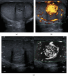
Figure 1
Figure 1
B-mode US, power Doppler imaging, and SMI of a capillary hemangioma. (a) Isoechogenic lesion in the central part of the left testicle with peripheral hypoechogenic rim (11 mm) and central feeder vessel. (b) Power Doppler imaging showed strong vascularization of the lesion compared to the surrounding tissue. (c) Monochromatic SMI in split screen mode determined stronger feeder vessels in the peripheral part of the lesion with high central vascular density. Abbreviations: US: ultrasound; SMI: superb microvascular imaging.
Figure 2
Figure 2
Elastographic assessment of the tumor lesion using 2D shear wave elastography (SWE) and strain elastography. (a) 2D shear wave elastography demonstrates higher and more inhomogenous stiffness of the tumor lesion (4.24 m/s) compared to the surrounding testicular tissue (1.49 m/s) with a SWE ratio of 2.85. (b) Tumor lesion represents blue (hard) in color-coded strain elastography compared to the surrounding testicular tissue (green-blue) presented by less compression capability indicating higher stiffness of the tumor. Abbreviations: SWE: shear wave elastography.
Figure 3
Figure 3
Arrival time imaging (ATI) and contrast-enhanced ultrasound (CEUS). (a) Parametric ATI depicted short arrival time in the central part of the tumor with feeder artery within 2 seconds (arrows; color-coded in red). (b, c) Accumulation mode and B-mode/CEUS image fusion demonstrates high peak intensity of the tumor compared to the surrounding testicular tissue. Abbreviations: ATI: arrival time imaging; CEUS: contrast-enhanced ultrasound.
Figure 4
Figure 4
Time intensity curve (TIC) measurement using a ROI in tumor lesion and regular testicular tissue. TIC measurements demonstrate a higher peak intensity, shorter mean transit time, and higher wash-in AUC of the tumor lesion (purple ROI) compared to regular surrounding tissue (blue ROI). Abbreviations: TIC: time intensity curve; ROI: region of interest; AUC: area under the curve.

References

    1. Garner M. J., Turner M. C., Ghadirian P., Krewski D. Epidemiology of testicular cancer: an overview. International Journal of Cancer. 2005;116(3):331–339. doi: 10.1002/ijc.21032. - DOI - PubMed
    1. Mazal P. R., Kratzik C., Kain R., Susani M. Capillary haemangioma of the testis. Journal of Clinical Pathology. 2000;53(8):641–642. doi: 10.1136/jcp.53.8.641. - DOI - PMC - PubMed
    1. Sidhu P. S. Multiparametric ultrasound (MPUS) imaging: terminology describing the many aspects of ultrasonography. Ultraschall in der Medizin - European Journal of Ultrasound. 2015;36(4):315–317. doi: 10.1055/s-0035-1553381. - DOI - PubMed
    1. Lock G., Schmidt C., Helmich F., Stolle E., Dieckmann K.-P. Early experience with contrast-enhanced ultrasound in the diagnosis of testicular masses: a feasibility study. Urology. 2011;77(5):1049–1053. doi: 10.1016/j.urology.2010.12.035. - DOI - PubMed
    1. Lock G., Schröder C., Schmidt C., Anheuser P., Loening T., Dieckmann K. Contrast-enhanced ultrasound and real-time elastography for the diagnosis of benign Leydig cell tumors of the testis – a single center report on 13 cases. Ultraschall in der Medizin - European Journal of Ultrasound. 2014;35(06):534–539. doi: 10.1055/s-0034-1385038. - DOI - PubMed

Publication types

LinkOut - more resources