Review
. 2020 Jan 18;13(1):163-175.
doi: 10.18240/ijo.2020.01.23. eCollection 2020.

Molecular pathobiology of scleritis and its therapeutic implications

Affiliations
Review

Molecular pathobiology of scleritis and its therapeutic implications

Undurti N Das. Int J Ophthalmol. .

Abstract

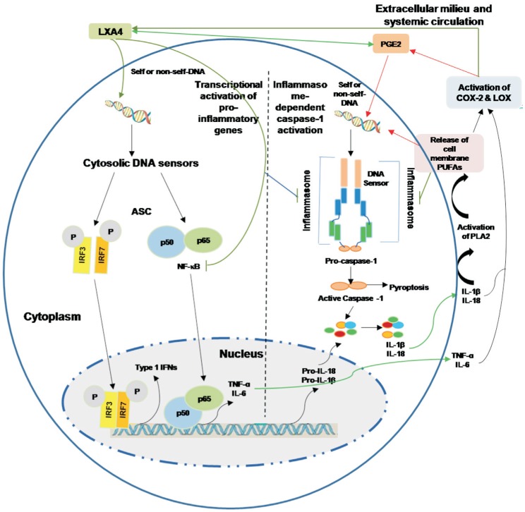
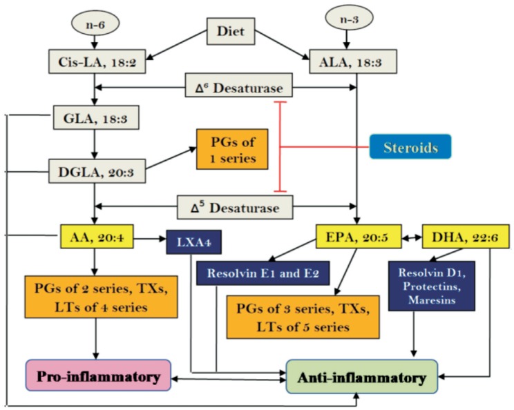
Scleritis and other autoimmune diseases are characterized by an imbalance in the levels of pro-inflammatory and anti-inflammatory molecules with the balance tilted more towards the former due to the failure of recognition of self. The triggering of inflammatory process could be ascribed to the presence of cytoplasmic DNA/chromatin that leads to activation of cytosolic DNA-sensing cGAS-STING (cyclic GMP-AMP synthase linked to stimulator of interferon genes) pathway and enhanced expression of NF-κB that results in an increase in the production of pro-inflammatory bioactive lipids. Bioactive lipids gamma-linolenic acid (GLA), dihomo-GLA (DGLA), prostaglandin E1 (PGE1), prostacyclin (PGI2) and lipoxin A4, resolvins, protectins and maresins have anti-inflammatory actions, bind to DNA to render it non-antigenic and are decreased in autoimmune diseases. These results suggest that efforts designed to enhance the production of anti-inflammatory bioactive lipids may form a new approach to autoimmune diseases. Local injection or infusion of lipoxins, resolvins, protectins and maresins or their precursors such as arachidonic acid may be exploited in the prevention and management of autoimmune diseases including scleritis, uveitis and lupus/rheumatoid arthritis.

Keywords: autoimmune diseases; bioactive lipids; cytokines; inflammation; micronucleus; resolution of inflammation; scleritis.


Figures

Figure 1
Figure 1. Innate immunity recognizes changes in cells induced by infection
NK cells have inhibitory receptors on their surface to differentiate “self” from “missing-self”. The lack of expression of MHC class I molecules (missing self) promotes the activation of NK cells and subsequent lysis of the target cells. NK cells also express activating receptors such as NKG2D that directly recognize ligands induced in response to infection (induced self). NK cell-mediated cytotoxicity against malignant target cells or infected cells is regulated by both activating and inhibitory cell surface immunoreceptors. In humans, such receptors are of three types: 1) the killer immunoglobulin receptors (KIRs), 2) natural cytotoxicity receptors (NCRs), and 3) the c-type lectin receptors. NKG2D is one member of the c-type lectin-activating receptor family that is evolutionarily conserved and is located within the NK gene complex on human chromosome 12p12-p13. NKG2D is expressed on all NK cells and is a promiscuous receptor that recognizes at least 6 counter ligands that include: the MHC class I-like molecules, MICA and MICB, and members of the ULBP family (ULPB1-4), named for the ability of some members to bind to the UL-16 protein of cytomegalovirus (CMV).
Figure 2
Figure 2. Cytoplasmic DNA triggers transcription of inflammatory genes and inflammasome-dependent proteolytic activation of caspase-1
Presence of DNA in the cytoplasm leads to the activation of two distinct signaling pathways 1) activation of IRF3, IRF7 and NF-κB that results in the transcriptional induction of type 1 IFN genes or pro-inflammatory genes IL-6 and TNF-α; 2) cytosolic DNA leads to the assembly of inflammasome leading to caspase-1 activation and subsequent cleavage of pro-IL-β and pro-IL-18 that results in the formation of biologically active and mature forms of IL-1β and IL-18. Caspase activation mediates the cell death under some very specific conditions (such as tumor cell death). Enhnced synthesis and secretion of pro-inflammatory cytokines leads to activation of COX-2 and LOX enzmes resulting in the produciton of pro-inflammatory bioactive lipids such as PGE2, LTs and TXs and a simultaenous decrease of anti-inflammatory LXa, resolvins, protectins and maresins from their respetive precursor PUFAs (AA, EPA and DHA). These PUFAs, LXA4, resolvins, protectins and maresins can suppress NF-κB and IRF3 and IRF7 activation and inhibit the production of pro-inflammatory cytokines. Under some very speific situations PGE2 may function as an anti-inflammatry molecule. PGE2 and IL-1β have been shown to trigger release of AA/EPA/DHA from the cell membrane pool by activating PLA2 that can lead to the formation of anti-inflammatory LXA4/resolvins/protectins/maresins that, in turn, suppress production of IL-6 and TNF-αto restore homeostasis. Thus, there is a very close and a positive and negative feed-back regulation between pro- and anti-inflammatory cytokines, bioactive lipids and respective signaling pathways.
Figure 3
Figure 3. Scheme showing metabolism of essential fatty acids and their pro- and anti-inflammatory products
Steroids block the activities of desaturases that leads to a decrease in the concentrations of GLA, DGLA, AA, EPA and DHA. This results in decreased formation of not only pro-inflammatory PGs, LTs and TXs but also lipoxins, resolvins, protectins and maresins (due to precursor deficiency). Hence, resolution of inflammation will be incomplete. This ultimately causes continuous of inflammation to chronic phase and failure of healing of wound due to deficiency of lipoxins, resolvins, protectins and maresins.
Figure 4
Figure 4. Scheme showing the relationship among pro- and anti-inflammatory cytokines, PGs, LTs, lipoxins, resolvins, protectins and maresins and steroids. Metabolism and actions of arachidonic acid is shown as a representative of various PUFAs (DGLA, EPA and DHA)
(+) Indicates increase in the synthesis/action or positive effect. (-) Indicates decrease in the synthesis/action or negative effect.

References

    1. Sainz de la Maza M, Foster CS, Jabbur NS. Scleritis-associated uveitis. Ophthalmology. 1997;104(1):58–63. - PubMed
    1. Dong ZZ, Gan YF, Zhang YN, Zhang Y, Li J, Zheng HH. The clinical features of posterior scleritis with serous retinal detachment: a retrospective clinical analysis. Int J Ophthalmol. 2019;12(7):1151–1157. - PMC - PubMed
    1. Cohn M. The common sense of the self-nonself discrimination. Springer Semin Immunopathol. 2005;27(1):3–17. - PubMed
    1. Roach JC, Glusman G, Rowen L, Kaur A, Purcell MK, Smith KD, Hood LE, Aderem A. The evolution of vertebrate Toll-like receptors. Proc Natl Acad Sci USA. 2005;102(27):9577–9582. - PMC - PubMed
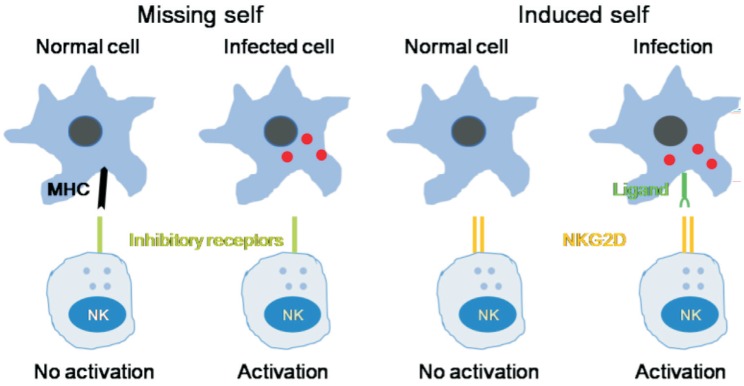
    1. Diefenbach A, Raulet DH. Strategies for target cell recognition by natural killer cells. Immunol Rev. 2001;181:170–184. - PubMed

LinkOut - more resources