

doi: 10.18240/ijo.2020.01.26.
eCollection 2020.
Ultrasound cyclo plasty for the management of glaucoma secondary to ocular irradiation for choroidal melanoma
Affiliations
- PMID: 31956588
- PMCID: PMC6942954
- DOI: 10.18240/ijo.2020.01.26
Item in Clipboard
Ultrasound cyclo plasty for the management of glaucoma secondary to ocular irradiation for choroidal melanoma
Int J Ophthalmol.
.
No abstract available
Figures
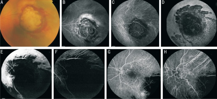
Fundus photography (A), fluorescein angiography (B) and indocyanine green angiography (C) showing the mushroom-shaped choroidal melanoma. Note the characteristic “double circulation” pattern of the choroidal melanoma consisting of normal retinal vessels overlying the internal circulation within the lesion. Fundus autofluorescence (D), fluorescein angiography (E) and indocyanine green angiography (F) showing the lesion 1y after PBT with atrophy of the surrounding retina and choroid. Fluorescein angiography (G) and indocyanine green angiography (H) showing radiation retinopathy involving the macular region, capillary leakage and retinal capillary dropout with foveal avascular zone enlargement.
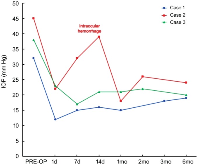
The IOP spike in Case 2 was found when intraocular hemorrhage occurred, with subsequent spontaneous resolution and IOP reduction.
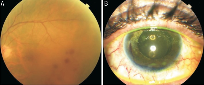
Fundus photography (A) showing radiation retinopathy with scattered dot and blot retinal hemorrhages in the temporal macular region. Anterior segment photography (B) showing the hyphema occurred 1wk after UCP procedure.
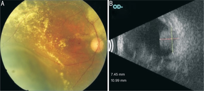
Fundus photography (A) showing the choroidal melanoma in the inferotemporal region with surrounding retinal exudation. B-scan ultrasonography (B) showing the solid hyperechoic choroidal mass protruding in the vitreous chamber.
References
-
- Bensoussan E, Thariat J, Maschi C, Delas J, Schouver ED, Hérault J, Baillif S, Caujolle JP. Outcomes after proton beam therapy for large choroidal melanomas in 492 patients. Am J Ophthalmol. 2016;165:78–87. - PubMed
-
- Floriani I, Quaranta L, Rulli E, Katsanos A, Varano L, Frezzotti P, Rossi GC, Carmassi L, Rolle T, Ratiglia R, Gandolfi S, Fossarello M, Uva M, Hollander L, Poli D, Grignolo F, Italian Study Group on QoL in glaucoma Health-related quality of life in patients with primary open-angle glaucoma. An Italian multicentre observational study. Acta Ophthalmol. 2016;94(5):e278–e286. - PubMed
-
- Riva I, Legramandi L, Katsanos A, Oddone F, Rulli E, Roberti G, Quaranta L, Italian Study Group on QoL in Glaucoma Influence of sociodemographic factors on disease characteristics and vision-related quality of life in primary open-angle glaucoma patients: the Italian primary open angle glaucoma study (IPOAGS) J Glaucoma. 2018;27(9):776–784. - PubMed
-
- Fabian ID, Tomkins-Netzer O, Stoker I, Arora AK, Sagoo MS, Cohen VM. Secondary enucleations for uveal melanoma: a 7-year retrospective analysis. Am J Ophthalmol. 2015;160(6):1104–1110.e1. - PubMed
LinkOut - more resources
Full Text Sources