

Schwartz-Matsuo syndrome: An important cause of secondary glaucoma
- PMID: 31956728
- PMCID: PMC6962651
- DOI: 10.1016/j.ajoc.2020.100586
Schwartz-Matsuo syndrome: An important cause of secondary glaucoma
Abstract
Purpose: We report a case of Schwartz-Matsuo syndrome that highlights the pathophysiology, diagnostic challenges, and management considerations of this rare disease.
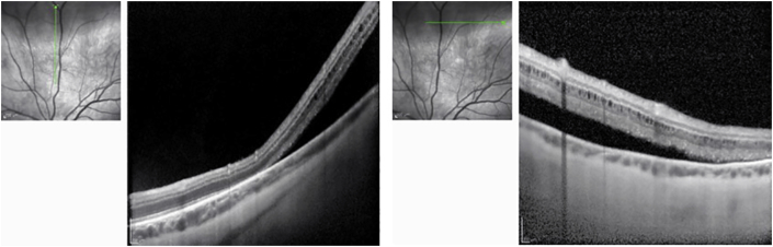
Observations: 31-year-old man with a history of left eye cataract presented with left eye photophobia and elevated intraocular pressure (IOP) of 64 mm Hg. Visual acuity 20/40. Open angles with an increased pigment of trabecular meshwork by gonioscopy, 2 + anterior chamber (AC) cell, superior retinal detachment, and 0.6 cup-to-disc ratio. Electron microscopy of AC fluid demonstrated outer segments of photoreceptors. IOP was lowered with oral and topical ophthalmic antihypertensives. Retinal detachment was treated with pars plana vitrectomy with endolaser, gas tamponade, and AC paracentesis. Follow-up VA 20/20 with normal IOP.
Conclusions and importance: Schwartz-Matsuo syndrome is characterized by elevated IOP with marked fluctuations, open angles, aqueous cells, and retinal detachment. Diagnosis is supported by electron microscopy of AC fluid with outer segments of photoreceptors. Treatment includes retinal detachment repair and antihypertensive therapy.
Keywords: Open-angle glaucoma; Retinal detachment; Schwartz-Matsuo syndrome.
© 2020 The Authors.
Conflict of interest statement
The following authors have no financial disclosures: T.E., J.C.L., T.M.N., A.C.M.
Figures


References
-
- Schwartz A. Chronic open-angle glaucoma secondary to rhegmatogenous retinal detachment. Am J Ophthalmol. 1973;75:205–211. - PubMed
-
- Matsuo N., Takabatake M., Ueno H., Nakayama T., Matsuo T. Photoreceptor outer segments in the aqueous humor in rhegmatogenous retinal detachment. Am J Ophthalmol. 1986;101:673–679. - PubMed
-
- Matsuo T., Muraoka N., Shiraga F., Matsuo N. Schwartz-Matsuo syndrome in retinal detachment with tears of the nonpigmented epithelium of the ciliary body. Acta Ophthalmol Scand. 1998;76:481–485. - PubMed
-
- Lambrou F.H., Vela M.A., Woods W. Obstruction of the trabecular meshwork by retinal rod outer segments. Arch Ophthalmol. 1989;107:742–745. - PubMed
-
- Netland P.A., Mukai S., Covington H.I. Elevated intraocular pressure secondary to rhegmatogenous retinal detachment. Surv Ophthalmol. 1994;39:234–240. - PubMed
Publication types
Grants and funding
LinkOut - more resources
Full Text Sources
Medical
Miscellaneous
