Review
. 2020 Jan 11;5(1):44-54.
doi: 10.1016/j.bioactmat.2019.12.004. eCollection 2020 Mar.

Additive manufacturing of magnesium alloys

Affiliations
Review

Additive manufacturing of magnesium alloys

Rakeshkumar Karunakaran et al. Bioact Mater. .

Abstract

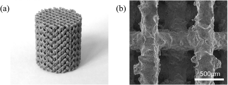
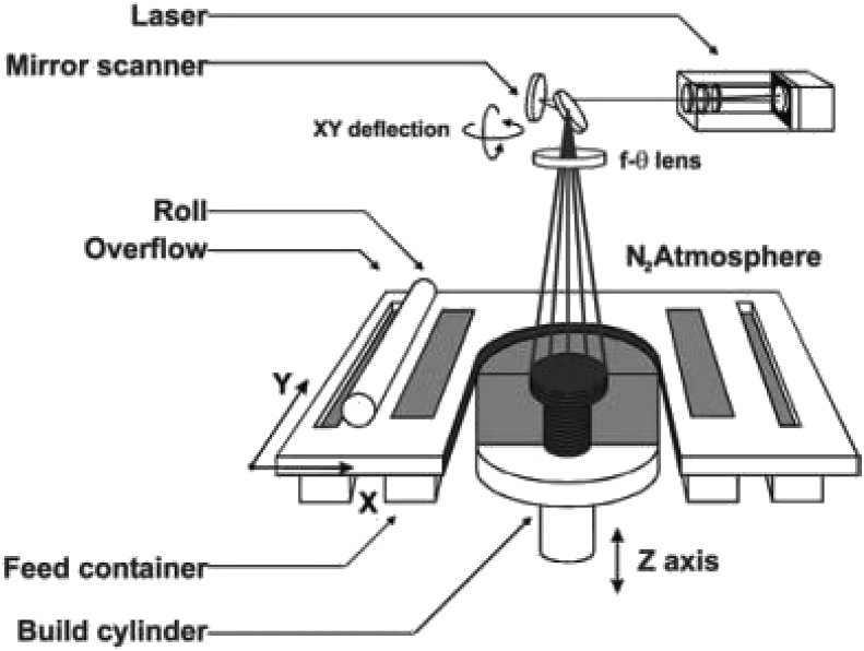
Magnesium alloys are a promising new class of degradable biomaterials that have a similar stiffness to bone, which minimizes the harmful effects of stress shielding. Use of biodegradable magnesium implants eliminates the need for a second surgery for repair or removal. There is a growing interest to capitalize on additive manufacturing's unique design capabilities to advance the frontiers of medicine. However, magnesium alloys are difficult to 3D print due to the high chemical reactivity that poses a combustion risk. Furthermore, the low vaporization temperature of magnesium and common biocompatible alloying elements further increases the difficulty to print fully dense structures that balance strength and corrosion requirements. The purpose of this study is to survey current techniques to 3D print magnesium constructs and provide guidance on best additive practices for these alloys.

Keywords: Additive manufacturing; Implants; Magnesium.


Conflict of interest statement

The authors declare that they have no known competing financial interests or personal relationships that could have appeared to influence the work reported in this paper.

Figures

Image 1
Graphical abstract
Fig. 1
Fig. 1
Schematic representation of (a) normal degradation of a plate/screw construct in one year and (b) premature catastrophic failure due to stress shielding and stress-corrosion cracking.
Fig. 2
Fig. 2
Technologies to slow Mg corrosion: (a) coatings, (b) alloying, and (c) surface treatments.
Fig. 3
Fig. 3
Schematic diagram showing the intersection point between failure of medical implant relative to bone recovery.
Fig. 4
Fig. 4
(a) As-printed WE43 scaffold and (b) surface morphology of as-polished strut [50].
Fig. 5
Fig. 5
CT-scans revealing evolution of corrosion products in a 3D printed WE43 scaffold over 28 days [50].
Fig. 6
Fig. 6
Flouroscopic images of cross-sections of a (a) degradable polymer and (b) and a magnesium rod with in vivo staining of newly formed bone [2,51].
Fig. 7
Fig. 7
Schematic diagram of a PBF System [53].
Fig. 8
Fig. 8
Influence of laser scanning speed on relative density of ZK60 [39].
Fig. 9
Fig. 9
Porosity of structures fabricated at (a) 40.6 J/mm3 produces dense structure and (b) 18.8 J/mm3 produces porous structure [57].
Fig. 10
Fig. 10
Grain size variation of Mg–9%Al powder as a function of laser power and scan speed [59].
Fig. 11
Fig. 11
Relative density obtained by PBF of AZ60 alloy [60].
Fig. 12
Fig. 12
Surface of deposited pure magnesium for thickness of (a) 0.25 mm non-preheat, (b) 0.25 mm preheat, (c) 0.30 mm non-preheat, and (d) 0.30 mm preheat [42].
Fig. 13
Fig. 13
Surface morphology for PBF deposition of pure magnesium with (a) 26 μm and (b) 43 μm powder particle sizes [65].
Fig. 14
Fig. 14
Defects in PBF of (a) Mg–1Zn and (b) Mg–2Zn. Modified from Ref. [66].
Fig. 15
Fig. 15
Magnesium phase diagram [37].
Fig. 16
Fig. 16
Mass gain due to oxidation of Mg powder for different rates of heating [67].
Fig. 17
Fig. 17
Surface of deposited magnesium for layer thickness of (a) 0.15 mm non-preheat, (b) 0.15 mm preheat, (c) 0.20 mm non-preheat, (d) 0.20 mm preheat [42].
Fig. 18
Fig. 18
Effect of preheating on roughness of deposition [42].
Fig. 19
Fig. 19
Material deposition for wire arc additive manufacturing [68].
Fig. 20
Fig. 20
Optical micrograph of fabricated material [68].
Fig. 21
Fig. 21
WAAM depositions of AZ31 at (a) 500 Hz, (b) 100 Hz, (c) 10 Hz, (d) 5 Hz, (e) 2 Hz, and (f) 1 Hz [45].
Fig. 22
Fig. 22
Microstructure of depositions at frequencies of (a) 500 Hz, (b) 100 Hz, (c) 10 Hz, (d) 5 Hz, (e) 2 Hz, and (f) 1 Hz [45].
Fig. 23
Fig. 23
Set-up for paste extrusion deposition [46].
Fig. 24
Fig. 24
Schematic of friction stir welding [47].
Fig. 25
Fig. 25
Principle of binder-less jetting: a) solvent deposition, b) Development of capillary bridges among wet particles, c) spreading of next powder layer, d) capillary action forms bridges between particles in new and previous layers, and e) fully developed solid structure is formed after drying and sintering [48].

References

    1. Chen Y., Xu Z., Smith C., Sankar J. Recent advances on the development of magnesium alloys for biodegradable implants. Acta Biomater. 2014;10:4561–4573. - PubMed
    1. Staiger M.P., Pietak A.M., Huadmai J., Dias G. Magnesium and its alloys as orthopedic biomaterials: a review. Biomaterials. 2006;27:1728–1734. - PubMed
    1. Witte F., Hort N., Vogt C., Cohen S., Kainer K.U., Willumeit R., Feyerabend F. Degradable biomaterials based on magnesium corrosion. Curr. Opin. Solid State Mater. Sci. 2008;12:63–72.
    1. Xin Y., Hu T., Chu P.K. In vitro studies of biomedical magnesium alloys in a simulated physiological environment: a review. Acta Biomater. 2011;7:1452–1459. - PubMed
    1. Erdmann N., Bondarenko A., Hewicker-Trautwein M., Angrisani N., Reifenrath J., Lucas A., Meyer-Lindenberg A. Evaluation of the soft tissue biocompatibility of MgCa0.8 and surgical steel 316L in vivo: a comparative study in rabbits. Biomed. Eng. Online. 2010;9:63. - PMC - PubMed

LinkOut - more resources