. 2020 Jan 2;5(1):369-377.
doi: 10.1021/acsomega.9b02800. eCollection 2020 Jan 14.

Insights into the Binding Recognition and Susceptibility of Tofacitinib toward Janus Kinases

Affiliations

Insights into the Binding Recognition and Susceptibility of Tofacitinib toward Janus Kinases

Kamonpan Sanachai et al. ACS Omega. .

Abstract

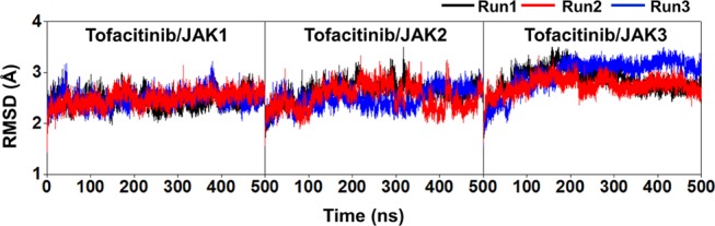
Janus kinases (JAKs) are enzymes involved in signaling pathways that affect hematopoiesis and immune cell functions. JAK1, JAK2, and JAK3 play different roles in numerous diseases of the immune system and have also been considered as potential targets for cancer therapy. In the present study, the susceptibility of the oral JAK inhibitor tofacitinib against these three JAKs was elucidated using the 500-ns molecular dynamics (MD) simulations and free energy calculations based on MM-PB(GB)SA, QM/MM-GBSA (PM3 and SCC-DFTB), and SIE methods. The obtained results revealed that tofacitinib could interact with all JAKs at the ATP-binding site via electrostatic attraction, hydrogen bond formation, and in particular van der Waals interaction. The conserved glutamate and leucine residues (E957 and L959 of JAK1, E930 and L932 of JAK2, and E903 and L905 of JAK3) located in the hinge region stabilized tofacitinib binding through strongly formed hydrogen bonds. Complexation with the incoming tofacitinib led to a closed conformation of the ATP-binding site and a decreased protein fluctuation at the glycine loop of the JAK protein. The binding affinities of tofacitinib/JAKs were ranked in the order of JAK3 > JAK2 ∼ JAK1, which are in line with the reported experimental data.


Conflict of interest statement

The authors declare no competing financial interest.

Figures

Figure 1
Figure 1
(A) Superimposition of JAK1, JAK2, and JAK3 crystal structures (tofacitinib and pyrrole-3-carboxamide represented in gray and blue stick models) in which the highly conserved sequences of the four important parts: catalytic loop, hinge region, glycine-rich loop (G loop), and activation loop (A loop), are shown by a large worm style structure, where their sequence alignment is given in (B). (C) Tofacitinib binding at the active site of JAKs and its chemical structure (D).
Figure 2
Figure 2
All-atom RMSD plot of the tofacitinib/JAK(s) complexes. The data are derived from the three independent simulations with different initial velocities.
Figure 3
Figure 3
(A) Per-residue decomposition free energy (ΔGbindresidue) of the domain of three JAKs for the binding of tofacitinib from three independent simulations and the binding orientation of tofacitinib inside the binding pocket drawn from the MD snapshot. The lowest and highest energies are ranged from dark magenta to yellow, respectively. The electrostatic and van der Waals (vdW) energy contributions are given in (B) using the data derived from the average three independent simulations.
Figure 4
Figure 4
Percentage of hydrogen bond occupation with the two important residues, glutamate, and leucine, in the hinge region of JAKs. The data were derived from the last 150 ns of the three different simulations determined using the criteria between the hydrogen bond donor (HD) and hydrogen acceptor (HA) as follows: (i) ≤3.5 Å for distance and (ii) ≥120° for the angle.
Figure 5
Figure 5
(A) SASA on the residues and (B) number of surrounding atoms within the 3.5 Å sphere of tofacitinib from the three independent simulations.
Figure 6
Figure 6
(A) PCA screen plot of PC modes and (B) the 2D projection of two PC modes, PC1 and PC2, derived from MD trajectories of the JAK3 apo form (left) and the tofacitinib/JAK3 complex (right). (C) PC1 Porcupine plot of the apo form and holo form of JAK3, where the arrow head indicates the direction of motion, while the length indicates the amplitude of motion.

References

    1. Schindler C.; Levy D. E.; Decker T. JAK-STAT signaling: from interferons to cytokines. J. Biol. Chem. 2007, 282, 20059–20063. 10.1074/jbc.R700016200. - DOI - PubMed
    1. Wang Y.; Huang W.; Xin M.; Chen P.; Gui L.; Zhao X.; Tang F.; Wang J.; Liu F. Identification of 4-(2-furanyl)pyrimidin-2-amines as Janus kinase 2 inhibitors. Bioorg. Med. Chem. 2017, 25, 75–83. 10.1016/j.bmc.2016.10.011. - DOI - PubMed
    1. Haan C.; Kreis S.; Margue C.; Behrmann I. Jaks and cytokine receptors - An intimate relationship. Biochem. Pharmacol. 2006, 72, 1538–1546. 10.1016/j.bcp.2006.04.013. - DOI - PubMed
    1. O’Shea J. J.; Plenge R. JAK and STAT signaling molecules in immunoregulation and immune-mediated disease. Immunity 2012, 36, 542–550. 10.1016/j.immuni.2012.03.014. - DOI - PMC - PubMed
    1. Yamaoka K.; Saharinen P.; Pesu M.; Holt V. E. T.; Silvennoinen O.; O’Shea J. J. The Janus kinases (Jaks). Genome Biol. 2004, 5, 25310.1186/gb-2004-5-12-253. - DOI - PMC - PubMed

LinkOut - more resources