. 2020 Apr 6;48(6):2880-2896.
doi: 10.1093/nar/gkaa012.

Dynamic changes in cis-regulatory occupancy by Six1 and its cooperative interactions with distinct cofactors drive lineage-specific gene expression programs during progressive differentiation of the auditory sensory epithelium

Affiliations

Dynamic changes in cis-regulatory occupancy by Six1 and its cooperative interactions with distinct cofactors drive lineage-specific gene expression programs during progressive differentiation of the auditory sensory epithelium

Jun Li et al. Nucleic Acids Res. .

Abstract

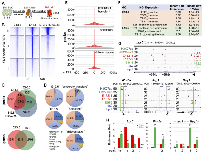
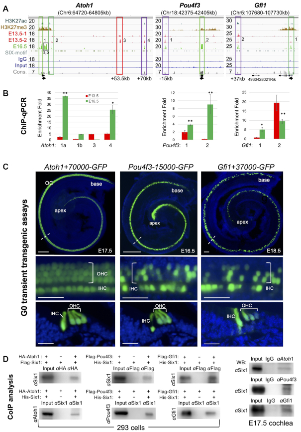
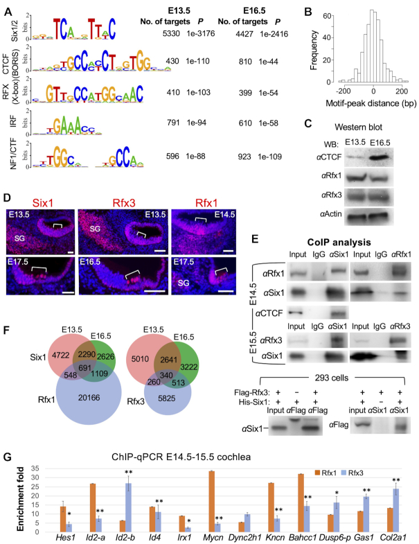
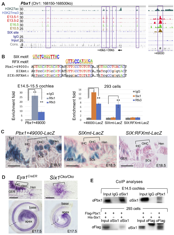
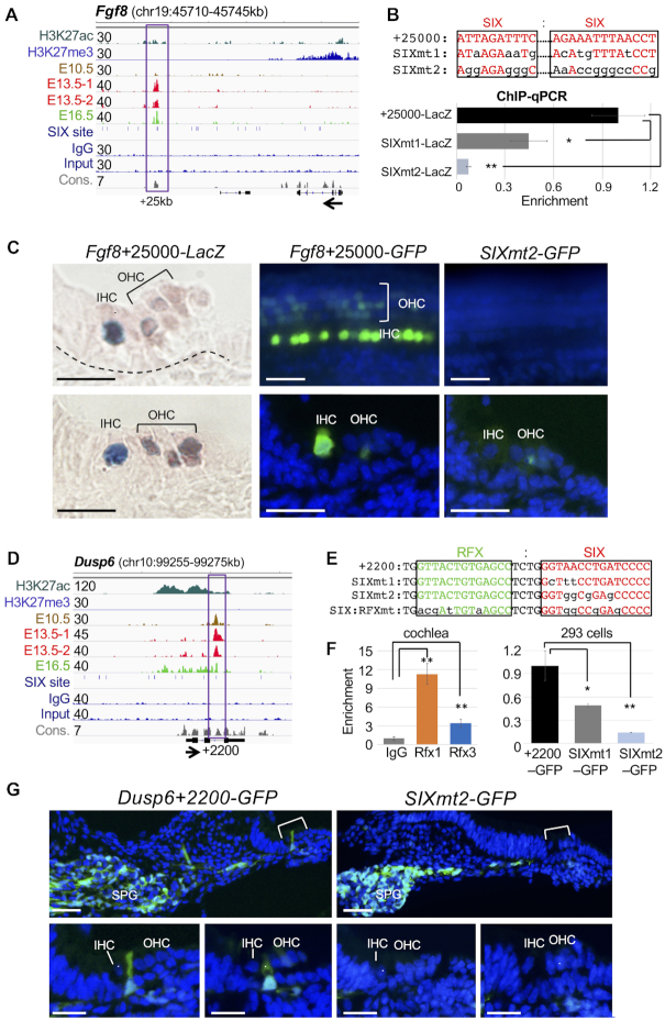
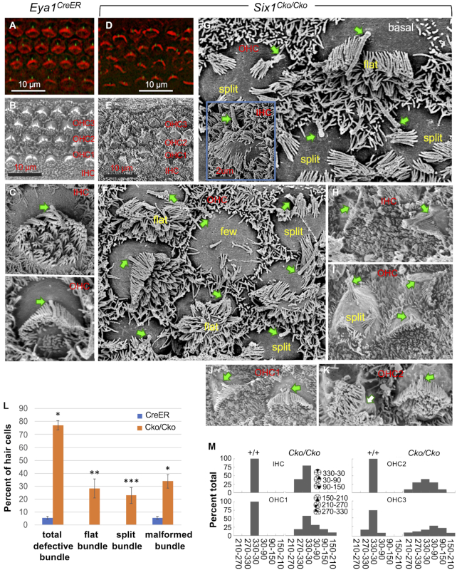
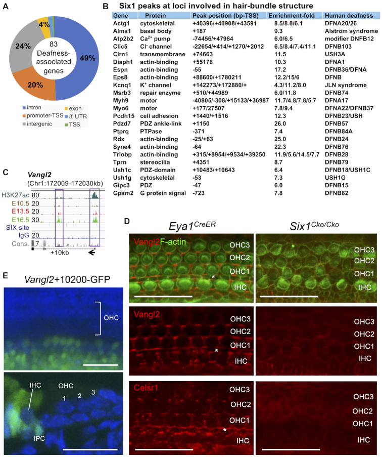
The transcription factor Six1 is essential for induction of sensory cell fate and formation of auditory sensory epithelium, but how it activates gene expression programs to generate distinct cell-types remains unknown. Here, we perform genome-wide characterization of Six1 binding at different stages of auditory sensory epithelium development and find that Six1-binding to cis-regulatory elements changes dramatically at cell-state transitions. Intriguingly, Six1 pre-occupies enhancers of cell-type-specific regulators and effectors before their expression. We demonstrate in-vivo cell-type-specific activity of Six1-bound novel enhancers of Pbx1, Fgf8, Dusp6, Vangl2, the hair-cell master regulator Atoh1 and a cascade of Atoh1's downstream factors, including Pou4f3 and Gfi1. A subset of Six1-bound sites carry consensus-sequences for its downstream factors, including Atoh1, Gfi1, Pou4f3, Gata3 and Pbx1, all of which physically interact with Six1. Motif analysis identifies RFX/X-box as one of the most significantly enriched motifs in Six1-bound sites, and we demonstrate that Six1-RFX proteins cooperatively regulate gene expression through binding to SIX:RFX-motifs. Six1 targets a wide range of hair-bundle regulators and late Six1 deletion disrupts hair-bundle polarity. This study provides a mechanistic understanding of how Six1 cooperates with distinct cofactors in feedforward loops to control lineage-specific gene expression programs during progressive differentiation of the auditory sensory epithelium.


Figures

Figure 1.
Figure 1.
Six1 binding is dynamic across the transition of prosensory precursors to hair-bundle development. (A) Schematic drawing of time course of cochleae for ChIP-seq analysis. (B) Clustered heatmaps of Six1 and H3K27ac within a −5 kb/+ 5 kb window centered on all 14 867 Six1 peaks in E13.5 and E16.5 cochlea and overlapping with the deposition of H3K27ac in E13.5 cochlear epithelium. Peaks were called with the MACS program with a P value cut-off of 1e–5. (C) A Venn diagram indicating overlap of Six1-binding sites of E13.5 and E16.5 and of H3K27ac-deposition at E13.5. Lower panel indicating overlap of Six1-associated genes between E13.5 and E16.5. (D) Genomic distribution of Six1-enriched regions. (E) Distribution of Six1 peaks relative to TSSs. (F) GREAT analysis showing association of Six1-enriched regions with terms in the mouse gene expression information (MGI) database. (G) Genome browser visualization of Six1 peaks at Lgr5, Wnt5a, Jag1 and Hey1. y-Axis numerical values in each track indicate track height scaling in read depth. The direction of transcription is shown by the arrow beginning at the TSS. (H) ChIP-qPCR analysis of the boxed peaks in (G) confirming stage-related changes in Six1-binding. IPs and mock IPs (IgGs) were normalized to inputs and the enrichment of mock IP was considered 1-fold (not shown). *P < 0.05, **P < 0.01.
Figure 2.
Figure 2.
Six1 occupies enhancer repertoire to induce sequential activation of Atoh1, Pou4f3 and Gfi1 that then engage in protein complexes. (A) Genome browser visualization of Six1 peaks at Atoh1, Pou4f3 and Gfi1. Note that the proximal peak-2 of Pou4f3 is in exon. y-Axis numerical values in each track indicate track height scaling in read depth. Sequence conservation (cons.) is indicated. The arrow at the TSS points to the direction of transcription. (B) ChIP-qPCR analysis of the boxed peaks in (A). IPs and mock IPs were normalized to inputs and the enrichment of mock IP was considered 1-fold (not shown). *P < 0.05, **P < 0.01. (C) Transient (G0) transgenic analysis of a 552-bp Six1-bound Atoh1+70000 (in 5/5 transgenic lines), Pou4f3-15000 (in 3/3 transgenic lines) or Gfi1+37000 (in 3/3 transgenic lines) driving GFP reporter showing the HC-restricted activity of these distal enhancers. Top panels, whole-cochlea images; middle panels, higher magnification of the areas indicated by dashed lines; lower panels, images of cochlear sections showing GFP+ hair cells in the organ of Corti. Scale bars: 100 μm for top panels and 30 μm for middle and bottom panels. (D) Co-immunoprecipitation (coIP) analysis of nuclear extracts from E17.5 cochleae or 293 cells cotransfected with indicated plasmids. Antibodies used for IP or for western detection are indicated. Anti-HA or -Flag was used for immunoprecipitating HA-Atoh1 or Flag-Pou4f3/Flag-Gfi1 fusion protein.
Figure 3.
Figure 3.
Motif analysis of Six1 peaks and physical interaction with RFX and CTCF. (A) Sequence logos of the most enriched top 5 motifs from Homer Known motif analysis. (B) Localization of Six1/2-motif within the peak sequence. (C) Western blot analysis of whole-cochlea extracts with indicated antibodies. (D) Immunostaining on cochlear sections showing Six1, Rfx3 and Rfx1 expression in the organ of Corti (brackets). Scale bars: 45 μm. (E) CoIP analysis using nuclear extracts from E14.5–15.5 cochleae or 293 cells cotransfected with Flag-Rfx3 and His-Six1 plasmids (Rfx1 expression plasmid is unavailable). Anti-Flag is used for precipitating and detecting Flag-Rfx3 fusion protein expressed in 293 cells. (F) Venn diagram indicating overlap of Six1-binding sites with Rfx1- or Rfx3-bound sites in the mouse Min6 cells (42). (G) ChIP-qPCR of 12 selected common peaks for Six1 and Rfx1/3 confirms binding of Rfx1 and/or Rfx3 to these regions. IPs and mock IPs were normalized to inputs and the enrichment of mock IP was considered 1-fold (not shown). *P < 0.05, **P < 0.01.
Figure 4.
Figure 4.
Dependence of a distal Pbx1 enhancer activity on co-binding of Six1 and Rfx1/3 through SIX:RFX motifs. (A) Genomic browser visualization of multiple Six1-bound regions at the Pbx1 and enlarged view of the intronic peak at ∼49-kb downstream from the TSS. (B) Pbx1+49 000 contains two SIX-motifs separated by a RFX-motif. ChIP-qPCR using chromatin from E14.5–E15.5 cochlear epithelium shows strong binding with Rfx1 and relative weaker binding with Rfx3. A 510-bp fragment of Pbx1+49000 driving LacZ reporter transgene and two mutant reporter transgenes were generated by introducing mutations into the predicted SIX-motifs or both SIX:RFX-motifs in combination. These reporters were assessed by ChIP-qPCR using chromatin prepared from 293 cells cotransfected with His-Six1 expression plasmid and reporter Pbx1+49 000, Pbx1+4900SIXmt or Pbx1+49000SIX:RFXmt. These mutations abolished Six1 or Rfx3 binding. Transfection was repeated three times and qPCR was performed in triplicates for each independent experiment. Input was used for normalization (see Materials and Methods) and the enrichment of mock IP was considered 1-fold. *P < 0.05, **P < 0.01. (C) G0 transgenic analysis of LacZ transgene driven by a 510-bp of Pbx1+49 000 showing activity in the sensory epithelium and flanking GER and Hensen's (Hen) ells (n = 7/7 transgenic embryos), while Pbx1+49 000SIXmt (n = 3/3 transgenic embryos) or Pbx1+49000SIX:RFXmt (n = 8/8 transgenic embryos). Brackets indicate the organ of Corti. (D) In situ hybridization of E17.5 Eya1CreER or Six1Cko/Cko (Eya1CreER;Six1fl/fl, tamoxifen given at E12.5). Top panels, sections of whole-cochlea shown in bottom panels indicated by dashed lines. Brackets indicate the organ of Corti. (B) Genomic browser visualization of multiple Six1-bound regions at the Pbx1 and enlarged view of the intronic peak at ∼49-kb downstream from the TSS. (E) CoIP analysis of nuclear extracts from E14.5 cochleae or 293 cells transfected with Flag-Pbx1/His-Six1. Other abb.: GER, greater epithelial ridge; IHC, inner hair cell; Hen, Hensen's cells; OHC, outer hair cell; SCs, supporting cells. GER, greater epithelial ridge. Scale bars: 30 μm.
Figure 5.
Figure 5.
Six1 directly regulates cell-type-specific genes/effectors of the Fgf signaling. (A) Genome browser visualization of Six1 peak ∼25 kb downstream from the TSS of Fgf8. Note: no enrichment of Six1 in E10.5 otocyst. (B) Two mutant enhancers SIXmt1 and SIXmt2 driving LacZ or GFP reporter were generated respectively by introducing different mutations into the Six1/2-motifs and assessed by ChIP-qPCR using chromatin prepared from 293 cells cotransfected with His-Six1 expression plasmid and reporter Fgf8+25000, Fgf8+25000SIXmt1 or Fgf8+25000SIXmt2. **P < 0.01 and ***P < 0.001. (C) G0 transgenic analysis of LacZ or GFP transgene driven by Fgf8+25000 or Fgf8+25000SIXmt2. Left panels, images of two cochlear sections showing strong activity in inner hair cells and very weak in outer hair cells (n = 9/9 transgenic embryos), which was disrupted by SIXmt2 (n = 7/7 transgenic embryos). Middle and left upper panels, images of whole-cochlea showing the organ of Corti; middle and right bottom panels, images of sections of upper panels. (D) Genome browser visualization of intronic Six1 peak ∼2200 bp downstream of the TSS of Dusp6, which contains RFX-motif next to the SIX motif with high conservation (con.). (E) Sequences indicate distinct mutations introduced into the SIX motif or both SIX:RFX motifs. (F) ChIP-qPCR analysis of chromatin prepared from E14.5–E15.5 cochleae showing stronger enrichment for Rfx1 and relatively weaker enrichment for Rfx3 at Dusp6+2200, while ChIP-qPCR of chromatin prepared from 293 cells cotransfected with His-Six1 expression plasmid and reporter driven by Dusp6+2200, Dusp6+2200SIXmt1 or SIXmt2. **P < 0.01. (G) G0 transgenic analysis of the Dusp6+2200 or Dusp6+2200SIXmt2. Images of cochlear sections showing GFP transgene expression in spiral ganglion (SPG) and inner-pillar-cells in the sensory epithelium at E18.5 (n = 8/8 transgenic embryos). SIXmt2 disrupted enhance activity specifically in the inner-pillar-cells in the sensory epithelium (n = 5/6 transgenic embryos). Scale bars: 20 μm.
Figure 6.
Figure 6.
Late conditional inactivation of Six1 in differentiating hair cells results in both structural polarity and PCP defects of hair-bundles. (AD) Hair-bindle structure and orientation at P0 was visualized by F-actin (red) and anti-acetylated tubulin (green, for kinocilium). (BK) SEM images from basal or middle cochlear duct showing surface views of the organ of Corti in Eya1CreER control and Six1Cko/Cko mutant. Arrows indicate kinocilium (C–J), which is absent in panel K. (L) Percentage of hair cells from the basal region of the cochlea of control (n = 645; 3 embryos) and Rac1 and CKO (n = 622; 3 embryos) with the indicated stereocilia. *P < 0.05, ** P < 0.01, *** P < 0.001. (M) Graphs showing distribution of hair cell orientation from wild-type and Six1 CKO mutant animals. The orientation of hair cells was determined by measuring the angle formed between the medial-to-lateral axis of the cochlea and the line bisecting the stereociliary bundle from the center of the hair cell to the vertex of the hair-bundle.
Figure 7.
Figure 7.
Six1 occupies key loci that are responsible for different forms of human deafness syndromes and for development of primary hair-bundle and orientation. (A) Genomic distribution of the 186 Six1 peaks that are mapped to 83 of the 152 deafness-associated genes collected in the Deafness Variation Database using UCSC liftOver. (B). List of Six1-enrichments and peak locations in the mouse genome for 23 homologs of the 83 deafness-causing genes. For a complete list of peak locations in the 83 human genes, see Supplementary file 4. (C). Genome browser visualization of Six1 peak at proximal-promoter and intronic regions of Vangl2. Six1-binding to both regions increases by E16.5. (D). Surface views of the organ of Corti stained with both anti-Vangl2 and F-actin or anti-Celsr1 alone. Asterisk indicates the position of pillar-cells between inner and outer hair cells. (E). G0 transgenic analysis of a 552-bp of the intronic Vangl2+10200 showing enhancer activity in inner hair cell and surrounding SCs on the medial region of the sensory epithelium as well as in the GER (in 3/4 transgenic embryos/lines). IPC, inner-pillar-cell. Scale bars: 30 μm.

References

    1. Kumar J.P. The sine oculis homeobox (SIX) family of transcription factors as regulators of development and disease. Cell. Mol. Life Sci. 2009; 66:565–583. - PMC - PubMed
    1. Oliver G., Wehr R., Jenkins N. A., Copeland N. G., Cheyette B. N., Hartenstein V., Zipursky S. L., Gruss P.. Homeobox genes and connective tissue patterning. Development. 1995; 121:693–705. - PubMed
    1. Xu P.X., Zheng W., Huang L., Maire P., Laclef C., Silvius D.. Six1 is required for the early organogenesis of mammalian kidney. Development. 2003; 130:3085–3094. - PMC - PubMed
    1. Nie X., Sun J., Gordon R.E., Cai C.L., Xu P.X.. SIX1 acts synergistically with TBX18 in mediating ureteral smooth muscle formation. Development. 2010; 137:755–765. - PMC - PubMed
    1. Kingsbury T.J., Kim M., Civin C.I.. Regulation of cancer stem cell properties by SIX1, a member of the PAX-SIX-EYA-DACH network. Adv. Cancer Res. 2019; 141:1–42. - PubMed

Publication types

MeSH terms