. 2020 Apr;9(8):e1901214.
doi: 10.1002/adhm.201901214. Epub 2020 Jan 20.

Relaxation of Extracellular Matrix Forces Directs Crypt Formation and Architecture in Intestinal Organoids

Affiliations

Relaxation of Extracellular Matrix Forces Directs Crypt Formation and Architecture in Intestinal Organoids

Ella A Hushka et al. Adv Healthc Mater. 2020 Apr.

Abstract

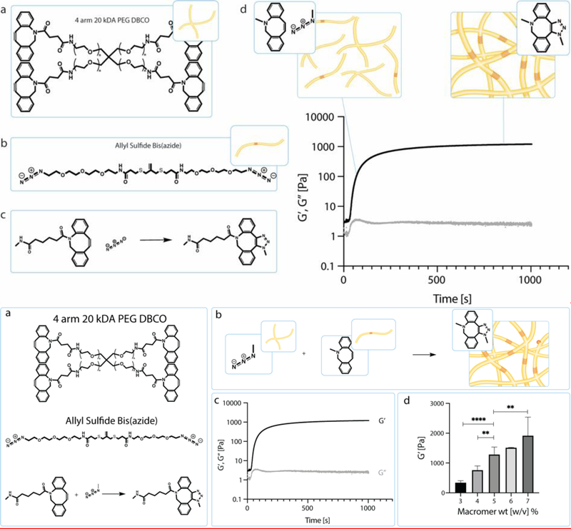
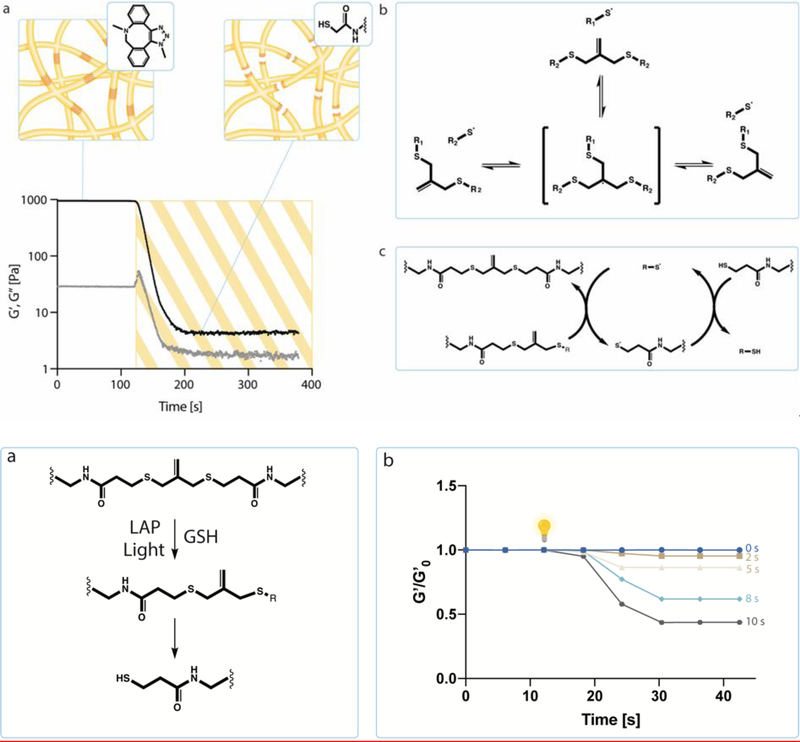
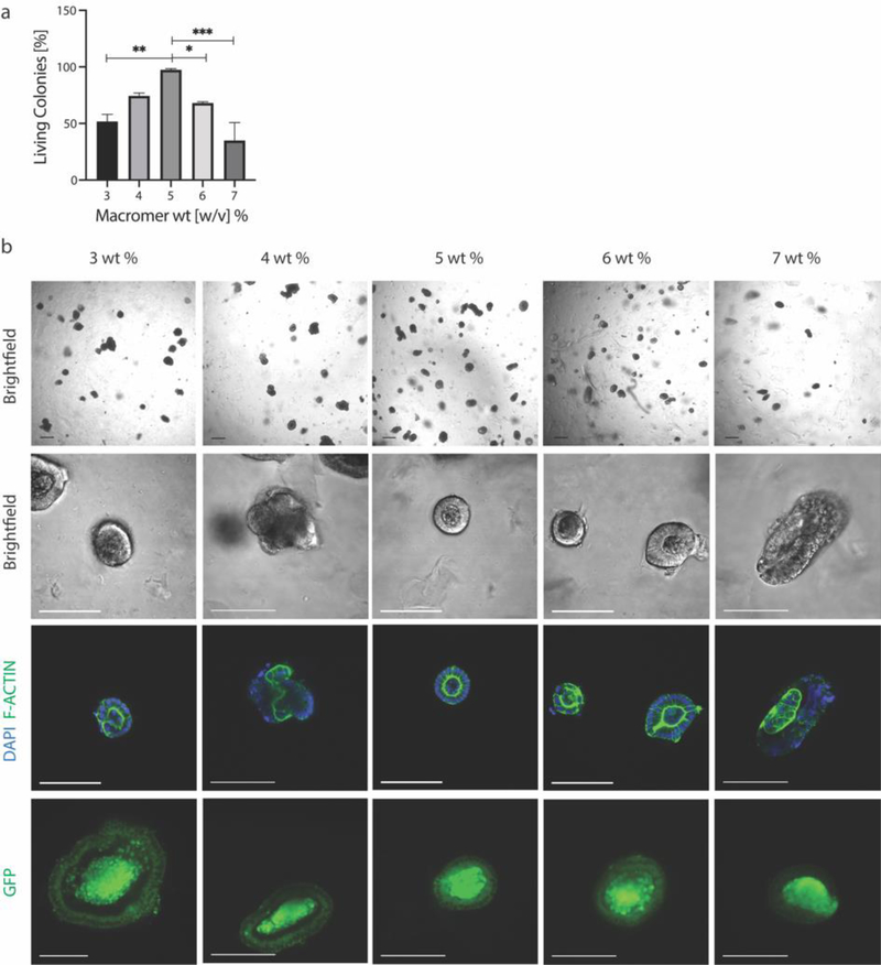
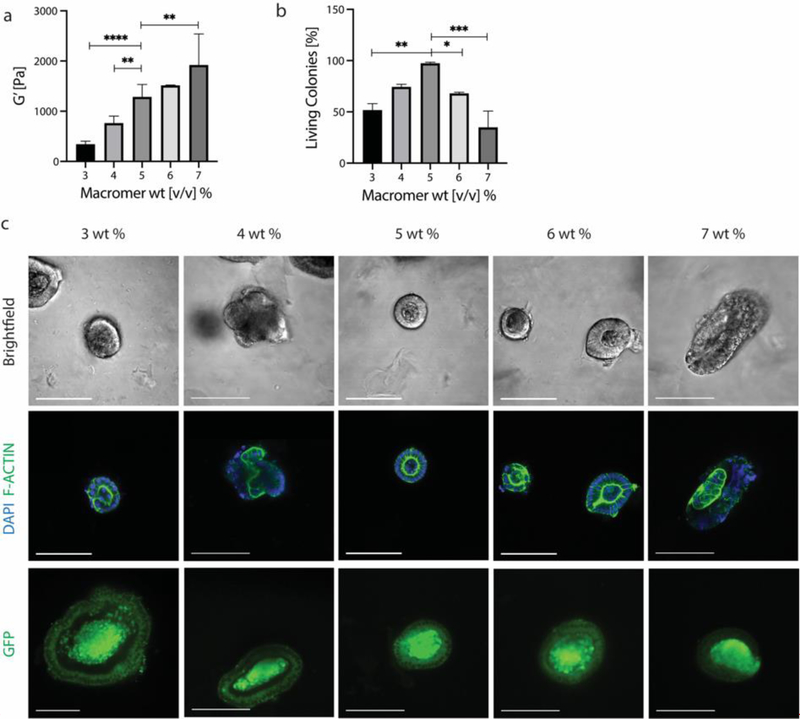
Intestinal organoid protocols rely on the use of extracellular scaffolds, typically Matrigel, and upon switching from growth to differentiation promoting media, a symmetry breaking event takes place. During this stage, the first bud like structures analogous to crypts protrude from the central body and differentiation ensues. While organoids provide unparalleled architectural and functional complexity, this sophistication is also responsible for the high variability and lack of reproducibility of uniform crypt-villus structures. If function follows form in organoids, such structural variability carries potential limitations for translational applications (e.g., drug screening). Consequently, there is interest in developing synthetic biomaterials to direct organoid growth and differentiation. It has been hypothesized that synthetic scaffold softening is necessary for crypt development, and these mechanical requirements raise the question, what compressive forces and subsequent relaxation are necessary for organoid maturation? To that end, allyl sulfide hydrogels are employed as a synthetic extracellular matrix mimic, but with photocleavable bonds that temporally regulate the material's bulk modulus. By varying the extent of matrix softening, it is demonstrated that crypt formation, size, and number per colony are functions of matrix softening. An understanding of the mechanical dependence of crypt architecture is necessary to instruct homogenous, reproducible organoids for clinical applications.

Keywords: adaptable materials; hydrogels; intestinal organoids; mechanosensing; photodegradation.


Figures

Figure 1.
Figure 1.
a) Schematic of the chemical structure of 4 arm 20 kDa PEG DBCO and the chemical structure of allyl sulfide bis(azide). Upon mixing the PEG DBCO with the allyl sulfide bis(azide), the strained octyne will react with the azide group through a strain-promoted azide alkyne cycloaddition (SPAAC). b) Through this SPAAC formation, a hydrogel network is formed. The red cross hatch region of the network is the photodegradable region. c) Shear storage modulus (black) and loss modulus (gray) were measured over time as the network polymerizes. Polymerization is rapid and occurs in under 5 min. d) Hydrogel moduli increase with increasing macromer wt%. Data is represented as means +/− standard deviation. An n of 3 was used and analyzed using a one-way Anova with multiple comparisons. **p<0.01, ****p<0.0001
Figure 2.
Figure 2.
a) Hydrogel network degradation initiated by free radicals. Allyl sulfide bis(azide) reacts with thiyl radicals to form a radical intermediate that undergoes beta scission. This results in the fragmentation of crosslinks and the regeneration of the thiyl radical and allyl sulfide bis(azide). A soluble thiol species (glutathione) exchanges with the allyl sulfide bis(azide), therefore producing a cleaved crosslink and a network tethered thiyl radical. The released network thiyl radical undergoes chain transfer to a soluble thiol species, which can initiate further exchanges, creating an amplified degradation process. b) Hydrogels that were swollen with glutathione and LAP were irradiated with 365 nm light, creating a range of soft conditions that varied from around 500 Pa to 1.3 kPa.
Figure 3.
Figure 3.
a) ISC colony survival is dependent on macromer wt%, and therefore modulus, with the highest viability seen at 5 wt% and 1.3 kPa. b) ISC colonies were marked as viable if they maintained stemness, as marked by GFP expression, and were polarized, as visualized by F-Actin staining. Scale bar 100 μm. Data is represented as means +/− standard deviation. At least three hydrogels were analyzed per condition and data was analyzed using a one-way Anova with multiple comparisons. *p<0.05, **p<0.01, ***p<0.001
Figure 3.
Figure 3.
a) ISC colony survival is dependent on macromer wt%, and therefore modulus, with the highest viability seen at 5 wt% and 1.3 kPa. b) ISC colonies were marked as viable if they maintained stemness, as marked by GFP expression, and were polarized, as visualized by F-Actin staining. Scale bar 100 μm. Data is represented as means +/− standard deviation. At least three hydrogels were analyzed per condition and data was analyzed using a one-way Anova with multiple comparisons. *p<0.05, **p<0.01, ***p<0.001
Figure 4.
Figure 4.
a) When encapsulated in hydrogels that were subsequently softened, colony viability was maintained and unaffected by irradiation for less than 10 seconds of exposure. b) To study crypt formation as a function of hydrogel moduli, ISC colonies were released from Matrigel and encapsulated in photodegradable hydrogels. After 24 hours, hydrogels were swollen with LAP and Glutathione in Flurobrite media for 30 minutes and then irradiated with 365 nm light. After irradiation, colonies were switched to differentiation media. 48 hours after softening, colonies were fixed and immunostained. c) Cell laden hydrogels were softened for 0, 2, 5, 8 or 10 seconds and crypt forming efficiency was quantified as the percentage of living colonies that formed crypts. d) Resulting crypts were measured by quantifying the distance from the main colony body to the tip of the crypt. e) Crypt length was quantified for all softening conditions and showed moduli dependence, f) as did the number of crypts that formed per colony. g) Crypts were marked by the presence of lysozyme producing Paneth cells. Scale bar 100 μm. Data is represented as means +/− standard deviation. At least three hydrogels were analyzed per condition and data was compared using a one-way Anova with multiple comparisons. *p<0.05, **p<0.01, ***p<0.001, ****p<0.0001
Figure 5.
Figure 5.
Organoids produced in all four soft hydrogel conditions were immunostained for the presence of differentiated cell types commonly found in the native intestine. Specifically, organoids were immunostained for E-cadherin, goblet cells (mucin 2), enterocytes (L-FABP) and enteroendocrine cells (Chromogranin A).

References

    1. Sato T, Vries RG, Snippert HJ, van de Wetering M, Barker N, Stange DE, van Es JH, Abo A, Kujala P, Peters PJ, et al., Nature 2009, 459, 262. - PubMed
    1. Serra D, Mayr U, Boni A, Lukonin I, Rempfler M, Challet Meylan L, Stadler MB, Strnad P, Papasaikas P, Vischi D, et al., Nature 2019, 569, 66. - PMC - PubMed
    1. Kong S, Zhang YH, Zhang W, Biomed Res. Int. 2018, 2018, DOI 10.1155/2018/2819154. - DOI - PMC - PubMed
    1. Barker N, Nat. Rev. Mol. Cell Biol. 2014, 15, 19. - PubMed
    1. Cruz-Acuña R, Quirós M, Farkas AE, Dedhia PH, Huang S, Siuda D, García-Hernández V, Miller AJ, Spence JR, Nusrat A, et al., Nat. Cell Biol. 2017, 19, 1326. - PMC - PubMed

Publication types