. 2020 Jan 20:9:e52651.
doi: 10.7554/eLife.52651.

Altered corticolimbic connectivity reveals sex-specific adolescent outcomes in a rat model of early life adversity

Affiliations

Altered corticolimbic connectivity reveals sex-specific adolescent outcomes in a rat model of early life adversity

Jennifer A Honeycutt et al. Elife. .

Abstract

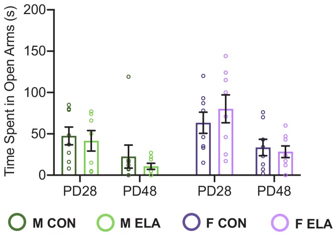
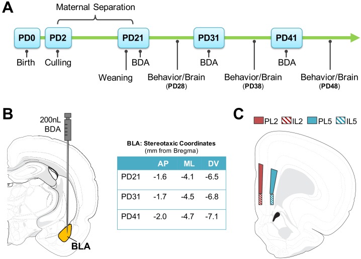
Exposure to early-life adversity (ELA) increases the risk for psychopathologies associated with amygdala-prefrontal cortex (PFC) circuits. While sex differences in vulnerability have been identified with a clear need for individualized intervention strategies, the neurobiological substrates of ELA-attributable differences remain unknown due to a paucity of translational investigations taking both development and sex into account. Male and female rats exposed to maternal separation ELA were analyzed with anterograde tracing from basolateral amygdala (BLA) to PFC to identify sex-specific innervation trajectories through juvenility (PD28) and adolescence (PD38;PD48). Resting-state functional connectivity (rsFC) was assessed longitudinally (PD28;PD48) in a separate cohort. All measures were related to anxiety-like behavior. ELA-exposed rats showed precocial maturation of BLA-PFC innervation, with females affected earlier than males. ELA also disrupted maturation of female rsFC, with enduring relationships between rsFC and anxiety-like behavior. This study is the first providing both anatomical and functional evidence for sex- and experience-dependent corticolimbic development.

Keywords: adolescence; basolateral amygdala; behavior; developmental biology; innervation; maternal separation; neuroscience; prefrontal cortex; rat.

Plain language summary

Having a traumatic childhood increases the risk a person will develop anxiety disorders later in life. Early life adversity affects men and women differently, but scientists do not yet know why. Learning more could help scientists develop better ways to prevent or treat anxiety disorders in men and women who experienced childhood trauma. Anxiety occurs when threat-detecting brain circuits turn on. These circuits begin working in infancy, and during childhood and adolescence, experiences shape the brain to hone the body’s responses to perceived threats. Two areas of the brain that are important hubs for anxiety-related brain circuits include the basolateral amygdala (BLA) and the prefrontal cortex (PFC). Now, Honeycutt et al. show that rats that experience early life adversity develop stronger connections between the BLA and PFC, and these changes occur earlier in female rats. In the experiments, one group of rats was repeatedly separated from their mothers and littermates (an early life trauma), while a second group was not. Honeycutt et al. examined the connections between the BLA and PFC in the two groups at three different time periods during their development: the juvenile stage, early adolescence, and late adolescence. The experiments showed stronger connections between the BLA and PFC begin to appear earlier in juvenile traumatized female rats. But these changes did not appear in their male counterparts until adolescence. Lastly, the rats that developed these strengthened BLA-PFC connections also behaved more anxiously later in life. This may mean that the ideal timing for interventions may be different for males and females. More work is needed to see if these results translate to humans and then to find the best times and methods to help people who experienced childhood trauma.


Conflict of interest statement

JH, CD, SP, MS, XC, PK, MC, HB No competing interests declared, CF has a financial interest in Animal Imaging Research, the company that makes the rat imaging system

Figures

Figure 1.
Figure 1.. BLA volume and BDA anterograde tracer bolus sites.
The average volume of the BLA significantly increased with age (A) as quantified via Cresyl Violet staining of adjacent tissue sections (B). As there were no significant differences as a function of sex or rearing condition on BLA volume estimates at each age group, data were collapsed for each age with a final number of n = 16 per age group. Anatomical coordinates located below Nissl-stained sections (B) indicate the approximate distance (in mm) from Bregma. There were no significant differences in the percent of BLA filled by the BDA anterograde tracer bolus between ages (C), nor was there any significant difference in the amount of bolus located outside of the BLA structure (D). n = 6–9 per group (C; D) before collapsing within each age due to a lack of differences as a function of sex or rearing condition at each age (therefore, the number of data points per age group for analyses in C and D were 24–36). Panel (E) shows representative photomicrographs of BDA bolus across the extent of the BLA for each age examined. Atlas modified from Swanson (2018).
Figure 1—figure supplement 1.
Figure 1—figure supplement 1.. No effects of sex or rearing condition on BLA volume.
There were no significant effects of sex or rearing on basolateral amygdala (BLA)volume as estimated via Cavalieri probe across the rostral-caudal extent of the structure in PD28 (A), PD38 (B), or PD48 (C) rats. n = 4 per group. As such, data presented in panels A, B, and C were collapsed across sex and rearing condition for each age group to determine average BLA volume (n = 16 per age). n.s. (non-significant).
Figure 1—figure supplement 2.
Figure 1—figure supplement 2.. PFC innervation is not driven by percentage of BLA filled or bolus size in included cases.
All included cases for each age (PD28, PD38, and PD48) were collapsed across sex and rearing condition to determine whether the percentage of BLA that was filled, and/or the total bolus size (both within and outside of the BLA structure) was related to total PFC axonal innervation in included cases (n = 29–32 per age). There were no observed relationships between these measures at any age, suggesting no need for correcting innervation based on bolus characteristics measured. Bolus size and location (i.e. % in and outside of BLA structure) were determined via Cavalieri probe volume estimate across the rostral-caudal extent of the biotinylated dextran amine (BDA) anterograde tracer bolus.
Figure 2.
Figure 2.. ELA leads to precocial BLA-PFC axonal innervation earlier in female than in male rats.
BLA-PFC axonal innervation of anterogradely biotinylated dextran amine (BDA)-labeled fibers were visualized with diaminobenzidine (DAB) staining, quantified via unbiased stereology, and are displayed here as an estimate of total axon terminal length (μm) in male and female rats in both PL (A) and IL (B). Top panel (A) indicates PL quantification location, as well as the representative photomicrographs, with dark axonal fibers clearly observed in layer 2 (L2; left side of each photomicrograph) and layer 5 (L5; right side of each photomicrograph). In the PL there was consistent innervation across CON groups at all ages, with transient ELA-induced spikes of innervation occurring at PD28 and PD48 in female rats, and at PD38 in male rats. This pattern of innervation appears to be driven by PL5 axon terminal length, particularly in ELA females. Bottom panel (B) indicates IL quantification location, as well as representative photomicrographs similar to those seen in PL. In the IL, female rats showed a precocial pattern of axonal innervation by PD28 that was comparable to adolescents and older adolescents, while male rats didn’t show an ELA-driven increase in IL innervation until PD38, with these findings appearing to be driven by IL2 axon terminal length, particularly in ELA animals. Data is presented as a function of sex (male, female), age at brain collection (PD28, PD38, PD48), and rearing condition (CON, ELA), with lines inside each group bar indicating group mean, and lines outside of the bars indicating the maximum and minimum observed data points within that group. n = 6–9 per group. The left graph for both PL (A) and IL (B) displays collapsed L2 and L5 data for the entire quantified region, with the middle and right graphs displaying layer-specific data (alpha adjusted to 0.025 significance threshold to account for multiple comparisons in subsequent two-way ANOVAs). Photomicrographs were imaged at 10x magnification. Atlas modified from Swanson (2018). *p<0.05 (for IL/PL Innervation graphs); *p<0.025 (for individual L2 and L5 innervation graphs); **p<0.01; ***p<0.001.
Figure 2—figure supplement 1.
Figure 2—figure supplement 1.. No differences in mounted thickness or probe volume.
Mounted thickness (µm) was assessed across the Z plane for each quantified tissue section for each animal, and this measure was averaged for each animal, and an average tissue thickness for each group within each age was computed (A). There were no significant differences in mounted thickness across groups at any age, as well as no significant differences as a function of age. The average total probe volume (PL+IL) for StereoInvestigator analysis of BLA-PFC axon fiber length for each brain region per animal was also computed for each age (B), and no significant differences were found. n = 6–9 per group. n.s. (non-significant).
Figure 3.
Figure 3.. Anxiety-like behavior following ELA is sex- and age-specific.
Anxiety-like behavior was assessed in the elevated plus maze (EPM) in both CON male (dark green bard) and female (dark purple bars) and ELA male (light green bars) and female (light purple bars) groups. Anxiety-like behavior in the EPM was assessed via time spent in open arms (seconds; A), number of head dips (B), and number of arm crossings (C). ELA animals overall displayed more anxiety-like behavior (measured via less time spent in open arms). Female, but not male, rats showed more anxiety-like behavior, exclusively at PD38 if they had been exposed to ELA (A). Analysis of the number of arm crossings (C) revealed that only males showed a significant effect of age, with PD48 males showing less arm crossings (indicative of increased anxiety) than those at PD28 and PD38. Each circle indicates the data from a single animal, and data shown is for behavior collected from all subjects, including those that did not meet neuroanatomical inclusion criteria. n = 16-28 per group.*p < 0.05; **p < 0.01; ***p < 0.001".
Figure 4.
Figure 4.. More BLA-derived axonal innervation into the PFC is correlated with increased anxiety-like behavior in a sex- and age-dependent manner.
Results of linear regression analyses to determine the relationship between BLA-PFC innervation (measured as an estimate of BDA-labeled axon terminal length) and performance in the EPM (time spent in open arms (seconds) as an index of anxiety-like behavior). At PD28, there were no significant correlations between axon terminal length in the PL and time spent in open arms of the EPM (A) in either males or females, regardless of rearing condition. However, females, but not males, showed a significant correlation between IL innervation and time spent in open arms at PD28 (B), suggesting that more axonal innervation of the IL was related to increased anxiety-like behavior (via decreased time spent in open arms). Conversely, at PD38, females no longer show behavior correlated with innervation in either PL (C) or IL (D), whereas males generally show that increased axonal innervation is correlated with increased anxiety in both of these regions (C; D). Regression lines are reflective of the overall relationship across both groups (CON and ELA), and individual subjects from anatomically included cases are signified by either solid circles (CON) or open circles (ELA). Bold lines/regression statements indicate a significant correlation. n = 6–9 per group. *p<0.05; **p<0.01.
Figure 5.
Figure 5.. Effects of ELA on BLA-PFC rsFC are sex-specific and endure in females.
Resting state functional connectivity (rsFC) was assessed in a longitudinal manner across development in male and female rats with a history of either CON or ELA rearing with the basolateral amygdala (BLA) as the seeded region (A). The rsFC between the BLA and the mPFC – specifically the prelimbic (PL) and infralimbic (IL) cortices (B) – was assessed. (C) Shows results from comparative analyses comparing CON and ELA groups at each age point (PD28 or PD48) in both male and female groups. Colored regions correspond to computed Z values and indicate specific regions within either the PL or IL that met criteria for significance with a minimum cluster size of 30 voxels. Generally, male rats show no group effects of ELA, with the exception of a finding of decreased BLA-PL rsFC in ELA compared to CON groups at PD28. Female rats showed no effect of ELA at PD28 but show striking differences in rsFC in both the PL and IL at PD48 (C). Effect size (Cohen’s D) maps for corresponding sections are illustrated in Figure 5—figure supplement 1, and a full collection of maps are available on Dryad https://doi.org/10.5061/dryad.jdfn2z371. Female, but not male, rats exposed to ELA showed a lack of typical maturation of the IL, evidenced by significantly reduced ΔrsFC from PD28 to PD48 compared to CON females (D). Conversely, there were no group changes in ΔrsFC observed within the PL (E). n = 7–8 per group. *p<0.05.
Figure 5—figure supplement 1.
Figure 5—figure supplement 1.. Effect size maps for cortical sections comparing CON to ELS in males and females at PD28 or PD48.
Effect sizes in the regions highlighted in Figure 5 range from 1.4 to 2.8. Full set of effect size maps generated are available on Dryad https://datadryad.org/stash/share/5u-rMFOwFrQvAHLThMjzn-rTA7Ne3HDELZbvjfTR0Ks.
Figure 6.
Figure 6.. ELA significantly impacts the relationship between BLA-IL rsFC and anxiety-like behavior only in females; with PD28 rsFC predictive of PD48 behavior.
Results of linear regression analyses to determine the relationship between BLA-IL rsFC correlation coefficients and performance in the EPM (time spent in open arms (seconds) as an index of anxiety-like behavior) are shown for PD28 in (A). Only females with a history of ELA showed a significant correlation, with higher rsFC correlation coefficients correlating with more time spent in the open arms of the EPM. As this data was conducted over development in a within-subjects manner, we could further explore predictive relationships between these variables. In line with other rsFC data described here, only female rats showed an overarching predictive effect of early/juvenile (PD28) rsFC correlation coefficients on later (PD48) behavior (B). Indeed, females with a higher rsFC correlation coefficients at PD28 exhibited less anxiety-like behavior (as evidenced by increased time spent in open arms). Furthermore, in the IL females showed a relationship between PD48 rsFC and time spent in open arms at PD48 (C). Individual data points for each animal can be seen on the graphs, with solid circles representing CON cases, and open circles representing ELA cases, with solid regression lines indicative of significant correlations. n = 7-8 per group.*p < 0.05; **p < 0.01".
Figure 6—figure supplement 1.
Figure 6—figure supplement 1.. Similar to findings in Figure 3 (main article), no effect of rearing on anxiety-like behavior was seen at PD28 or PD48.
Figure 7.
Figure 7.. Timeline and methodology for study 1.
(A) Methodological timeline for Study one indicating maternal separation (for ELA groups), weaning timeline, and surgical/behavioral milestones. Biotinylated Dextran Amine (BDA) microinfusions were performed at PD21, PD31, or PD41. Behavior (elevated plus maze; EPM) was performed at PD28, PD38, or PD48, and was followed by brain collection. (B) Stereotaxic coordinates for surgeries at each developmental time point and anatomical map of basolateral amygdala (BLA) injection site where 200 nL of BDA was infused via Hamilton Neuros syringe. (C) Neuroanatomical map of quantified regions of BLA-PFC axonal innervation. Quantification was conducted via unbiased stereology within the prelimbic (PL) and infralimbic (IL) in layers 2 and 5. Atlas modified from Swanson (2018). AP (anterior-posterior); ML (medial-lateral); DV (dorsal-ventral).

References

    1. Alarcón G, Cservenka A, Rudolph MD, Fair DA, Nagel BJ. Developmental sex differences in resting state functional connectivity of amygdala sub-regions. NeuroImage. 2015;115:235–244. doi: 10.1016/j.neuroimage.2015.04.013. - DOI - PMC - PubMed
    1. Andersen SL. Trajectories of brain development: point of vulnerability or window of opportunity? Neuroscience & Biobehavioral Reviews. 2003;27:3–18. doi: 10.1016/S0149-7634(03)00005-8. - DOI - PubMed
    1. Andersen SL, Teicher MH. Stress, sensitive periods and maturational events in adolescent depression. Trends in Neurosciences. 2008;31:183–191. doi: 10.1016/j.tins.2008.01.004. - DOI - PubMed
    1. Arruda-Carvalho M, Wu WC, Cummings KA, Clem RL. Optogenetic examination of Prefrontal-Amygdala synaptic development. The Journal of Neuroscience. 2017;37:2976–2985. doi: 10.1523/JNEUROSCI.3097-16.2017. - DOI - PMC - PubMed
    1. Arruda-Carvalho M, Clem RL. Pathway-selective adjustment of prefrontal-amygdala transmission during fear encoding. Journal of Neuroscience. 2014;34:15601–15609. doi: 10.1523/JNEUROSCI.2664-14.2014. - DOI - PMC - PubMed

Publication types