. 2020 May:199:105600.
doi: 10.1016/j.jsbmb.2020.105600. Epub 2020 Jan 17.

VDR in salivary gland homeostasis and cancer

Affiliations

VDR in salivary gland homeostasis and cancer

Kara A DeSantis et al. J Steroid Biochem Mol Biol. 2020 May.

Abstract

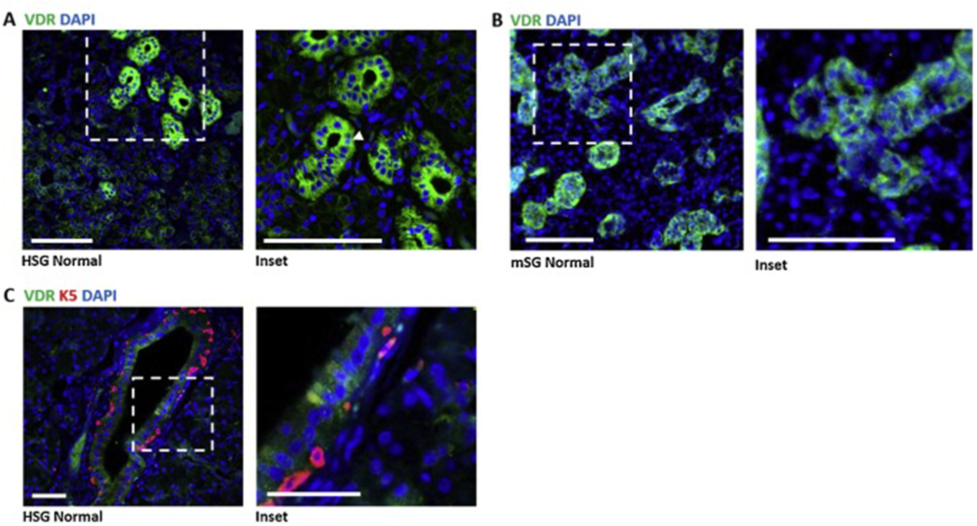
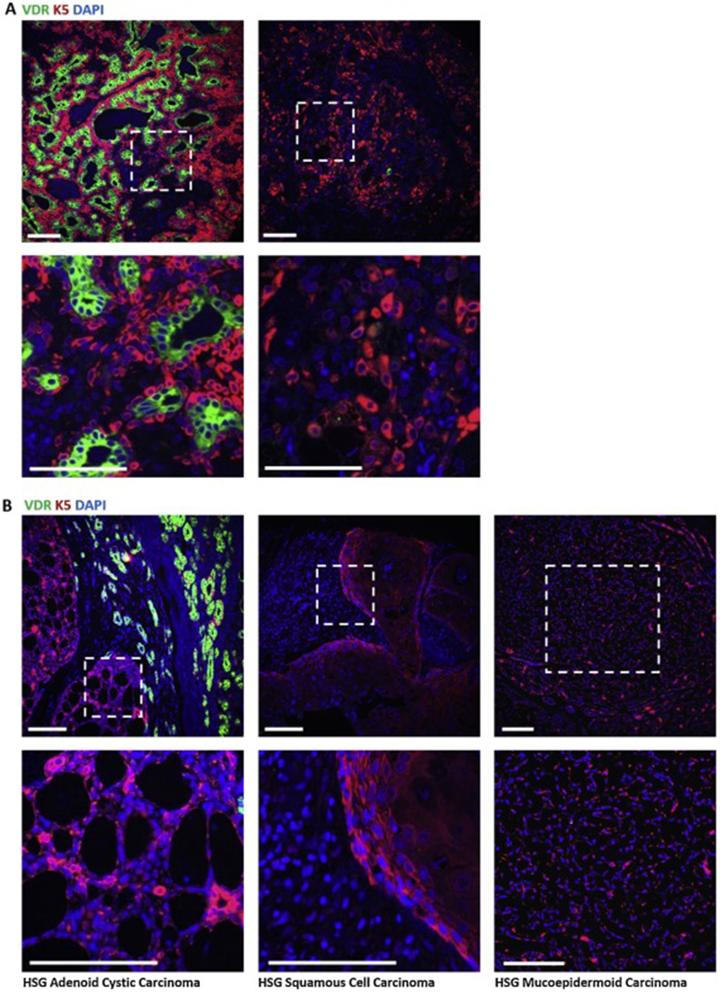
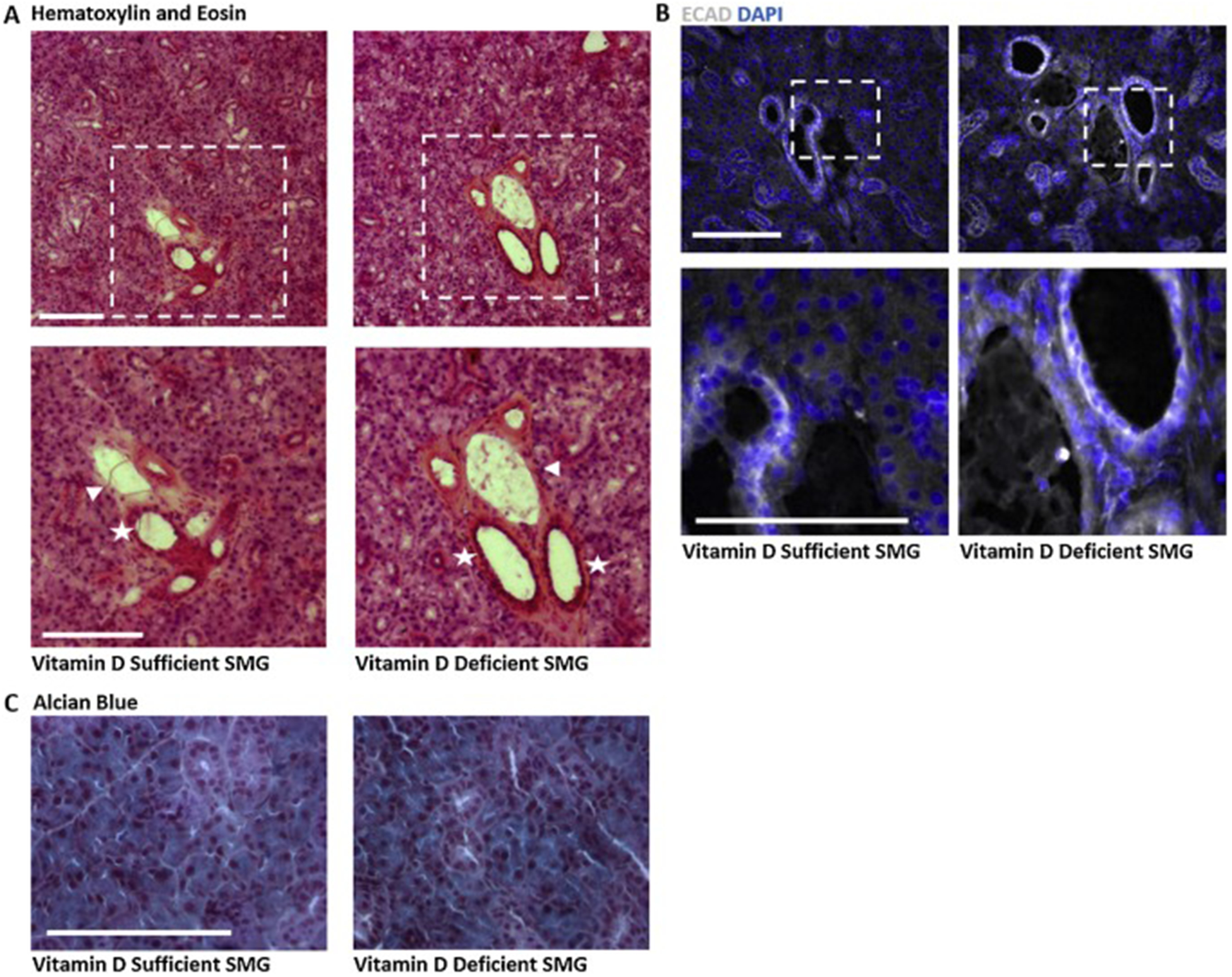
The vitamin D receptor (VDR) and its ligand 1,25(OH)2D3 (1,25D) impact differentiation and exert anti-tumor effects in many tissues, but its role in salivary gland has yet to be defined. Using immunohistochemistry (IHC), we have detected strong VDR expression in murine and human salivary gland ducts. Compared to normal gland, VDR protein expression was retained in differentiated human pleomorphic adenoma (PA) but was undetectable in undifferentiated PA and in carcinomas, suggesting deregulation of VDR during salivary cancer progression. To gain insight into the potential role of VDR in salivary cancer, we assessed the effects of vitamin D in vivo and in vitro. Despite the presence of VDR in salivary gland, chronic dietary vitamin D restriction did not alter morphology of the salivary epithelium in C57/Bl6 mice. The localization of VDR in ductal epithelium prompted us to examine the effects of 1,25D in an established cell line (mSGc) derived from normal murine submandibular gland (SMG). This previously characterized cell line consists of multiple stem, progenitor and differentiated cell types as determined by mutually exclusive cellular expression of basal, ductal and myoepithelial markers. We demonstrated VDR expression and regulation of VDR target genes Vdr and Postn by 1,25D in mSGc, indicating functional ligand-mediated transcriptional activity. The effect of VDR signaling on epithelial differentiation markers was assessed by qPCR and IHC in mSGc cells treated with 1,25D. We found that 1,25D reduced mRNA expression of the basal cell progenitor marker keratin 5 (K5) and increased expression of the differentiated ductal cell marker keratin 7 (K7). Further, we found that 1,25D significantly decreased the number of proliferating cells, including proliferating K5+ cells. Characterization of cell cycle by Muse cytometry indicated 1,25D treatment decreased cells in S, G2, and M phase. The inhibition of K5+ cell proliferation by 1,25D is of particular interest because K5+ basal cells contribute to a wide variety of salivary tumor types. Our studies suggest that 1,25D alters cancer-relevant progenitor and differentiation markers in the salivary gland.

Keywords: Cancer; Salivary gland; VDR; Vitamin D.


Conflict of interest statement

Declaration of Competing Interest The authors declare no financial or non-financial conflicts of interest.

Figures

Fig. 1
Fig. 1. Localization of VDR in human and murine salivary glands.
A) Immunohistochemistry of 4μm thick FFPE sections of normal human salivary gland shows VDR (green) reactivity primarily in ductal cells but also in other cell types. Expression appears mainly cytoplasmic, however nuclear expression marked by DAPI (blue) was also observed (inset, arrowheads, n=2 patients). Scale bars 100μm. B) Immunohistochemistry of 8μm thick FFPE sections of normal adult mouse SMG tissue shows similar localization for VDR (green), staining mainly in the ducts. Scale bars 100μm. C) Normal human parotid salivary gland immunostained for VDR (green) and basal cell marker K5 (n=2 patients). Scale bars 50μm.
Fig. 2
Fig. 2. VDR expression is altered in human salivary cancer.
A) In benign pleomorphic adenoma (PA), VDR staining (green) is intense in areas retaining glandular structure yet is absent in areas lacking structural elements (n=8 patients, see Supp Fig. 2). B) Immunostaining for VDR in three types of metastatic salivary cancer, adenoid cystic carcinoma (AdCC), squamous cell carcinoma (SCC), and mucoepidermoid carcinoma (MEC). AdCC samples show positive staining for VDR outside of cystic areas and loss of VDR signal within cystic areas (n=3 patients). SCC and MEC do not show appreciable VDR signal (n=1 patient each). Scale bars 100μm.
Fig. 3
Fig. 3. Effect of vitamin D depletion on murine submandibular salivary gland.
Submandibular glands were harvested, frozen and sectioned for histological analysis. Images representative of 3–6 mice are shown. A) Hematoxylin and eosin staining of SMGs highlights increased intensity of eosin surrounding large ducts (stars) and accompanying vasculature (arrows) in glands from vitamin D deficient compared to vitamin D sufficient mice. Scale bar 100μm. B) IHC for epithelial cadherin (ECAD) shows no visible change in the epithelial compartment in glands from vitamin D deficient mice compared to controls. Scale bar 50μm. C) Alcian blue staining showing secretory mucins in salivary glands from vitamin D deficient mice as compared to controls. Scale bar 100μm.
Fig. 4
Fig. 4. VDR expression and function in murine salivary gland cell culture.
A) Western blot for VDR in murine submandibular salivary gland cells (mSGc) demonstrates VDR expression in vitro. B,C) RNA isolated from mSGc cultures treated with 100nM 1,25D or vehicle for 24 hrs was used for qPCR analysis with primers specific for mVdr and mPostn. Data were normalized to 18S mRNA and expressed as fold change in 1,25D treated samples relative to control values, shown +/− SD. VDR transcript was not significantly altered in 1,25D treated cells (Student’s unpaired two-tailed t test, p=0.16, mean +/− SD, n=3). Periostin (Postn) was significantly decreased in cells treated with 1,25D. (Student’s unpaired two-tailed t test, p<0.0001, mean +/− SD, n=3). D) Phase contrast images show morphology of mSGc treated with 100nM 1,25D or vehicle for 24, 48 and 72 hrs. Whereas vehicle treated cells were flat and round, 1,25D treated cells appeared raised and formed spindle-like projections (arrows). Scale bar 100μm. E, F) Cells were stained with DAPI nuclear dye and manually counted after 72h treatment with 100nM 1,25D or vehicle. Three representative fields were counted and averaged per sample. 1,25D decreased total cell number by approximately 50% compared to control cultures. (Student’s unpaired two-tailed t test, p=0.008, mean ± SEM, n=3 biological replicates, 3 representative fields counted and averaged for each sample). Scale bars 50μm.
Fig. 5
Fig. 5. 1,25D increases differentiation markers and decreases proliferation of mSMGc in vitro.
A) RNA isolated from mSGc cultures treated with 100nM 1,25D or vehicle for 24 hrs was used for qPCR analysis with primers specific for mCdh1. Data were normalized to 18S mRNA and expressed as fold change in 1,25D treated samples relative to control values, shown +/− SD. Epithelial cadherin (Cdh1) expression increases with 100nM 1,25D after 24 hours exposure in culture. (Student’s unpaired two-tailed t test, p=0.18, mean +/− SD, n= 3 biological replicates). B) Immunocytochemistry for ECAD (green) after 72 hours suggests increased ECAD protein in 1,25D treated samples (n= 3 biological replicates). Scale bar 50μm. C) Quantitative PCR using RNA isolated from mSGc cultures treated with 100nM 1,25D or vehicle for 24 hrs with primers specific for mKrt5 or mKrt7. Data were normalized to 18S mRNA and expressed as fold change in 1,25D treated samples relative to control values, shown +/− SD. Krt5 and Krt7 indicate basal and mature ductal cells respectively, with analysis showing decreased Krt5 expression with increased Krt7 expression (Unweighted ANOVA with Tukey’s HSD, ** p<0.01, +/− SD, n= 3). D) Immunocytochemistry for Ki67 (white) in K5 positive cells (green) shows a global decrease in proliferation in 1,25D treated cells. Scale bar 50μm. E) Cell count of Ki67+ cells shows a significant decrease in percent of Ki67+ cells per area after 72 hours in culture with 100nM 1,25D (Student’s unpaired two-tailed t test, * p=0.02, +/− avg SEM, n= 3 independent biological replicates, with 3 representative fields counted, normalized to total DAPI+ cells and averaged per sample). F) Further characterization of Ki67+ cells co-positive for K5 indicates 1,25D significantly decreases the percentage of Ki67+K5+ cells per field. (Student’s unpaired two-tailed t test, * p=0.02, +/− avg SEM, n= 3 independent biological replicates, with 3 representative fields counted, normalized to total K5+ cells and averaged per sample). Scale bars 50μm. G) Flow cytometry was used to identify the percentage of cells in G1/G0 versus S+M phases of the cell cycle. 1,25Ddecreased the percentage of cells in S+M phases relative to control samples. (Unweighted ANOVA with Tukey’s HSD, ** p≤0.01, +/− avg SEM, n=4 biological replicates with n=2 averaged technical replicates each)

References

    1. van Leeuwen JP, van Driel M, van den Bemd GJ, Pols HA. Vitamin D control of osteoblast function and bone extracellular matrix mineralization. Crit Rev Eukaryot Gene Expr 2001;11:199–226. - PubMed
    1. Bikle DD. Vitamin D and Bone. Curr Osteoporos Rep 2012;10:151–9. 10.1007/s11914-012-0098-z. - DOI - PMC - PubMed
    1. Zinser GM, Suckow M, Welsh J. Vitamin D receptor (VDR) ablation alters carcinogen-induced tumorigenesis in mammary gland, epidermis and lymphoid tissues. J Steroid Biochem Mol Biol 2005;97:153–64. 10.1016/j.jsbmb.2005.06.024. - DOI - PubMed
    1. Krishnan AV, Trump DL, Johnson CS, Feldman D. The Role of Vitamin D in Cancer Prevention and Treatment. Endocrinol Metab Clin North Am 2010;39:401–contents 10.1016/j.ecl.2010.02.011. - DOI - PMC - PubMed
    1. Welsh J. Vitamin D and breast cancer: Past and present. J Steroid Biochem Mol Biol 2018;177:15–20. 10.1016/j.jsbmb.2017.07.025. - DOI - PMC - PubMed

Publication types

MeSH terms